

Clinical classification of age-related macular degeneration
- PMID: 23332590
- PMCID: PMC11551519
- DOI: 10.1016/j.ophtha.2012.10.036
Clinical classification of age-related macular degeneration
Abstract
Objective: To develop a clinical classification system for age-related macular degeneration (AMD).
Design: Evidence-based investigation, using a modified Delphi process.
Participants: Twenty-six AMD experts, 1 neuro-ophthalmologist, 2 committee chairmen, and 1 methodologist.
Methods: Each committee member completed an online assessment of statements summarizing current AMD classification criteria, indicating agreement or disagreement with each statement on a 9-step scale. The group met, reviewed the survey results, discussed the important components of a clinical classification system, and defined new data analyses needed to refine a classification system. After the meeting, additional data analyses from large studies were provided to the committee to provide risk estimates related to the presence of various AMD lesions.
Main outcome measures: Delphi review of the 9-item set of statements resulting from the meeting.
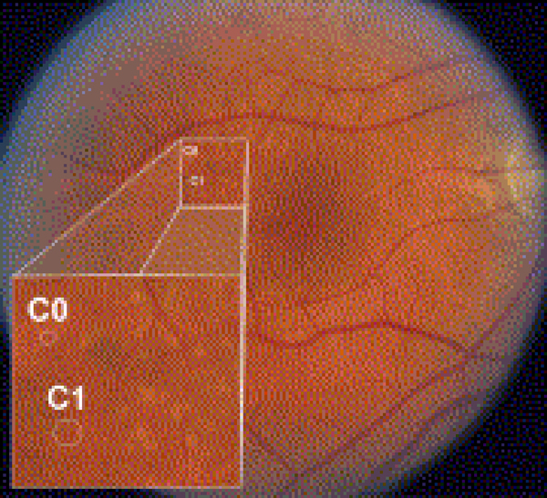
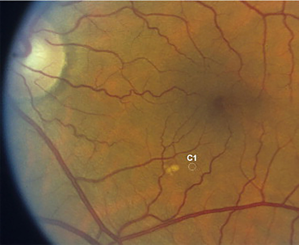
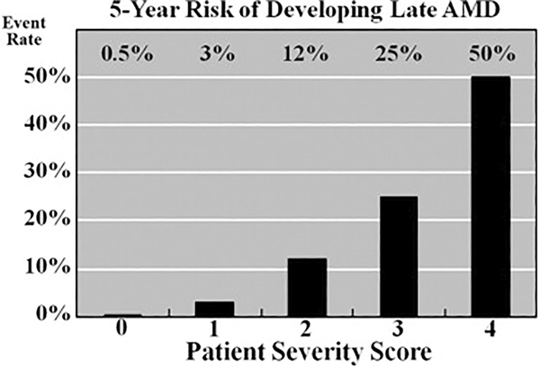
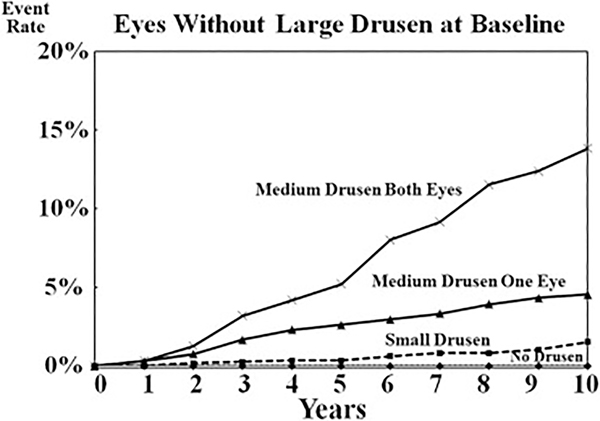
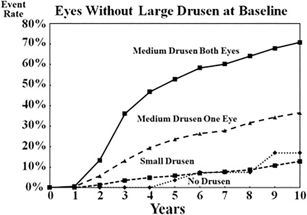
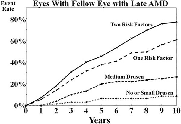
Results: Consensus was achieved in generating a basic clinical classification system based on fundus lesions assessed within 2 disc diameters of the fovea in persons older than 55 years. The committee agreed that a single term, age-related macular degeneration, should be used for the disease. Persons with no visible drusen or pigmentary abnormalities should be considered to have no signs of AMD. Persons with small drusen (<63 μm), also termed drupelets, should be considered to have normal aging changes with no clinically relevant increased risk of late AMD developing. Persons with medium drusen (≥ 63-<125 μm), but without pigmentary abnormalities thought to be related to AMD, should be considered to have early AMD. Persons with large drusen or with pigmentary abnormalities associated with at least medium drusen should be considered to have intermediate AMD. Persons with lesions associated with neovascular AMD or geographic atrophy should be considered to have late AMD. Five-year risks of progressing to late AMD are estimated to increase approximately 100 fold, ranging from a 0.5% 5-year risk for normal aging changes to a 50% risk for the highest intermediate AMD risk group.
Conclusions: The proposed basic clinical classification scale seems to be of value in predicting the risk of late AMD. Incorporating consistent nomenclature into the practice patterns of all eye care providers may improve communication and patient care.
Copyright © 2013 American Academy of Ophthalmology. Published by Elsevier Inc. All rights reserved.
Figures
Comment in
-
Klinische Klassifikation der AMD.Ophthalmologe. 2015 Nov;112(11):889. Ophthalmologe. 2015. PMID: 26894247 German. No abstract available.
References
-
- Eye Diseases Prevalence Research Group. Prevalence of age-related macular degeneration in the United States. Arch Ophthalmol 2004;122:564–72. - PubMed
-
- International ARM Epidemiological Study Group. An international classification and grading system for age-related maculopathy and age-related macular degeneration. Surv Ophthalmol 1995;39:367–74. - PubMed
-
- Seddon JM, Sharma S, Adelman RA. Evaluation of the clinical age-related maculopathy staging system. Ophthalmology 2006;113:260–6. - PubMed
Publication types
MeSH terms
Grants and funding
LinkOut - more resources
Full Text Sources
Other Literature Sources
Medical
Miscellaneous
