. 2013 Jan 17;152(1-2):196-209.
doi: 10.1016/j.cell.2012.12.001.

The cotranslational function of ribosome-associated Hsp70 in eukaryotic protein homeostasis

Affiliations

The cotranslational function of ribosome-associated Hsp70 in eukaryotic protein homeostasis

Felix Willmund et al. Cell. .

Abstract

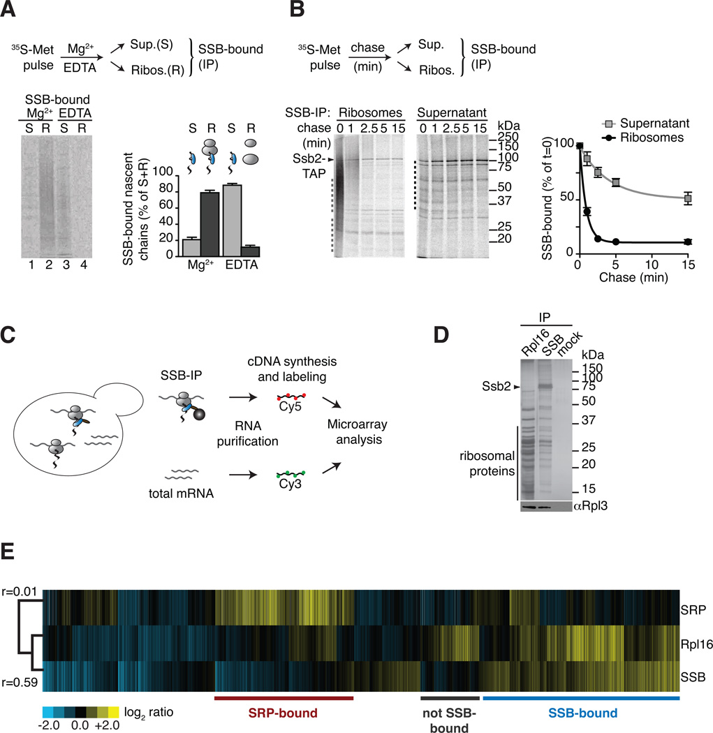
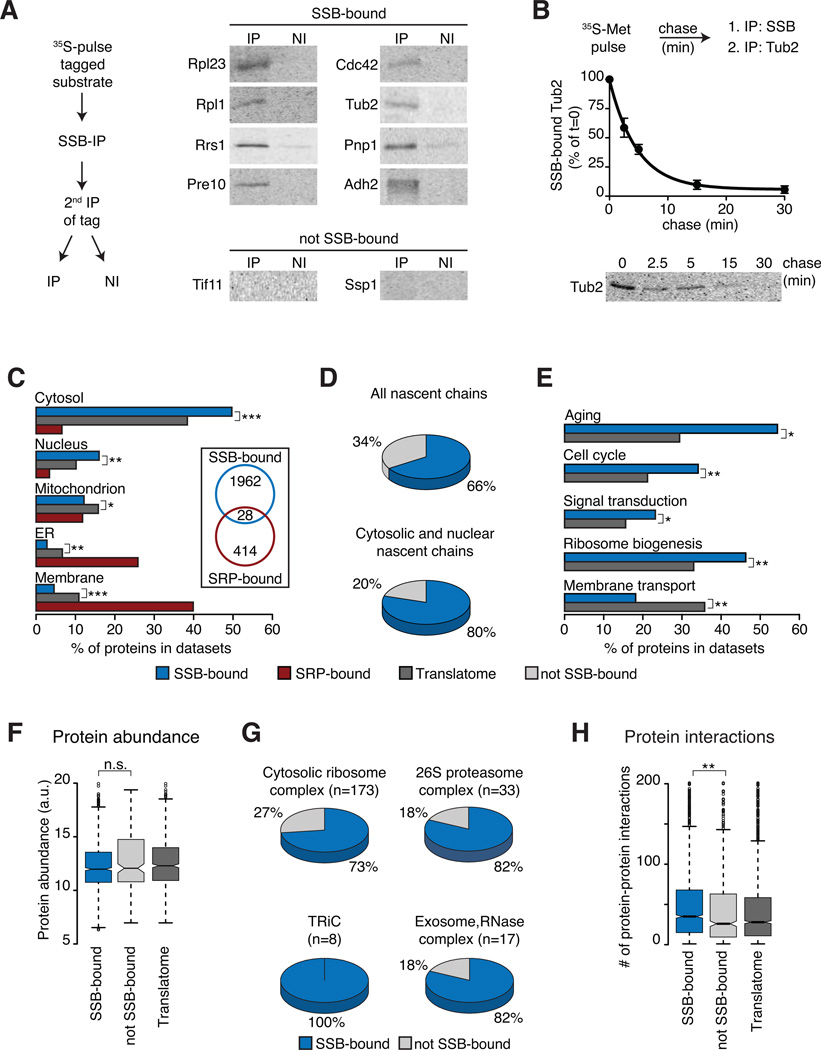
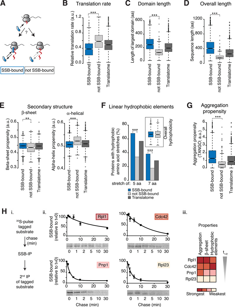
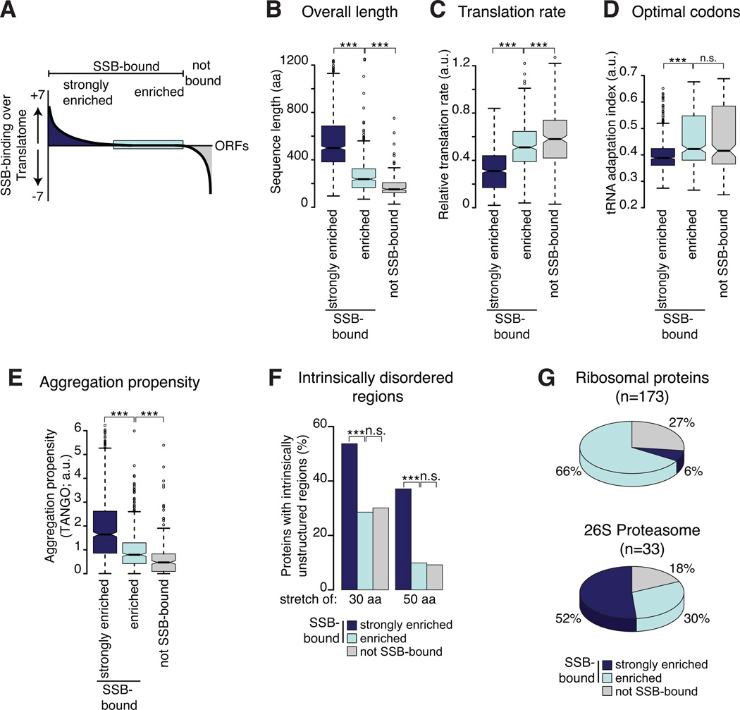
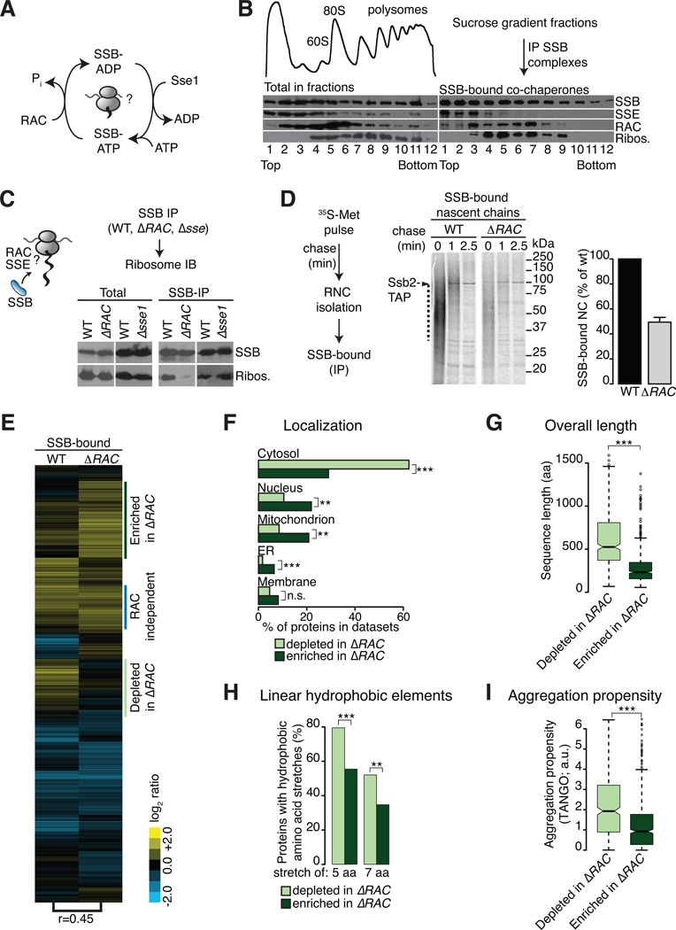
In eukaryotic cells a molecular chaperone network associates with translating ribosomes, assisting the maturation of emerging nascent polypeptides. Hsp70 is perhaps the major eukaryotic ribosome-associated chaperone and the first reported to bind cotranslationally to nascent chains. However, little is known about the underlying principles and function of this interaction. Here, we use a sensitive and global approach to define the cotranslational substrate specificity of the yeast Hsp70 SSB. We find that SSB binds to a subset of nascent polypeptides whose intrinsic properties and slow translation rates hinder efficient cotranslational folding. The SSB-ribosome cycle and substrate recognition is modulated by its ribosome-bound cochaperone, RAC. Deletion of SSB leads to widespread aggregation of newly synthesized polypeptides. Thus, cotranslationally acting Hsp70 meets the challenge of folding the eukaryotic proteome by stabilizing its longer, more slowly translated, and aggregation-prone nascent polypeptides.


Figures

Figure 1
Figure 1. Global identification of physiological SSB-associated nascent polypeptides
(A) SSB directly binds nascent polypeptides. Nascent polypeptides were pulse labeled with 35S-methionine (35S-Met), ribosome-nascent chain complexes (RNC) were either stabilized (Mg2+) or dissociated using 25 mM EDTA (EDTA) and fractionated by centrifugation in ribosome-bound (R) and soluble (S). SSB-nascent chain interactions in each fraction were determined after Ssb2-TAP immunopurification (IP), SDS-PAGE and autoradiography, followed by quantification of SSB-bound labeled nascent chains (mean ± SEM, n=3). (B) Kinetics of newly translated polypeptide flux through SSB. Nascent polypeptides were 35S-labeled and chased with cold methionine, samples at indicated time points were processed as in (A). Quantification of SSB-bound polypeptides in ribosome and soluble fractions reflects their co-/post-translational flux through SSB (mean ± SEM, n=4). For totals of (A) and (B) see Figure S1. (C) Scheme: Global identification of cotranslational SSB substrates. SSB-bound RNCs and total mRNAs were isolated, reverse transcribed, and labeled for subsequent microarray analysis. (D) Top: SDS-PAGE and silver staining of IPs of TAP-tagged Rpl16, Ssb2 and untagged cells (mock). Bottom: Immunoblot of ribosomal Rpl3. Ssb2-TAP IP depleted all Ssb2 from the lysate (see Figure S1D). (E) Translation profile of SRP-bound and SSB-bound mRNAs compared to the total Translatome identified by ribosome isolation (Rpl16). Hierarchically clustered heat map showis the average values of three individual experiments in rows; columns represent genes. mRNAs enriched over total RNAs in yellow, mRNAs depleted in blue. Pearson correlation coefficients are shown next to the tree. Comparison with the Translatome shows that some RNCs are preferentially enriched for SSB binding (blue) others are enriched in SRP binding (red). See also Figure S1.
Figure 2
Figure 2. Selectivity of SSB for specific nascent polypeptides
(A) Direct biochemical interaction of SSB with candidate substrates from Figure 1. N-terminally tagged substrates were briefly 35S-labeled, SSB-bound, labeled substrates were isolated by SSB-IP, and enrichment through a 2nd IP for the tag (IP). Non-immune controls were done in parallel (NI). Samples were analyzed by SDS-PAGE and autoradiography. (B) Flux of identified substrates through SSB. N-terminally tagged Tub2 was 35S-labeled and chased for the indicated times. Samples were processes as in (A) and quantified (mean ± SEM, n=3). (C) Subcellular localization of SSB and SRP substrates versus the Translatome is plotted as fraction of the datasets (%). Insert highlights low overlap between SSB and SRP substrates. (D) SSB substrates within the Translatome (top) and fractions of cytosolic and nuclear proteins. (E) SSB substrates are enriched in key cytoplasmic regulatory functions, but not in membrane proteins (Aging n=68; Cell-Cycle n=533; Sign. transd. n=224; Rib. biog. n=406; Mem. transp. n=187), plotted as fraction of the datasets (%). (F) Protein abundance of SSB-bound and not SSB-bound proteins. (G) Many subunits of large oligomeric complexes are substrates of SSB. (H) Enrichment of protein-protein interactions among SSB substrates. *p ≤ 0.01; **p≤ 10−4; ***p ≤10−10, n.s. not significant. See also Figure S2.
Figure 3
Figure 3. Underlying properties characterize SSB association with nascent polypeptides
(A) Specific nascent chain properties determine cotranslational SSB binding or lack thereof. (B–G) Analysis of intrinsic properties of the SSB substrates compared to those in the “not SSB-bound” dataset. The Translatome serves as reference. Only protein properties of cytosolic and nuclear localized proteins are shown since they represent the majority of SSB-bound substrates and they undergo maturation in the cellular compartment where SSB is localized. However, the conclusions were generally applicable for all SSB substrates. SSB substrates were found to differ significantly from non-SSB-bound nascent polypeptides for the indicated properties (B–G). (H) Association kinetics of SSB with substrates correlates with intensities of substrate properties i. N-terminally tagged substrates were 35S-Met pulse labeled and chased for the indicated times. SSB-bound, labeled substrates were isolated by SSB-IP, and enrichment through a 2nd IP for the tag. ii. SDS-PAGE and autoradiography shows SSB-bound 35S-labeled substrates; flux through SSB was measured by quantification (mean ± SEM, n=3). iii. Heat map represents the intensities of intrinsic sequence features. *p ≤ 0.01; **p≤ 10−4; ***p ≤10−10. See also Figure S3.
Figure 4
Figure 4. Nascent chain properties modulate the strength of SSB association
(A) The degree of enrichment of SSB substrates over the Translatome was determined by statistical analysis comparing the enrichment of each mRNA in the RNC of SSB-bound to the Translatome. mRNAs with positive enrichment scores were termed “strongly enriched” (dark blue), mRNAs with negative scores were termed “not SSB-bound” (grey), and equally enriched mRNAs in both dataset were classified “enriched” (light blue). (B)– (F) SSB substrate properties were compared to those of the “not SSB-bound” set. The Translatome serves as reference. As in Figure 3, only protein properties of cytosolic and nuclear proteins are shown. (G) Small and rapidly translated subunits of abundant complexes contain SSB-bound, enriched and not bound subunits. *p ≤ 0.01; **p≤ 10−4; ***p ≤10−10, n.s. not significant. See also Figure S4.
Figure 5
Figure 5. A co-chaperone network regulates the cotranslational substrate cycle of SSB
(A) Postulated function of RAC and Sse1 in SSBs nucleotide cycle. (B) Association of RAC and Sse1 with SSB on/off ribosomes. Top left: OD254 reading of polysome profiles after sucrose gradient fractionation. Bottom left: Immunoblot of ribosomes and distribution of chaperones in fractions. Right panel: SSB-IP, SDS-PAGE and immunoblot analysis from each gradient fraction examining the association of RAC and Sse1 with soluble and ribosome-bound SSB. As described, larger polysomal complexes are less stable during IP due to the very high molecular weight of the complex (Inada et al., 2002). (C) RAC but not Sse1 stimulate SSB-ribosome-association. SSB-IPs from wild type (WT), ΔRACzuo1/Δssz1) and Δsse1 cells and immunoblot for SSB-bound ribosomal protein Rpl3. Left: Totals; right: Immunoblot of IPs. For controls see Figure S5A. (D) Loss of RAC decreases the cotranslational flux of nascent polypeptides through SSB. SSB binding to RNC complexes was assessed by 35S-pulse-chase analysis as in Figure 1B (scheme left), autoradiography (middle) and quantification of SSB-bound radiolabeled nascent chains as indicated by the dotted line (right panel). Time=0 values were adjusted over Totals (Figure S5B) and plotted relative to WT (mean ± SEM, n=3). (E) RAC modulates the cotranslational specificity of SSB. Hierarchically clustered heat map of SSB-bound mRNAs in WT and ΔRAC cells (column=average of three experiments; row=single genes). Enriched SSB-bound mRNAs are in yellow, blue displays depleted mRNAs. Pearson correlation coefficients between experiments are indicated at the bottom of the tree. (F)–(I) Comparison of cotranslational substrate properties that are depleted or enriched for SSB binding in ΔRAC. Statistical analysis determined SSB-substrates less enriched or lost in ΔRAC cells (“depleted in ΔRAC”, light green) and those enriched in SSB binding in ΔRAC cells (“enriched in ΔRAC”, dark green). Importantly, enrichment or depletion for SSB-binding in ΔRAC cells was not due to up or down regulation of mRNAs on a transcriptional level. *p ≤ 0.01; **p≤ 10−4; ***p ≤10−10, n.s. not significant. See also Figure S5.
Figure 6
Figure 6. SSB maintains solubility of aggregation-prone nascent polypeptides
(A) We hypothesize that SSB prevents aggregation of newly synthesized proteins. (B) Loss of SSB or RAC leads to widespread and loss of Sse1 leads to partial aggregation. The presence of insoluble proteins in WT, mutant cells was examined by SDS-PAGE and silver-staining (left panel: Totals; right panel: aggregates). (C) Loss of SSB leads to rapid aggregation of newly synthesized proteins. Newly made proteins of WT and ΔSSB cells were 35S-Met pulse labeled followed by a chase with cold methionine. Aggregates were isolated and analyzed by SDS-PAGE and autoradiography. (D) Proteins aggregated in ΔSSB cells are ubiquitylated as shown by SDS-PAGE and immunoblotting for Ub. (E) Global identification of aggregates in WT and ΔSSB cells and mass spectrometry. (F–G) Comparison of intrinsic properties between aggregates in ΔSSB cells, SSB substrates and non-SSB-bound proteins. The Translatome serves as reference. Only protein properties of cytosolic and nuclear localized proteins are shown. Proteins that aggregate in ΔSSB cells have similar intrinsic properties as cotranslational substrates of SSB. (H) Distinct enrichment of protein-protein interactions in protein aggregates of ΔSSB cells. *p ≤ 0.01; **p≤ 10−4; ***p ≤10−10. See also Figure S6.
Figure 7
Figure 7. The cotranslational function of ribosome-associated Hsp70 in eukaryotic protein homeostasis
(A) The role of cotranslational acting Hsp70s in protecting nascent polypeptides. Hsp70 associates with approximately 70% of newly translated polypeptides with a strong enrichment of cytosolic and nuclear proteins. The cotranslational specificity of Hsp70 for its substrates is modulated by the co-chaperone RAC. Early sorting of SSB and SRP results in mutually exclusive binding to nascent chains at the ribosomes. Maturation of not Hsp70-bound proteins is likely facilitated by other chaperone like NAC. (B) Co-translationally acting Hsp70 meets the challenge of folding the eukaryotic proteome by protecting newly translated polypeptides challenged in co-translational folding. (C) Loss of co-translational acting Hsp70 leads to widespread aggregation of newly made polypeptides with properties hindered in efficient co-translational folding.

References

    1. Agashe VR, Guha S, Chang H-C, Genevaux P, Hayer-Hartl M, Stemp M, Georgopoulos C, Hartl FU, Barral JM. Function of trigger factor and DnaK in multidomain protein folding: increase in yield at the expense of folding speed. Cell. 2004;117:199–209. - PubMed
    1. Albanese V, Reissmann S, Frydman J. A ribosome-anchored chaperone network that facilitates eukaryotic ribosome biogenesis. The Journal of cell biology. 2010;189:69–81. - PMC - PubMed
    1. Albanese V, Yam AY-W, Baughman J, Parnot C, Frydman J. Systems Analyses Reveal Two Chaperone Networks with Distinct Functions in Eukaryotic Cells. Cell. 2006;124:75–88. - PubMed
    1. Arava Y, Wang Y, Storey JD, Liu CL, Brown PO, Herschlag D. Genome-wide analysis of mRNA translation profiles in Saccharomyces cerevisiae. Proceedings of the National Academy of Sciences. 2003;100:3889–3894. - PMC - PubMed
    1. Beckmann RP, Mizzen LE, Welch WJ. Interaction of Hsp 70 with newly synthesized proteins: implications for protein folding and assembly. Science. 1990;248:850–854. - PubMed

Publication types

MeSH terms

Substances