Review
. 2013 Apr;36(4):218-26.
doi: 10.1016/j.tins.2012.12.004. Epub 2013 Jan 17.

Spatial organization of ubiquitin ligase pathways orchestrates neuronal connectivity

Affiliations
Review

Spatial organization of ubiquitin ligase pathways orchestrates neuronal connectivity

Tomoko Yamada et al. Trends Neurosci. 2013 Apr.

Abstract

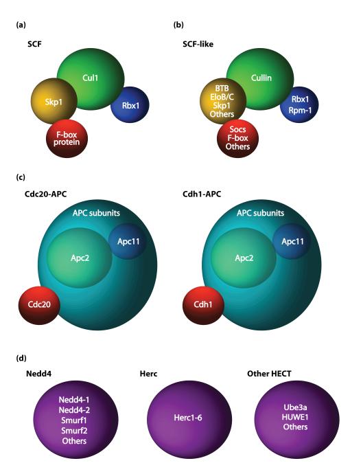
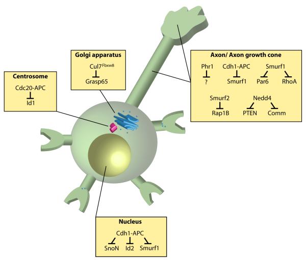
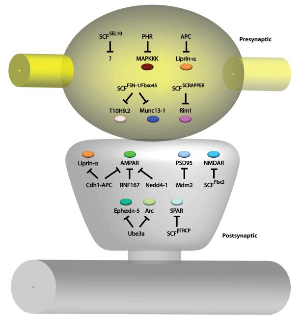
Recent studies have revealed that E3 ubiquitin ligases have essential functions in the establishment of neuronal circuits. Strikingly, a common emerging theme in these studies is that spatial organization of E3 ubiquitin ligases plays a critical role in the control of neuronal morphology and connectivity. E3 ubiquitin ligases localize to the nucleus, centrosome, Golgi apparatus, axon and dendrite cytoskeleton, and synapses in neurons. Localization of ubiquitin ligases within distinct subcellular compartments may facilitate neuronal responses to extrinsic cues and the ubiquitination of local substrates. Here, we review the functions of neuronal E3 ubiquitin ligases at distinct subcellular locales and explore how they regulate neuronal morphology and function in the nervous system.


Figures

Figure 1
Figure 1. RING and HECT domain E3 ubiquitin ligase complexes
SCF, SCF-like, and APC RING finger E3 ligases are structurally similar [11-14]. (a-b) The SCF (a) and SCF-like (b) E3 ubiquitin ligases contain a cullin protein (green), and variable RING finger (blue), adaptor (yellow), and substrate receptor proteins (red) [12-13]. (c) The APC contains the cullin-like scaffold protein Apc2 (green), RING finger protein Apc11 (blue), and substrate recognition protein Cdh1 or Cdc20 (red) [11, 14]. (d) HECT-domain E3 ubiquitin ligases (purple) are subdivided into the Nedd4, Herc, or other HECT protein families [15].
Figure 2
Figure 2. E3 ubiquitin ligases localized to distinct subcellular compartments control neuronal morphogenesis
E3 ubiquitin ligases operate in the nucleus, centrosome, Golgi apparatus, and axon and dendrite cytoskeleton in neurons. Nuclear Cdh1-APC regulates the abundance of SnoN, Id2, and Smurf1 [23, 25-26] in the control of axon growth and patterning, while centrosomal Cdc20-APC targets Id1 for degradation to promote dendrite growth and arborization [22]. A pool of Cdh1-APC may also function in the cytoplasm to regulate Smurf1 levels to inhibit axon growth [26]. Cul7Fbxw8 localizes at the Golgi apparatus and induces the degradation of Grasp65 to regulate Golgi morphology and dendrite growth [40]. Smurf1 and Smurf2 operate locally at the axon to induce the degradation of Par6, RhoA, and Rap1B to regulate axon specification [45, 47]. The E3 ubiquitin ligase Nedd4 operates at the axon growth cone to ubiquitinate the proteins PTEN and Comm in the control of axon morphogenesis [48-50].
Figure 3
Figure 3. E3 ubiquitin ligases locally regulate synapse development and function
E3 ubiquitin ligases are localized to presynaptic sites (yellow) and postsynaptic sites (gray) to coordinate the wiring of neuronal circuits. In the presynaptic compartment, the E3 ubiquitin ligases APC and SCFSCRAPPER ubiquitinates Liprin-α and Rim1, respectively, to regulate presynaptic morphology and neurotransmission [19-20, 67]. PHR downregulates the MAPKKK pathway [58, 61-62, 64], while SCFFSN-1/Fbxo45 targets the kinase T10H9.2 and the active zone protein Munc13-1 [65, 68] for degradation to control presynaptic differentiation and function. The E3 ubiquitin ligase SCFSEL10 is necessary for presynaptic elimination, but the substrate is unknown [66]. In the postsynaptic compartment, the E3 ubiquitin ligases Cdh1-APC, RNF167, and Nedd4-1 recognize AMPARs for ubiquitination [19-20, 70, 77, 80-81], and Cdh1-APC also regulates postsynaptic Liprin-α abundance [71]. The scaffold protein PSD95 is targeted by the E3 ubiquitin ligase Mdm2 [78], while retrotranslocated NMDARs are degraded by SCFFbx2 [75]. SCFβTRCP controls levels of the GTPase SPAR [72]. The E3 ubiquitin ligase Ube3A recognizes the substrates Ephexin-5 and Arc to control synapse number and maturation [83-84]. In dendrites, SCFLIN-23 controls the levels of BAR-1 and decreases GLR-1 abundance [74].

References

    1. Finley D. Recognition and processing of ubiquitin-protein conjugates by the proteasome. Annu Rev Biochem. 2009;78:477–513. - PMC - PubMed
    1. Hicke L, Dunn R. Regulation of membrane protein transport by ubiquitin and ubiquitin-binding proteins. Annual review of cell and developmental biology. 2003;19:141–172. - PubMed
    1. Huang TT, D’Andrea AD. Regulation of DNA repair by ubiquitylation. Nat Rev Mol Cell Biol. 2006;7:323–334. - PubMed
    1. Kawabe H, Brose N. The role of ubiquitylation in nerve cell development. Nat Rev Neurosci. 2011;12:251–268. - PubMed
    1. Ang XL, Harper JW. Interwoven ubiquitination oscillators and control of cell cycle transitions. Sci STKE. 2004;2004:pe31. - PubMed

Publication types

Substances

LinkOut - more resources