

Clinical examination of the rotator cuff
- PMID: 23332909
- PMCID: PMC3826176
- DOI: 10.1016/j.pmrj.2012.08.019
Clinical examination of the rotator cuff
Abstract
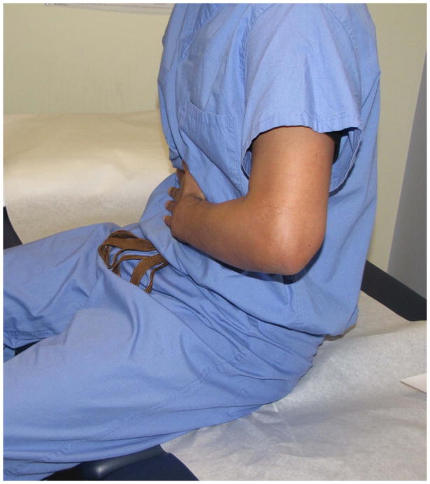
Rotator cuff tears are the leading cause of shoulder pain and shoulder-related disability and account for 4.5 million physician visits in the United States annually. A careful history and structured physical examination are often sufficient for diagnosing rotator cuff disorders. We are not aware of a clinical review article that presents a structured physical examination protocol of the rotator cuff for the interested clinician. To fill this void, we present a physical examination protocol developed on the basis of review of prior literature and our clinical experience from dedicated shoulder practices. Our protocol includes range of motion testing by using a goniometer, strength testing by using a dynamometer, and select special tests. Among the many tests for rotator cuff disorders that have been described, we chose ones that have been more thoroughly assessed for sensitivity and specificity. This protocol can be used to isolate the specific rotator cuff tendon involved. The protocol can typically be completed in 15 minutes. We also discuss the clinical implications and limitations of the physical examination maneuvers described in our protocol. This protocol is thorough yet time efficient for a busy clinical practice. It is useful in the diagnosis of rotator cuff tears, impingement syndrome, and biceps pathology.
Copyright © 2013 American Academy of Physical Medicine and Rehabilitation. Published by Elsevier Inc. All rights reserved.
Figures


Comment in
-
Re: teaching rounds on clinical examination of the rotator cuff.PM R. 2013 May;5(5):445-6. doi: 10.1016/j.pmrj.2013.03.001. PM R. 2013. PMID: 23701982 No abstract available.
-
Reply: To PMID 23332909.PM R. 2013 May;5(5):446-7. doi: 10.1016/j.pmrj.2013.03.002. PM R. 2013. PMID: 23701983 No abstract available.
References
-
- Oh LS, Wolf BR, Hall MP, Levy BA, Marx RG. Indications for rotator cuff repair: a systematic review. Clin Orthop Relat Res. 2007 Feb;455:52–63. - PubMed
-
- Meislin RJ, Sperling JW, Stitik TP. Persistent shoulder pain: epidemiology, pathophysiology, and diagnosis. Am J Orthop (Belle Mead NJ) 2005 Dec;34(12 Suppl):5–9. - PubMed
-
- Yamaguchi K, Ditsios K, Middleton WD, Hildebolt CF, Galatz LM, Teefey SA. The demographic and morphological features of rotator cuff disease. A comparison of asymptomatic and symptomatic shoulders. J Bone Joint Surg Am. 2006 Aug;88(8):1699–1704. - PubMed
-
- Parker BJ, Zlatkin MB, Newman JS, Rathur SK. Imaging of shoulder injuries in sports medicine: current protocols and concepts. Clin Sports Med. 2008 Oct;27(4):579–606. - PubMed
-
- Hegedus EJ, Goode A, Campbell S, et al. Physical examination tests of the shoulder: a systematic review with meta-analysis of individual tests. Br J Sports Med. 2008 Feb;42(2):80–92. discussion 92. - PubMed
Publication types
MeSH terms
Grants and funding
LinkOut - more resources
Full Text Sources
Other Literature Sources
Medical
