

A new method to quantify male pelvic floor displacement from 2D transperineal ultrasound images
- PMID: 23332998
- PMCID: PMC4123322
- DOI: 10.1016/j.urology.2012.11.034
A new method to quantify male pelvic floor displacement from 2D transperineal ultrasound images
Abstract
Objective: To develop a method to quantify displacement of pelvic structures during contraction of the pelvic floor muscles from transperineal ultrasound images in men and investigate the reliability of the method between days.
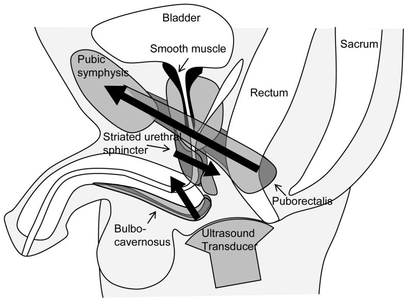
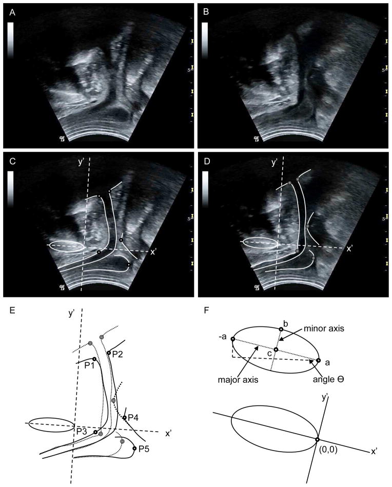
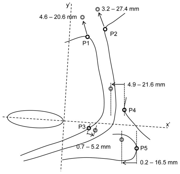
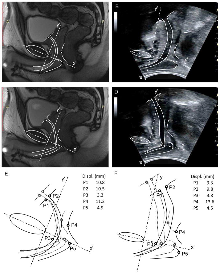
Methods: Ten healthy male volunteers (aged 28-41 years) attended 2 separate data collection sessions. Ultrasound images were recorded during voluntary pelvic floor muscle contractions in cine-loop (video) format with the transducer aligned in the midsagittal plane on the perineum. Five anatomic points were defined to represent contraction from striated urethral sphincter (SUS), levator ani (LA), and bulbocavernosus (BC) muscles. Displacement of each point was calculated between the relaxed and contracted-state images. Intraclass correlation coefficient (ICC) values were calculated from displacement data to assess reliability of the method between days.
Results: Displacements of the 5 anatomic points closely matched predictions based on anatomic considerations of the male pelvic musculature. ICC values for displacement data calculated from 1, 2, and 3 repetitions ranged between 0.82 and 0.95 for ICC (2,1), 0.85 and 0.97 for ICC (2,2), and 0.86 and 0.97 for ICC (2,3), respectively.
Conclusion: The new method reliably calculates displacements of points previously validated for women (ano-rectal junction and bladder base) in addition to new measures of muscle actions (SUS and BC) specific to men. Future use might include assessment of clinical populations to understand how these displacements relate to symptoms of incontinence.
Copyright © 2013 Elsevier Inc. All rights reserved.
Figures
References
-
- Peng Q, et al. 2D Ultrasound image processing in identifying responses of urogenital structures to pelvic floor muscle activity. Annals of Biomedical Engineering. 2006;34:477–93. - PubMed
-
- Constantinou CE, et al. Displacement sequence and elastic properties of anterior prostate/urethral interface during micturition of spinal cord injured men. Ultrasound in Medicine and Biology. 2002;28:1157–63. - PubMed
-
- Strasser H, et al. Transurethral ultrasound: evaluation of anatomy and function of the rhabdosphincter of the male urethra. J Urol. 1998;159:100–4. discussion 04–5. - PubMed
-
- Strasser H, et al. Three-dimensional transrectal ultrasound of the male urethral rhabdosphincter. World J Urol. 2004;22:335–8. - PubMed
Publication types
MeSH terms
Grants and funding
LinkOut - more resources
Full Text Sources
Other Literature Sources
