Review
. 2013 Feb;23(1):134-43.
doi: 10.1016/j.sbi.2013.01.003. Epub 2013 Jan 16.

PARP-1 mechanism for coupling DNA damage detection to poly(ADP-ribose) synthesis

Affiliations
Review

PARP-1 mechanism for coupling DNA damage detection to poly(ADP-ribose) synthesis

Marie-France Langelier et al. Curr Opin Struct Biol. 2013 Feb.

Abstract

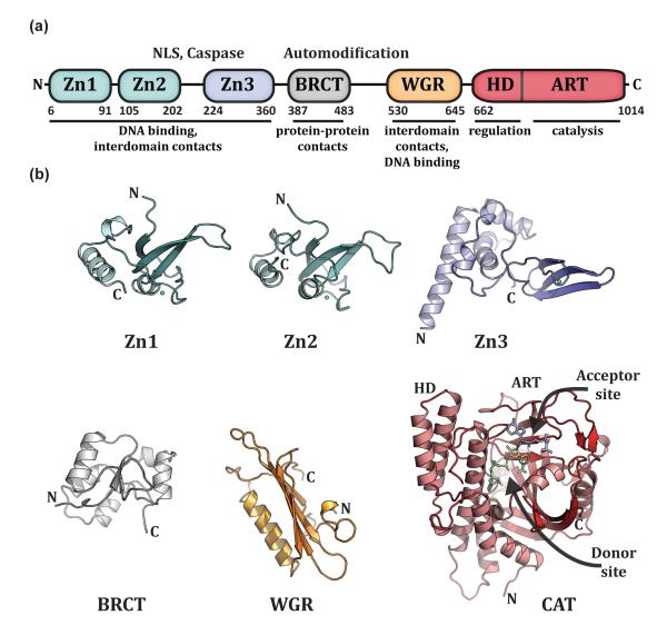
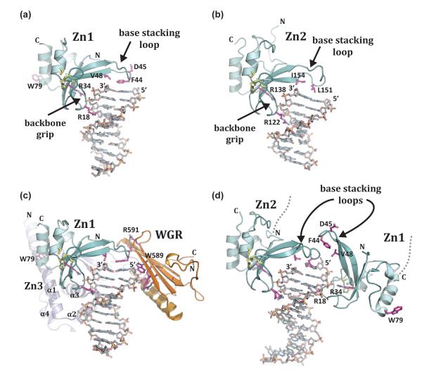
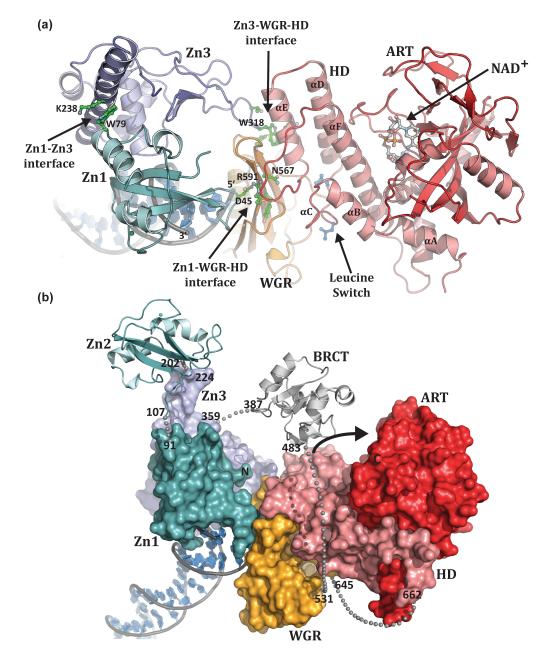
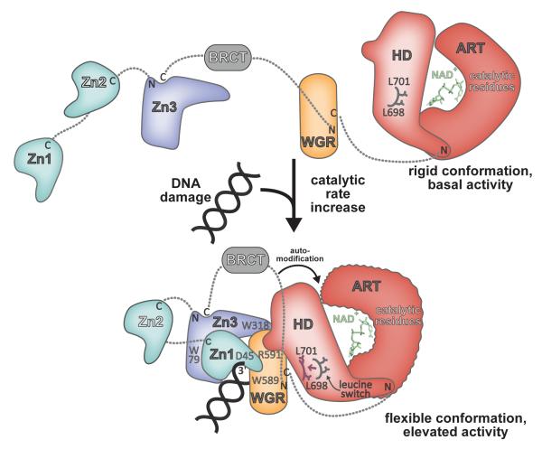
Poly(ADP-ribose) polymerase 1 (PARP-1) regulates gene transcription, cell death signaling, and DNA repair through production of the posttranslational modification poly(ADP-ribose). During the cellular response to genotoxic stress PARP-1 rapidly associates with DNA damage, which robustly stimulates poly(ADP-ribose) production over a low basal level of PARP-1 activity. DNA damage-dependent PARP-1 activity is central to understanding PARP-1 biological function, but structural insights into the mechanisms underlying this mode of regulation have remained elusive, in part due to the highly modular six-domain architecture of PARP-1. Recent structural studies have illustrated how PARP-1 uses specialized zinc fingers to detect DNA breaks through sequence-independent interaction with exposed nucleotide bases, a common feature of damaged and abnormal DNA structures. The mechanism of coupling DNA damage detection to elevated poly(ADP-ribose) production has been elucidated based on a crystal structure of the essential domains of PARP-1 in complex with a DNA strand break. The multiple domains of PARP-1 collapse onto damaged DNA, forming a network of interdomain contacts that introduce destabilizing alterations in the catalytic domain leading to an enhanced rate of poly(ADP-ribose) production.


Figures

Figure 1
Figure 1. PARP-1 has a highly modular, “beads-on-a-string” architecture
(a). Schematic representation of human PARP-1 domains. A bipartite nuclear localization signal (NLS) and a caspase 3 cleavage site (Caspase) are located between the Zn2 and Zn3 domains. A BRCA C-terminus (BRCT) fold is located within the region of PARP-1 that is primarily targeted for automodification. The catatlyic domain is composed of an alpha-helical subdomain (HD) and an ADP-ribosyl transferase subdomain (ART). (b). Crystal and/or NMR structures have been determined for each of the PARP-1 domains in the absence of DNA. Shown are the NMR structures of the homologous Zn1 and Zn2 domains (PDB code 2dmj and 2cs2, respectively [64], see also [8]), the NMR structure of the Zn3 domain (PDB code 2jvn, see also reference [9] for crystal structure), the NMR structure of the BRCT fold (PDB code 2cok [64], see also reference [11]), the NMR structure of the WGR domain (PDB code 2cr9 [64]), and the crystal structure of the catalytic domain (PDB code 1a26 [12]). Catalytic subdomains are labeled (HD and ART). The catalytic domain structure was determined in the presence of an NAD+ analogue that has defined the “acceptor site” for poly(ADP-ribose) formation. NAD+ is modeled in the “donor site” based on the related structure of diphthetia toxin [19]. All structure depictions were made using PYMOL (www.pymol.org).
Figure 2
Figure 2. PARP-1 recognizes exposed nucleotide bases as a signature of DNA damage
(a). Crystal structure of human Zn1 domain in complex with a DNA double strand break (PDB code 3od8 [13]). Together with panel b, these structures illustrate the features of damage DNA that are recognized by PARP-1 zinc finger domains: a continuous phosphate backbone engaged by conserved Arg residues on the “backbone grip”, and exposed nucleotide bases engaged by hydrophobic residues on the “base stacking loop”. D45 and W79 are key Zn1 residues involved in contacts with other PARP-1 domains. (b). Crystal structure of human Zn2 in complex with a DNA double strand break (PDB code 3odc [13]). (c). The Zn1, Zn3, and WGR domains collectively assemble on a DNA double strand break, with each domain forming specific protein-DNA, and protein-protein contacts with adjacent domains [41] (see also Figure 3a). Zn1 has the same orientation as in panel a. Residues critical for PARP-1 DNA-dependent activity are labeled: WGR residues W589 and R591, and Zn1 residue W79. (d). Crystal structure of the tandem Zn1–Zn2 domains of PARP-1 bound to a DNA double strand break with a single 5’ nucleotide overhang [40]. Zn2 forms DNA contacts similar to that seen for the isolated domain bound to DNA in panel b (the same conserved residues are shown). Zn1 binds to DNA with opposite polarity to that seen in panel a, with Arg18 positioned in the major groove rather than the minor groove, and the “base stacking loop” forming protein-protein contacts rather than protein-DNA contacts.
Figure 3
Figure 3. PARP-1 domains collectively assemble on DNA damage
(a). Human PARP-1 domains Zn1, Zn3, and WGR–CAT were crystallized in complex with a DNA double-strand break (PDB code 4dqy [41]). The PARP-1/DNA complex illustrates how DNA damage detection is coupled to structural transitions in the catalytic domain that elevate poly(ADP-ribosyl)ation activity [41]. Three essential interdomain contact regions form upon PARP-1 interaction with DNA: Zn1–WGR–HD, Zn3–WGR–HD, and Zn1–Zn3. Mutations that target has captured interdomain contacts that are relevant to PARP-1 regulation. Interdomain communication with the HD displaces conserved Leu residues from the hydrophobic interior of the HD, leading to destabilization of the CAT that correlates with an elevation in PARP-1 catalytic activity. (b). A model for the approximate positioning of the Zn2 and BRCT domains within the PARP-1/DNA complex. Zn1, Zn3, and WGR-CAT are shown as surfaces, labeled, and colored as in Figure 1a. The Zn2 and BRCT domains are drawn in schematic representation. Their positioning is based on the relative location of the termini of adjacent domains in the structure. The numbering and location of linker residues are shown. The arrow indicates the location of the PARP-1 automodification region near the catalytic active site.
Figure 4
Figure 4. Model for DNA damage-dependent activation of PARP-1
In the absence of DNA damage, PARP-1 domains exist in an extended, “beads-on-a-string” conformation. The HD serves as a modulator of PARP-1 activity, holding the ART in a rigid conformation. Upon detecting DNA damage the Zn1, Zn3, and WGR domains collapse together, forming a network of interdomain contacts that perturb the structure of the HD, displacing a “leucine switch” that decreases the stability of the catalytic domain and increases the catalytic activity. A more flexible, dynamic ART conformation is more efficient to perform the multi-step synthesis of poly(ADP-ribose). The collapsed conformation positions the automodication region adjacent to the catalytic domain, providing substrate specificity and contributing to an enhanced rate of poly(ADP-ribose) production.

References

    1. Krishnakumar R, Kraus WL. The PARP side of the nucleus: molecular actions, physiological outcomes, and clinical targets. Mol Cell. 2010;39:8–24. - PMC - PubMed
    1. Rouleau M, Patel A, Hendzel MJ, Kaufmann SH, Poirier GG. PARP inhibition: PARP1 and beyond. Nat Rev Cancer. 2010;10:293–301. - PMC - PubMed
    1. Hassa PO, Hottiger MO. The diverse biological roles of mammalian PARPS, a small but powerful family of poly-ADP-ribose polymerases. Front Biosci. 2008;13:3046–3082. - PubMed
    1. Haince JF, McDonald D, Rodrigue A, Dery U, Masson JY, Hendzel MJ, Poirier GG. PARP1-dependent kinetics of recruitment of MRE11 and NBS1 proteins to multiple DNA damage sites. J Biol Chem. 2008;283:1197–1208. - PubMed
    1. Gottschalk AJ, Timinszky G, Kong SE, Jin J, Cai Y, Swanson SK, Washburn MP, Florens L, Ladurner AG, Conaway JW, et al. Poly(ADP-ribosyl)ation directs recruitment and activation of an ATP-dependent chromatin remodeler. Proc Natl Acad Sci U S A. 2009;106:13770–13774. - PMC - PubMed

Publication types

LinkOut - more resources