. 2013 Jan 31;3(1):23-9.
doi: 10.1016/j.celrep.2012.12.010. Epub 2013 Jan 17.

Influenza A virus utilizes suboptimal splicing to coordinate the timing of infection

Affiliations

Influenza A virus utilizes suboptimal splicing to coordinate the timing of infection

Mark A Chua et al. Cell Rep. .

Abstract

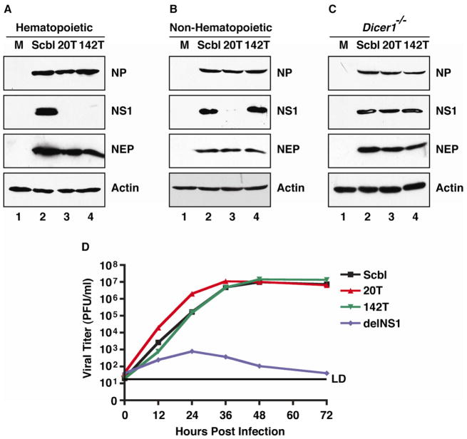
Influenza A virus is unique as an RNA virus in that it replicates in the nucleus and undergoes splicing. With only ten major proteins, the virus must gain nuclear access, replicate, assemble progeny virions in the cytoplasm, and then egress. In an effort to elucidate the coordination of these events, we manipulated the transcript levels from the bicistronic nonstructural segment that encodes the spliced virus product responsible for genomic nuclear export. We find that utilization of an erroneous splice site ensures the slow accumulation of the viral nuclear export protein (NEP) while generating excessive levels of an antagonist that inhibits the cellular response to infection. Modulation of this simple transcriptional event results in improperly timed export and loss of virus infection. Together, these data demonstrate that coordination of the influenza A virus life cycle is set by a "molecular timer" that operates on the inefficient splicing of a virus transcript.


Figures

Figure 1
Figure 1. Silencing of NS1 does not impact virus replication in vitro
(A-C) Western blot analysis of whole cell extract (WCE) derived from (A) Jurkat T cells, (B) murine embryonic fibroblasts (MEFs), and (C) MEFs deficient in Dicer1 (Dicer1 −/−) infected with Scbl, 20T, and 142T viruses. Immunoblots depict NP, NS1, NEP, and Actin. (D) Multicycle growth curves performed in A549 cells in response to Scbl, 20T, 142T, and delNS1 IAV strains. Error bars represent average ± SD of the mean of samples from independent growth curves analyzed in triplicate. LD indicates limit of detection. See also Figure S1.
Figure 2
Figure 2. Low levels of NS1 are sufficient for virus replication in vivo
Western blot analysis of WCE from (A) primary lung fibroblasts (pLFs) or (B) bone marrow-derived macrophages (BMDMs) from wild type (WT) C57BL/6 mice infected with Scbl, 20T, or 142T IAV or mock (M) treated for 24 hours. Western blot analysis of (C) Ifnar1−/−/Il28r−/− or (D) WT mouse lungs infected intranasally with either Scbl, 20T, or 142T IAV or mock (M) treated at 48 hpi. Immunoblots depict NP, NS1, and Actin. (E) Viral titers from WT C57BL/6 mice infected with Scbl, 20T, 142T or mock (PBS) treated. Data points represent individual mice assayed in triplicate. See also Figure S2.
Figure 3
Figure 3. NEP is essential in determining the levels of virus replication
(A) Western blot analysis of WCE from fibroblasts infected with either WT or 2A viruses or mock treated for 3, 6, 9, or 12 hours post infection (HPI). (B) Multicycle growth curve for WT and 2A IAV. (C) Western blot analysis of WCE from fibroblasts infected with IAV-like vectors (VLVs) containing either an extragenic scrambled (Scbl) sequence or NEP on segment four. (D) Multicycle growth curve of WT IAV coinfected with VLVs characterized in (C). (A and C) Immunoblots depict NP, NS1, NEP, and Actin. (B and D) Error bars represent mean ± SD of the mean of samples from independent multicycle growth curve analyzed in triplicate. LD indicates limit of detection. See also Figure S3.
Figure 4
Figure 4. NEP accumulation is essential in coordinating virus replication
(A) Western blot analysis of WCE from A549 cells infected with an NS encoding an optimal 5′ splice site (NSo), a parental splice NS (NSp) IAV, or mock treated for 3, 6, 9, or 12 hours post infection (HPI). Immunoblots depict NP, NS1, NEP, and Actin. (B) Multicycle growth curves for NSo and NSp IAV in A549 cells. (C) Same as (B) but performed in MDCK-NS1-GFP cells. Error bars represent mean ± SD of the mean of samples from independent multicycle growth curve analyzed in triplicate. LD indicates limit of detection. (D) Viral titers from WT C57BL/6 mouse lungs infected intranasally with NSo or NSp. (E) A549 cells stained for NP expression from NSo and NSp infection at 5 or 7 hours post infection and imaged by fluorescence microscopy. Hoechst dye used to visualize nuclei. See also Figure S4.

References

    1. Agris CH, Nemeroff ME, Krug RM. A block in mammalian splicing occurring after formation of large complexes containing U1, U2, U4, U5, and U6 small nuclear ribonucleoproteins. Mol Cell Biol. 1989;9:259–267. - PMC - PubMed
    1. Akarsu H, Burmeister WP, Petosa C, Petit I, Muller CW, Ruigrok RW, Baudin F. Crystal structure of the M1 protein-binding domain of the influenza A virus nuclear export protein (NEP/NS2) EMBO J. 2003;22:4646–4655. - PMC - PubMed
    1. Boulo S, Akarsu H, Ruigrok RW, Baudin F. Nuclear traffic of influenza virus proteins and ribonucleoprotein complexes. Virus Res. 2007;124:12–21. - PubMed
    1. Cros JF, Palese P. Trafficking of viral genomic RNA into and out of the nucleus: influenza, Thogoto and Borna disease viruses. Virus Res. 2003;95:3–12. - PubMed
    1. Egorov A, Brandt S, Sereinig S, Romanova J, Ferko B, Katinger D, Grassauer A, Alexandrova G, Katinger H, Muster T. Transfectant influenza A viruses with long deletions in the NS1 protein grow efficiently in Vero cells. J Virol. 1998;72:6437–6441. - PMC - PubMed

Publication types

LinkOut - more resources