. 2013 Feb 4;23(3):229-35.
doi: 10.1016/j.cub.2012.12.033. Epub 2013 Jan 17.

Mask is required for the activity of the Hippo pathway effector Yki/YAP

Affiliations

Mask is required for the activity of the Hippo pathway effector Yki/YAP

Leticia Sansores-Garcia et al. Curr Biol. .

Abstract

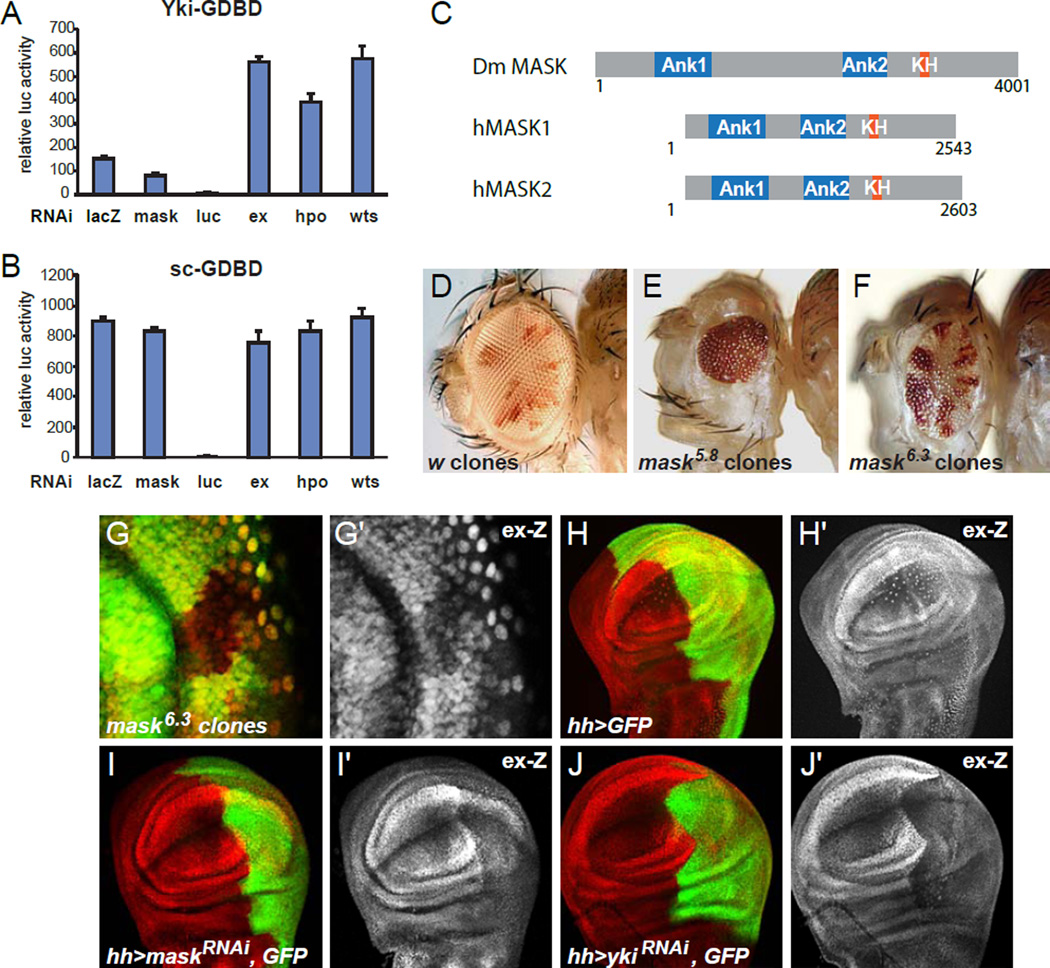
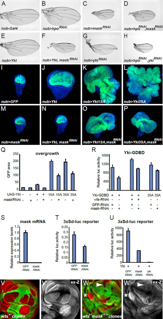
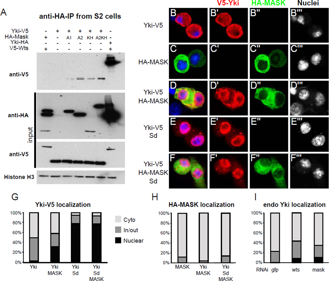
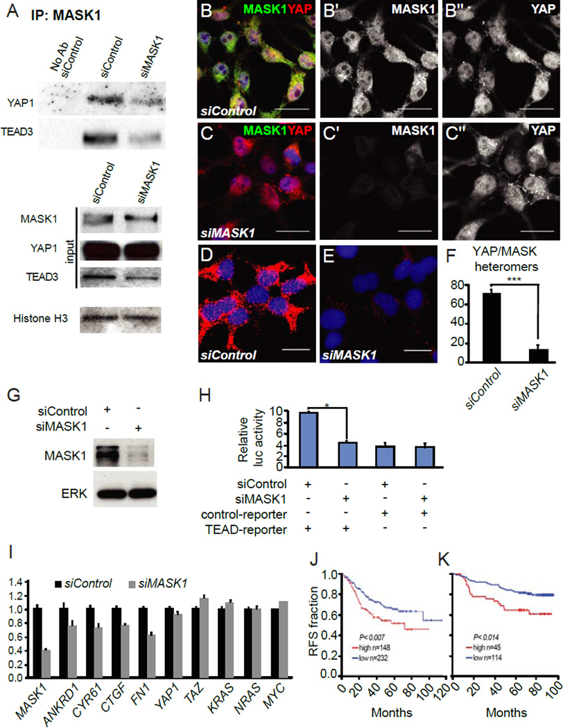
The Drosophila Yorkie (Yki) protein and its mammalian homolog Yes-associated protein (YAP) are potent growth promoters, and YAP overexpression is associated with multiple types of cancer. Yki and YAP are transcriptional coactivators and function as downstream effectors of the Hippo tumor suppressor pathway. The regulation of Yki and YAP by the Hippo signaling pathway has been extensively investigated; however, how they regulate gene expression is poorly understood. To identify additional regulators of Yki activity, we performed a genome-wide RNAi screen in Drosophila S2 cells. In this screen, we identified the conserved protein Mask (Multiple ankyrin repeats single KH domain) as a novel promoter of Yki activity in vitro and validated this function in vivo in Drosophila. We found that Mask is required downstream of the Hippo pathway for Yki to induce target-gene expression and that Mask forms complexes with Yki. The human Mask homolog MASK1 complexes with YAP and is required for the full activity of YAP. Additionally, elevated MASK1 expression is associated with worsened outcomes for breast cancer patients. We conclude that Mask is a novel cofactor for Yki/YAP required for optimal Yki/YAP activity during development and oncogenesis.


Figures

Figure 1
Figure 1. Mask is a positive regulator of Yki activity
(A) Luciferase (luc) activity assay to measure the activity of a Yki-GAL4-DNA-binding-domain (Yki-GDBD) fusion protein on a UAS-luc reporter plasmid normalized with Renilla Luc expressed from a constitutive promoter. S2 cells were transfected with UAS-luc, Yki-GDBD and HA-Ex expressing plasmids in the presence of dsRNAs as indicated. (B) Similar as (A) but using a Scute (Sc)-GDBD construct instead of Yki-GDBD. (C) Schematic of Drosophila and human Mask proteins indicating conserved domains. (D–F) Adult flies with (D) white control clones, (E) mask5.8 null clones, and (F) mask6.3 hypomorphic clones. (G) Confocal image of a third instar antennal imaginal disc showing mask6.3 hypomorphic clones marked by the lack of GFP and stained for ex-lacZ expression (red, gray in G’). (H–J) Confocal images of third instar wing imaginal discs expressing GFP alone or together with maskRNAi or ykiRNAi in the posterior compartment driven by hh-GAL4 and stained for ex-lacZ (red or grey in the ’ panels).
Figure 2
Figure 2. Mask is required for full Yki activity independent of Wts phosphorylation
(A–H) Pictures of wings from flies expressing the indicated constructs driven by the wing specific nubbin-GAL4 driver. “maskRNAi" refers to the VDRC v33396 maskRNAi line. (I–P) Confocal images of third instar wing discs with nubbin-GAL4 driving GFP plus the indicated constructs. GFP is in green and nuclei are stained with DAPI displayed in blue. (Q) Quantification of the GFP expressing area of imaginal discs with the genotypes shown in (I–P) (median area of GFP pixels ± S.E.M., n=5). (R) Luciferase assay on S2 cells transfected with plasmids expressing UAS-luc and wild-type Yki-GDBD or Yki3SA-GDBD fusion proteins in the presence of the indicated dsRNAs. (S) Quantification of mask mRNA levels by RT-qPCR from S2 cells transfected with dsRNA targeting GFP or mask. (T) Luciferase assay on S2 cells transfected with the Yki/Sd responsive 3xSd-luc reporter, which has multimerized Sd binding sites, in the presence of the indicated dsRNAs and normalized to beta-galactosidase activity expressed from a constitutive promoter. (U) Luciferase assay on S2 cells transfected with the 3X-Sd-luc reporter and Yki expressing plasmids in the presence of the indicated dsRNAs. (V) wtsx1 null mutant clones in a wing disc marked by lack of GFP and stained for ex-lacZ expression (red, gray in V’). (W) mask10.22 wtsx1 double mutant clones in a wing disc stained for ex-lacZ expression (red, gray in W’). The arrowhead indicates a clone with low levels of ex-lacZ expression and the asterisk indicates a clone with upregulated ex-lacZ expression.
Figure 3
Figure 3. Mask complexes with Yki but localizes independently of Yki
(A) Immunoprecipitation of lysates from S2 cells transfected with Yki-V5 and different HA-Mask or V5-Wts and HA-Yki constructs. Top shows an anti-V5 western blot from an anti-HA immunoprecipitation. Panels below that show anti-V5 and anti-HA western blots to detect expression of the input proteins, and an anti-Histone H3 western blot as a loading control. Lane 1: V5-Wts co-expressed with HA-tagged Ank1, Ank2 and KH domains of Mask. Lane 2: Yki-V5 alone. Lane 3–6: Yki-V5 co-expressed with the Ank1, Ank2, KH, or Ank2-KH constructs respectively. Lane 7: Yki-HA co-expressed with V5-Wts as a positive control. (B–F) Confocal images of S2 cells transfected with the constructs indicated on the left and stained for the antigens indicated on the top. Nuclei were labeled with DAPI shown in blue. (G) Quantification of the localization of Yki-V5 in (B–F). (H) Quantification of the localization of Mask in (C,D) and (F). (I) Quantification of endogenous Yki localization in S2 cells transfected with dsRNAs targeting GFP, wts or mask.
Figure 4
Figure 4. MASK1 interacts with YAP and TEAD3, regulates YAP activity and its expression levels correlate with breast cancer patient outcomes
(A) Lysates of HEK293T cells treated with non-targeting or MASK1 siRNAs were immunoprecipitated for endogenous MASK1 and immunoblotted for YAP1 or TEAD3. (B,C) Endogenous MASK1 (green, grey in B′ and C′) and YAP (red, grey in B′′ and C′′) localization in HEK293T cells treated with non-targeting (B) or MASK1 siRNAs (C). (D–E) MASK1/YAP heteromers (red) detected by in situ proximity ligation assay (PLA) using MASK1 and YAP antibodies in cells treated with non-targeting (D) or MASK1 siRNA (E). (F) Quantification of the PLA signals in (D) and (E). (G) Western blot demonstrating the level of siRNA knock down of MASK1 protein in HEK293T cells. (H) Relative luciferase activity of a luciferase reporter plasmid with TEAD binding sites and the control vector in HEK293T cells co-transfected with MASK1 siRNA and a non-targeting siRNA. The error bars represent mean ± SE (n=6) (* p > 0.005). (I) RT-qPCR analysis of mRNA expression of YAP target genes in HEK293T cells treated with non-targeting or MASK1 siRNAs. (J) Kaplan-Meier graphs showing a significant association of MASK1 expression levels with Relapse Free Survival (RFS) in independent datasets of breast cancer patients (p>0.007 and p>0.014, log-rank test). Scale bars are 20 µm.

Comment in

References

    1. Zhao B, Li L, Lei Q, Guan KL. The Hippo-YAP pathway in organ size control and tumorigenesis: an updated version. Genes & development. 2010;24:862–874. - PMC - PubMed
    1. Pan D. The hippo signaling pathway in development and cancer. Developmental cell. 2010;19:491–505. - PMC - PubMed
    1. Halder G, Johnson RL. Hippo signaling: growth control and beyond. Development. 2011;138:9–22. - PMC - PubMed
    1. Staley BK, Irvine KD. Hippo signaling in Drosophila: recent advances and insights. Developmental dynamics : an official publication of the American Association of Anatomists. 2012;241:3–15. - PMC - PubMed
    1. Huang J, Wu S, Barrera J, Matthews K, Pan D. The Hippo signaling pathway coordinately regulates cell proliferation and apoptosis by inactivating Yorkie, the Drosophila Homolog of YAP. Cell. 2005;122:421–434. - PubMed

MeSH terms