

Structure of glutaraldehyde cross-linked ryanodine receptor
- PMID: 23333333
- PMCID: PMC3587655
- DOI: 10.1016/j.jsb.2013.01.001
Structure of glutaraldehyde cross-linked ryanodine receptor
Abstract
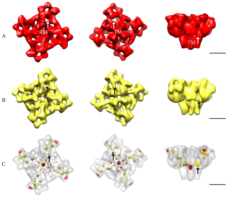
The ryanodine receptor (RyR) family of calcium release channels plays a vital role in excitation-contraction coupling (ECC). Along with the dihydropyridine receptor (DHPR), calsequestrin, and several other smaller regulatory and adaptor proteins, RyRs form a large dynamic complex referred to as ECC machinery. Here we describe a simple cross-linking procedure that can be used to stabilize fragile components of the ECC machinery, for the purpose of structural elucidation by single particle cryo-electron microscopy (cryo-EM). As a model system, the complex of the FK506-binding protein (FKBP12) and RyR1 was used to test the cross-linking protocol. Glutaraldehyde fixation led to complete cross-linking of receptor-bound FKBP12 to RyR1, and also to extensive cross-linking of the four subunits comprising RyR to one another without compromising the RyR1 ultrastructure. FKBP12 cross-linked with RyR1 was visualized in 2D averages by single particle cryo-EM. Comparison of control RyR1 and cross-linked RyR1 3D reconstructions revealed minor conformational changes at the transmembrane assembly and at the cytoplasmic region. Intersubunit cross-linking enhanced [(3)H]ryanodine binding to RyR1. Based on our findings we propose that intersubunit cross-linking of RyR1 by glutaraldehyde induced RyR1 to adopt an open like conformation.
Copyright © 2013 Elsevier Inc. All rights reserved.
Figures


Similar articles
-
The FKBP12 subunit modifies the long-range allosterism of the ryanodine receptor.J Struct Biol. 2019 Feb 1;205(2):180-188. doi: 10.1016/j.jsb.2018.12.007. Epub 2019 Jan 11. J Struct Biol. 2019. PMID: 30641143 Free PMC article.
-
Structural characterization of the RyR1-FKBP12 interaction.J Mol Biol. 2006 Mar 3;356(4):917-27. doi: 10.1016/j.jmb.2005.12.023. Epub 2005 Dec 27. J Mol Biol. 2006. PMID: 16405911
-
A cryo-EM-based model of phosphorylation- and FKBP12.6-mediated allosterism of the cardiac ryanodine receptor.Sci Signal. 2017 May 23;10(480):eaai8842. doi: 10.1126/scisignal.aai8842. Sci Signal. 2017. PMID: 28536302
-
Structural insights into excitation-contraction coupling by electron cryomicroscopy.Biochemistry (Mosc). 2004 Nov;69(11):1226-32. doi: 10.1007/s10541-005-0068-5. Biochemistry (Mosc). 2004. PMID: 15627376 Review.
-
Structural Basis for the Modulation of Ryanodine Receptors.Trends Biochem Sci. 2021 Jun;46(6):489-501. doi: 10.1016/j.tibs.2020.11.009. Epub 2020 Dec 22. Trends Biochem Sci. 2021. PMID: 33353849 Review.
Cited by
-
Functional Impact of Ryanodine Receptor Oxidation on Intracellular Calcium Regulation in the Heart.Rev Physiol Biochem Pharmacol. 2016;171:39-62. doi: 10.1007/112_2016_2. Rev Physiol Biochem Pharmacol. 2016. PMID: 27251471 Free PMC article. Review.
-
Denaturing mass photometry for rapid optimization of chemical protein-protein cross-linking reactions.Nat Commun. 2024 Apr 25;15(1):3516. doi: 10.1038/s41467-024-47732-4. Nat Commun. 2024. PMID: 38664367 Free PMC article.
References
-
- Aghdasi B, Zhang J, Wu Y, Reid MB, Hamilton SL. Multiple classes of sulfhydryls modulate the skeletal muscle Ca2+ release channel. J Biol Chem. 1997;272:3739–3748. - PubMed
-
- Bradford MM. A rapid and sensitive method for the quantitation of microgram quantities of protein utilizing the principle of protein-dye binding. Anal Biochem. 1976;72:248–54. - PubMed
Publication types
MeSH terms
Substances
Grants and funding
LinkOut - more resources
Full Text Sources
Other Literature Sources
Research Materials
