. 2013 May 1;98(2):286-96.
doi: 10.1093/cvr/cvt012. Epub 2013 Jan 19.

Flecainide reduces Ca(2+) spark and wave frequency via inhibition of the sarcolemmal sodium current

Affiliations

Flecainide reduces Ca(2+) spark and wave frequency via inhibition of the sarcolemmal sodium current

Markus B Sikkel et al. Cardiovasc Res. .

Abstract

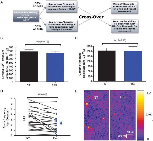
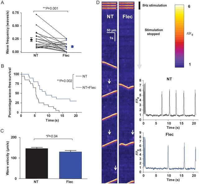
Aims: Ca(2+) waves are thought to be important in the aetiology of ventricular tachyarrhythmias. There have been conflicting results regarding whether flecainide reduces Ca(2+) waves in isolated cardiomyocytes. We sought to confirm whether flecainide inhibits waves in the intact cardiomyocyte and to elucidate the mechanism.

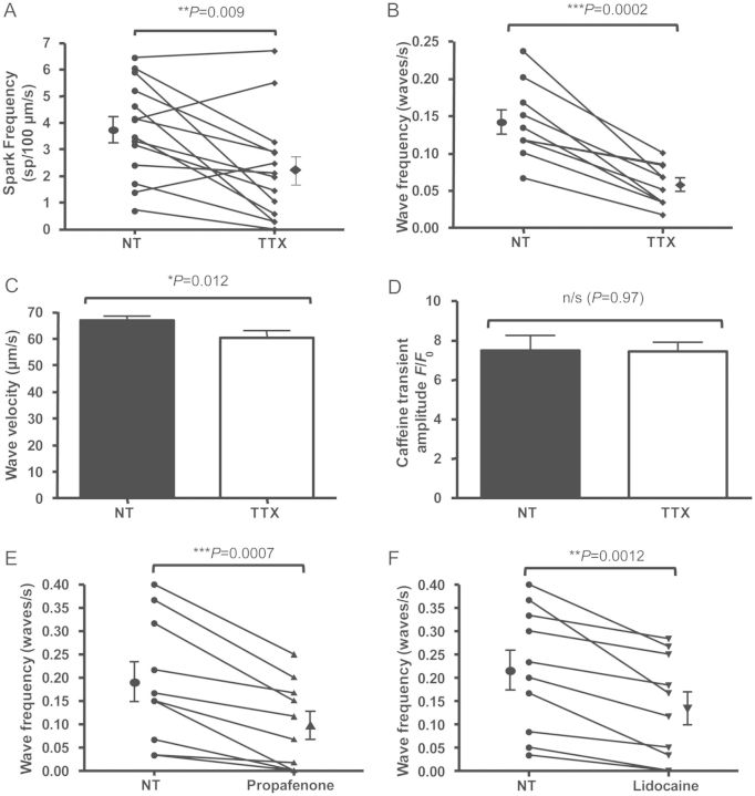
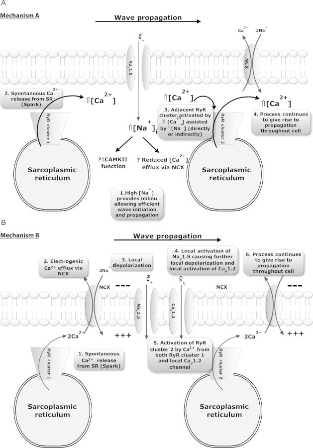
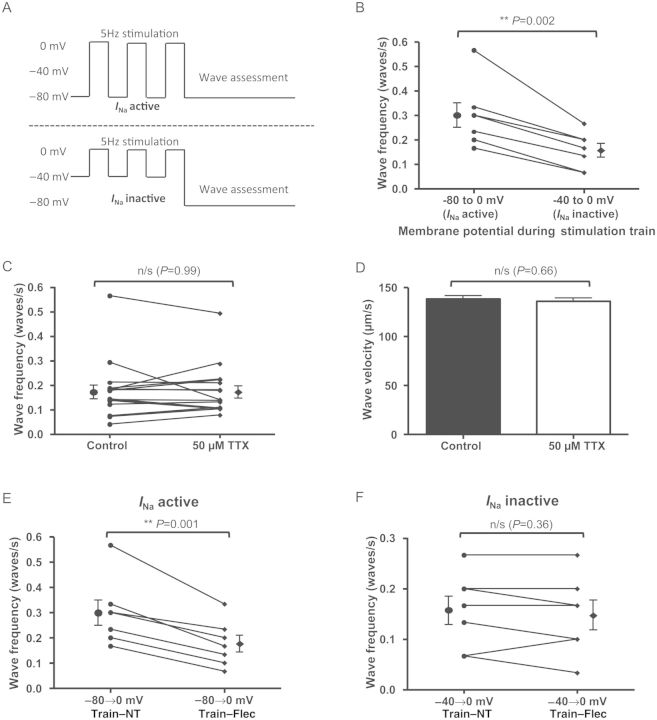
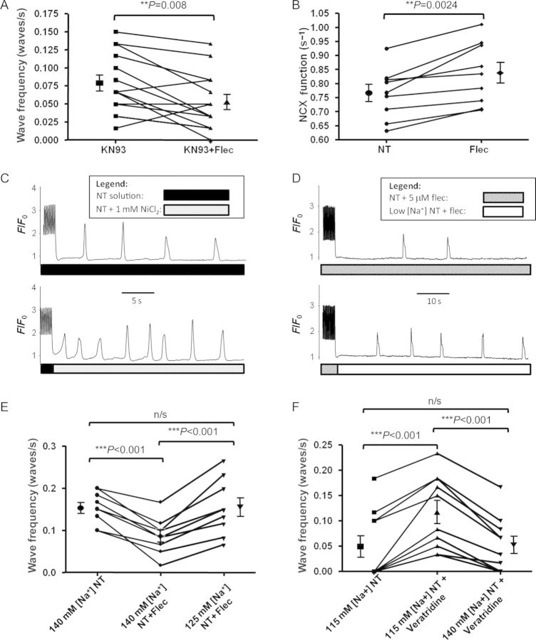
Methods and results: We imaged spontaneous sarcoplasmic reticulum (SR) Ca(2+) release events in healthy adult rat cardiomyocytes. Variation in stimulation frequency was used to produce Ca(2+) sparks or waves. Spark frequency, wave frequency, and wave velocity were reduced by flecainide in the absence of a reduction of SR Ca(2+) content. Inhibition of I(Na) via alternative pharmacological agents (tetrodotoxin, propafenone, or lidocaine) produced similar changes. To assess the contribution of I(Na) to spark and wave production, voltage clamping was used to activate contraction from holding potentials of -80 or -40 mV. This confirmed that reducing Na(+) influx during myocyte stimulation is sufficient to reduce waves and that flecainide only causes Ca(2+) wave reduction when I(Na) is active. It was found that Na(+)/Ca(2+)-exchanger (NCX)-mediated Ca(2+) efflux was significantly enhanced by flecainide and that the effects of flecainide on wave frequency could be reversed by reducing [Na(+)](o), suggesting an important downstream role for NCX function.

Conclusion: Flecainide reduces spark and wave frequency in the intact rat cardiomyocyte at therapeutically relevant concentrations but the mechanism involves I(Na) reduction rather than direct ryanodine receptor (RyR2) inhibition. Reduced I(Na) results in increased Ca(2+) efflux via NCX across the sarcolemma, reducing Ca(2+) concentration in the vicinity of the RyR2.


Figures

Figure 1
Figure 1
Experimental protocol and effects of flecainide on Ca2+ transients and Ca2+ sparks. (A) Experimental flowchart to explain cross-over protocol used in experiments. Fifty per cent of cells had NT applied first with drug wash-on, whereas 50% had drug applied first and subsequently washed-off. (B) Stimulated Ca2+ transients were assessed using the ratiometric dye fura-2 calibrated to give [Ca2+]i. Transient amplitude was not changed in the presence of 5 μM flecainide (n = 30 cells, P = 0.19). (C) 20 mM caffeine in 0Na+/0Ca2+ solution was used to assess SR Ca2+ load following a 5 Hz contraction train. The amplitude was unchanged in the presence of 5 µM flecainide (n = 13 cells in each group, P = 0.96 by Student's t-test). (D) Spark frequency was reduced following flecainide application (n = 24 cells, P = 0.002). (E) Representative line-scans showing a reduction in spark frequency with flecainide. The same cell is shown before and after flecainide application.
Figure 2
Figure 2
Effects of 5 µM flecainide on Ca2+ waves. (A) Flecainide was washed on or off via cross-over protocol for 5 min. In the presence of flecainide, wave frequency was significantly reduced (P = 0.001, n = 20 cells). (B) Latency period from the last transient to the first wave is shown in the Kaplan–Meier survival format (i.e. wave-free survival). Cells in the presence of flecainide have an increased wave-free survival period (P = 0.002 by log-rank test, n = 20 cells). (C) Wave velocity is reduced in the presence of flecainide (P = 0.04 by Student's t-test, NT: n = 81 waves; flec: n = 36 waves from 20 cells). (D) Representative line-scans from a cell assessed for waves pre- and post-flecainide application. The end of the 30 s period of 5 Hz stimulation evoking Ca2+ transients can be seen at the top of the scans with subsequent quiescent phase during which waves are observed. Areas of increased spark activity prior to waves are highlighted with white arrows and are more prominent in the absence of flecainide. Inset: line-scans converted into F/F0 plots—reduction of wave frequency and increased latency is apparent.
Figure 3
Figure 3
Effects of INa inhibition by tetrodotoxin (TTX), propafenone and lidocaine on SR Ca2+ release events. (A) 5 µM TTX applied via similar cross-over protocol to flecainide experiments induced a similar reduction in Ca2+ spark frequency (P = 0.009, n = 14 cells). (B) 5 µM TTX reduced wave frequency (P = 0.0002, n = 10 cells). (C) Wave velocity is significantly reduced in the presence of TTX (P = 0.012 by Student's t-test, NT: n = 84 waves; TTX: n = 34 waves from 10 cells). (D) Similar to flecainide experiments, no significant change in SR load was seen in the presence of 5 µM TTX (P = 0.97 by Student's t-test, n = 20 cells from three isolations). (E) 5 μM propafenone reduced Ca2+ wave frequency in a similar manner (P = 0.0007, n = 10 cells), as did (F) 200 μM lidocaine (P = 0.0012, n = 10 cells).
Figure 4
Figure 4
Possible hypotheses to explain how INa can contribute to wave initiation and propagation. (A) Entry of Na+ ions occurs via INa and an alteration of wave properties may result from changes in [Na+]i, particularly in the sub-sarcolemmal space. In this proposed mechanism (1) increased fuzzy space [Na+] provides a milieu that enhances the probability of (2) Ca2+ sparks leading to (3) the activation and firing of an adjacent RyR cluster to result in (4) wave initiation and propagation throughout the cell. (B) Alternatively Nav1.5 channels may be involved in wave propagation per se in the intact cardiomyocyte. Such involvement could comprise (1) spontaneous SR Ca2+ release in the form of a spark resulting in (2) local Ca2+ efflux by NCX causing (3) local depolarization of the sarcolemma, which (4) subsequently results in local activation of INa and ICa assisting the rise in local (‘fuzzy space’) [Ca2+]i that can lead to (5) adjacent RyR clusters firing and (6) wave propagation.
Figure 5
Figure 5
Elucidation of Mechanism A as most likely cause for reduction in Ca2+ waves due to INa blockade. (A) Voltage clamp stimulation trains used to assess wave frequency with and without INa activity. Stimulation was induced by stepping from −80 to 0 mv (INa active) or −40 to 0 mV (INa inactive). Pulse duration was 100 ms and pulses were applied at 5 Hz. Waves were assessed in a subsequent 30 s interval during which membrane potential was held at −80 mV. (B) With INa inactive during the stimulation train (but available during the quiescent phase of the experiment), wave frequency was reduced (P = 0.002, n = 7 cells). (C) High-dose (50 µM) TTX was rapidly applied to cells to terminate stimulation following a period of external field stimulation at 5 Hz and compared with the control arm in which stimulation was terminated in the usual fashion at 30 s (see Supplementary material online, Figure S4 for further explanation). This produced the opposite situation to the previous experiment with INa active during the stimulation train but Nav1.5 channels unavailable for stimulation during the quiescent phase. This produced no change in wave frequency (P = 0.99, n = 17 cells). (D) Similarly, there was no change in wave velocity (P = 0.66 by Student's t-test. Control: n = 88 waves; 50 µM TTX: n = 89 waves from 17 cells). (E) Voltage clamp experiments showing effects of flecainide on wave frequency with INa active vs. inactive. With INa active, flecainide reduces wave frequency (P = 0.001, n = 7 cells). (F) However, with INa inactive, no reduction in wave frequency was observed (P = 0.36, n = 7 cells).
Figure 6
Figure 6
Role of CaMKII and NCX in wave reduction by flecainide. (A) Despite incubation of cells with 1 μM CaMKII inhibitor KN-93, flecainide was still able to significantly reduce Ca2+ wave frequency. Magnitude of reduction was similar in the presence of inactive analogue KN-92 (see Supplementary material online, Figure S5A), suggesting CaMKII inhibition is not the mechanism of wave reduction with flecainide. (B) NCX function in terms of Ca2+ efflux efficacy was significantly improved following a 5 Hz contraction train in the presence of flecainide. (C) Direct partial inhibition of NCX by 1 mM Ni2+ applied after the contraction train increased Ca2+ wave frequency. (D) Reduction of [Na+]o after the contraction train can reverse the reduction in wave frequency seen with flecainide. (E) Pooled data from experimental protocol shown in (D) revealing that a reduction in wave frequency induced by flecainide can be reversed by reducing [Na+]o to 125 mM. (F) 0.5 μM veratridine can increase Ca2+ wave frequency via enhancing INa. This effect was abolished by increasing [Na+]o from 115 to 140 mM.

Comment in

Similar articles

Cited by

References

    1. Zhou Q, Xiao J, Jiang D, Wang R, Vembaiyan K, Wang A, et al. Carvedilol and its new analogs suppress arrhythmogenic store overload-induced Ca2+ release. Nat Med. 2011;17:1003–1009. doi:10.1038/nm.2406. - DOI - PMC - PubMed
    1. Shannon TR, Pogwizd SM, Bers DM. Elevated sarcoplasmic reticulum Ca2+ leak in intact ventricular myocytes from rabbits in heart failure. Circ Res. 2003;93:592–594. doi:10.1161/01.RES.0000093399.11734.B3. - DOI - PubMed
    1. Lyon AR, Bannister ML, Collins T, Pearce E, Sepehripour AH, Dubb SS, et al. SERCA2a gene transfer decreases sarcoplasmic reticulum calcium leak and reduces ventricular arrhythmias in a model of chronic heart failure. Circ Arrhythm Electrophysiol. 2011;4:362–372. doi:10.1161/CIRCEP.110.961615. - DOI - PMC - PubMed
    1. MacLennan DH, Chen SR. Store overload-induced Ca2+ release as a triggering mechanism for CPVT and MH episodes caused by mutations in RYR and CASQ genes. J Physiol. 2009;587:3113–3115. doi:10.1113/jphysiol.2009.172155. - DOI - PMC - PubMed
    1. Kushnir A, Marks AR. The ryanodine receptor in cardiac physiology and disease. Adv Pharmacol. 2010;59:1–30. doi:10.1016/S1054-3589(10)59001-X. - DOI - PMC - PubMed

Publication types

MeSH terms

Substances