. 2014 Jan 30;33(5):599-610.
doi: 10.1038/onc.2012.628. Epub 2013 Jan 21.

The mitotic kinase Aurora--a promotes distant metastases by inducing epithelial-to-mesenchymal transition in ERα(+) breast cancer cells

Affiliations

The mitotic kinase Aurora--a promotes distant metastases by inducing epithelial-to-mesenchymal transition in ERα(+) breast cancer cells

A B D'Assoro et al. Oncogene. .

Abstract

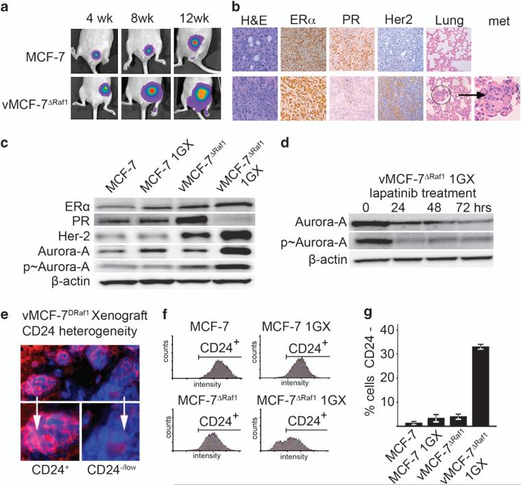
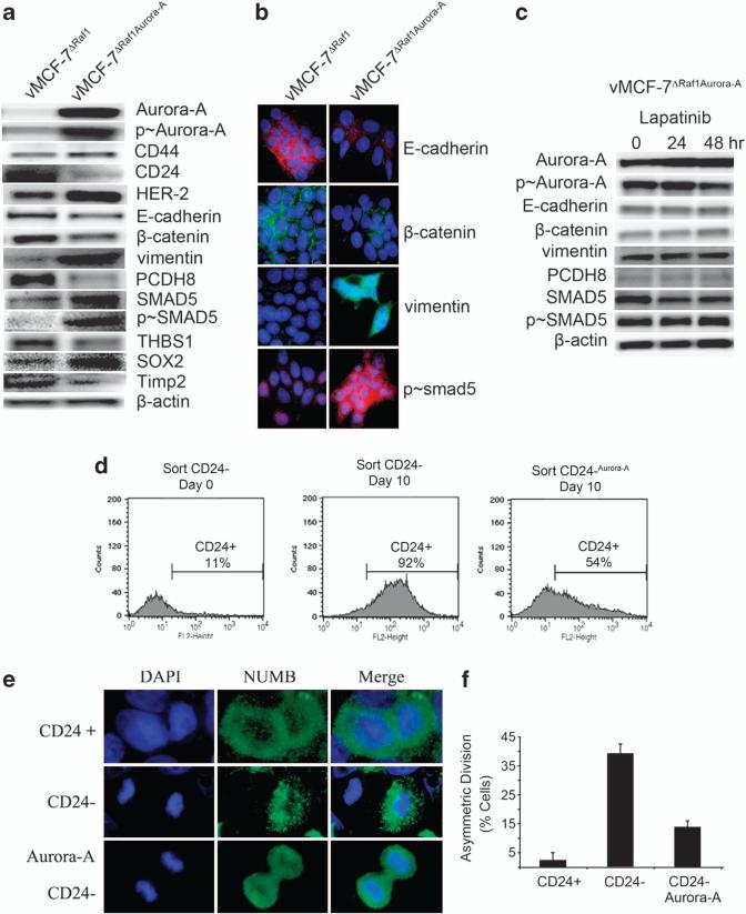
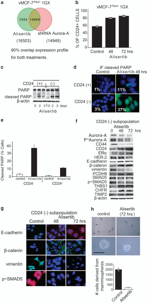
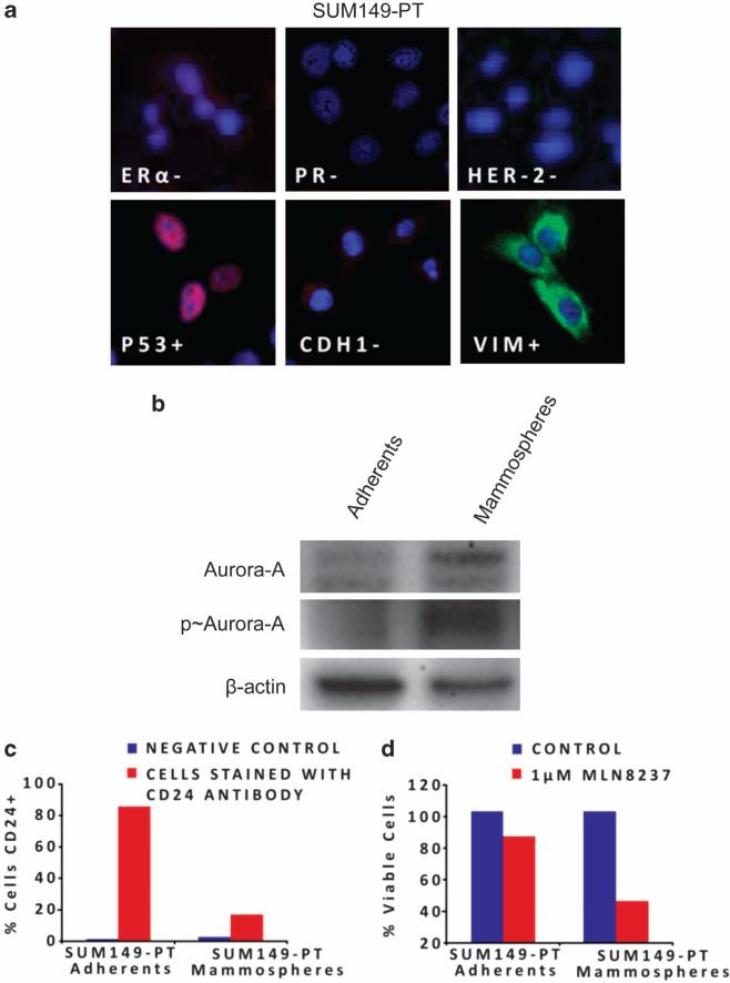
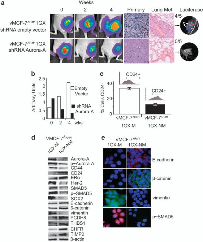
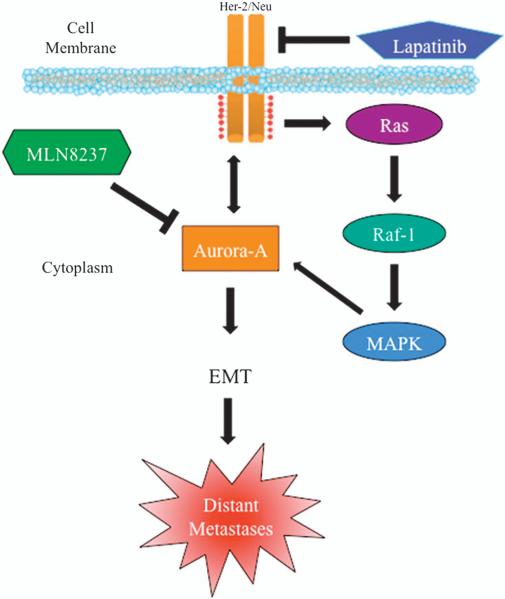
In this study, we demonstrate that constitutive activation of Raf-1 oncogenic signaling induces stabilization and accumulation of Aurora-A mitotic kinase that ultimately drives the transition from an epithelial to a highly invasive mesenchymal phenotype in estrogen receptor α-positive (ERα(+)) breast cancer cells. The transition from an epithelial- to a mesenchymal-like phenotype was characterized by reduced expression of ERα, HER-2/Neu overexpression and loss of CD24 surface receptor (CD24(-/low)). Importantly, expression of key epithelial-to-mesenchymal transition (EMT) markers and upregulation of the stemness gene SOX2 was linked to acquisition of stem cell-like properties such as the ability to form mammospheres in vitro and tumor self-renewal in vivo. Moreover, aberrant Aurora-A kinase activity induced phosphorylation and nuclear translocation of SMAD5, indicating a novel interplay between Aurora-A and SMAD5 signaling pathways in the development of EMT, stemness and ultimately tumor progression. Importantly, pharmacological and molecular inhibition of Aurora-A kinase activity restored a CD24(+) epithelial phenotype that was coupled to ERα expression, downregulation of HER-2/Neu, inhibition of EMT and impaired self-renewal ability, resulting in the suppression of distant metastases. Taken together, our findings show for the first time the causal role of Aurora-A kinase in the activation of EMT pathway responsible for the development of distant metastases in ERα(+) breast cancer cells. Moreover, this study has important translational implications because it highlights the mitotic kinase Aurora-A as a novel promising therapeutic target to selectively eliminate highly invasive cancer cells and improve the disease-free and overall survival of ERα(+) breast cancer patients resistant to conventional endocrine therapy.


Figures

Figure 1
Figure 1
Establishment of MCF-7 and vMCF-7ΔRaf-1 breast cancer xenografts. (a) Tumor xenografts imaging in live animals of MCF-7 (upper row) and vMCF-7ΔRaf-1 (lower row) expressing the firefly luciferase reporter lentivector at 4, 8 and 12 weeks after mammary fat pad injection. (b) Paraffin sections of xenograft tumors (12 weeks) showing: hematoxylin and eosin (H&E) staining of low-grade tubular tumors for MCF-7 (upper row) and high-grade vMCF-7ΔRaf-1 tumors (lower row); expression of ER in both xenografts; loss of progesterone receptor (PR) and HER-2/Neu expression in vMCF-7ΔRaf-1 xenografts; and H&E staining of lungs showing development of metastases in vMCF-7Raf-1 xenografts. (c) Immunoblot analysis of parental and cancer cells re-cultured from tumor xenografts (1GX) showing that vMCF-7ΔRaf-1 1GX cells retain the expression of ER, lack expression of progesterone receptor and overexpress HER-2/Neu and Aurora-A. (d) Immunoblot analysis of vMCF-7ΔRaf-1 1GX cells treated with 1 μM lapatinib showing reduced expression of total and p-Aurora-A. (e) Immunofluorescence analysis showing tumor cell heterogeneity for the luminal marker CD24 in vMCF-7ΔRaf-1 xenografts. CD24 receptor was labeled in red and DNA was labeled in blue with Hoechst dye. (f) FACS analysis showing that only vMCF-7ΔRaf-1 1GX cells developed a subpopulation of CD24–/low cells (~30%), while MCF-7, MCF-7 1GX and vMCF-7ΔRaf-1 displayed a CD24+ phenotype. (g) Graph showing the percentage of MCF-7 and variant cells displaying a CD24–/low phenotype from three independent experiments (±s.d.).
Figure 2
Figure 2
Molecular characterization of CD24+ and CD24–/low breast cancer cells. (a) Lung metastases imaging in live animals of MDA-MB 231, MCF-7 and vMCF-7ΔRaf-1 cells expressing the firefly luciferase reporter lentivector 1 week after tail vein injection. (b) Heat map representing the unsupervised cluster analysis of global gene expression in CD24+ and CD24–/low subpopulations derived from vMCF-7ΔRaf-1 1GX cells and identification of an invasive transcriptome signature. (c) Immunoblot analysis showing that CD24–/low cells overexpress HER-2/Neu, Aurora-A, EMT and cancer stem cell markers. (d) Immunofluorescence analysis showing activation of EMT in CD24–/low cells characterized by loss of E-cadherin and β-catenin, expression of vimentin and nuclear localization of p-SMAD5. (e) Light microscopy analysis showing development of mammospheres from CD24–/low cells grown under non-adherent conditions at 0, 2, 8 and 24 days (three passages). (f) FACS analysis showing the percentage of CD24+ cells in CD24+ and CD24–/low subpopulations at day 0 and after 10 days following sorting analysis from three independent experiments (±s.d). (g) Tumor xenografts imaging in live animals of CD24+ and CD24–/low cancer cells expressing the firefly luciferase reporter lentivector at 4 weeks after mammary fat pad injection. CD24+ and CD24–/low cells were injected into the mammary fat pad of nude mice and only CD24 cells showed tumorigenic activity at a concentration of 100 000 cells.
Figure 3
Figure 3
Overexpression of Aurora-A induces EMT and a cancer stem cell-like phenotype. (a) Immunoblot analysis of vMCF-7ΔRaf-1 and vMCF-7ΔRaf-1 cells engineered to overexpress Aurora-A (vMCF-7ΔRaf-1/Aurora-A) showing that vMCF-7ΔRaf-1/Aurora-A cells overexpress CD44, HER-2/Neu and Aurora-A, CD24 downregulation, and expression of EMT and cancer stem cell markers. (b) Immunofluorescence analysis of vMCF-7ΔRaf-1 and vMCF-7ΔRaf-1/Aurora-A cells showing activation of EMT in vMCF-7ΔRaf-1/Aurora-A cells characterized by loss of E-cadherin and β-catenin, expression of vimentin and nuclear localization of p-SMAD5. (c) Immunoblot analysis of vMCF-7ΔRaf-1/Aurora-A cells treated with 1 μM lapatinib showing that constitutive activation of Aurora-A kinase activity is essential for the development of EMT regardless inhibition of HER-2/Neu signaling. (d) FACS analysis showing that sorted CD24–/low cells infected with a lentivector overexpressing Aurora-A maintain a higher percentage of cells carrying a CD24–/low phenotype after 10 days in culture compared with control CD24–/low cells that give rise to a CD24+ subpopulation. (e) Asymmetric division in CD24–/low cells, and symmetric division in CD24+ and CD24–/low cells overexpressing Aurora-A (CD24–/low/Aurora-A). The marker of asymmetric mitotic divisions NUMB was labeled in green and DNA was labeled in blue with Hoechst dye. (f) Graph showing the percentage of cells with asymmetric divisions in CD24+, CD24–/low and CD24–/low/Aurora-A cells from three independent experiments (±s.d.).
Figure 4
Figure 4
Molecular inhibition of Aurora-A kinase activity in vitro reverses EMT and suppresses self-renewal ability of CD24–/low breast cancer cells. (a) Venn diagram of the unsupervised cluster analysis of global gene expression between vMCF-7ΔRaf-1 1GX cells treated with Alisertib or with an shRNA Aurora-A vector showing 90% overlap expression profile. (b) Graph showing FACS analysis of vMCF-7ΔRaf-1 1GX cells treated with 1 μM Alisertib for 48 and 72 h displaying a CD24+ phenotype from three independent experiments (±s.d). (c) Immunoblot analysis of CD24+ and CD24–/low cells showing that treatment with 1 μM Alisertib induced cleaved PARP mainly in the CD24–/low subpopulation. (d) Immunofluorescence analysis of CD24+ and CD24–/low cells treated with 1 μM Alisertib for 48 h showing activation of apoptosis mainly in CD24–/low cells. Cleaved PARP was labeled in green and DNA was labeled in blue with Hoechst dye. (e) Graph showing the percentage of CD24+ and CD24–/low cells displaying cleaved PARP before and after treatment with Alisertib. Experiments were performed in triplicate (±s.d.). (f) Immunoblot analysis of CD24–/low cells treated with Alisertib for 48 and 72 h showing reversion of EMT and restoration of an epithelial CD44/CD24+ phenotype. (g) Immunofluorescence analysis of CD24–/low cells treated with1 μM MLN8237 for 48 and 72 h showing reversion of EMT. E-cadherin and p-SMAD5 were labeled in red, β-catenin and vimentin were labeled in green, and DNA was labeled in blue with Hoechst dye. (h) Light microscopy analysis showing inhibition of mammospheres growth derived from CD24–/low cells after treatment with 0.1 μM Alisertib for 72 h. Upper panels: low magnification; lower panels: high magnification. Graph showing the percentage of cells derived from mammospheres before and after treatment with Alisertib. Experiments were performed in triplicate (±s.d.).
Figure 5
Figure 5
Chemosensitivity of SUM149-PT breast cancer cells to Alisertib. (a) Immunofluorescence showing a triple-negative and fibroblastoid-like phenotype of SUM149-PT cells. ER, PR, P53, CDH1 were labeled in red, HER-2 and Vimentin were labeled in green and DNA was labeled in blue with Hoechst dye. (b) Immublotting assay showing increased expression and activity of Aurora-A kinase in SUM149-PT mammospheres. (c) FACS analysis showing loss of CD24 receptor in SUM149-PT mammospheres. (d) 3-[4,5-Dimethylthiazol-2-yl]-2,5-diphenyl tetrazolium bromide (MTT) assay showing chemosensitivity of SUM149-PT mammospheres to Alisertib.
Figure 6
Figure 6
Molecular targeting of Aurora-A kinase activity in vivo restores an epithelial phenotype and suppresses breast cancer metastases. (a) Tumor imaging in live animals of vMCF-7ΔRaf-1 xenografts expressing the firefly luciferase reporter lentivector. Following 8 weeks growth, tumor xenografts were treated with an empty shRNA vector (upper row) and an shRNA targeting Aurora-A (lower row) and imaged at 0, 2 and 4 weeks after treatment. Paraffin sections of xenograft tumors showing hematoxylin and eosin (H&E) staining of primary tumors and lung tissue. (b) Graph showing the area of tumor xenografts growth using NIH Image J program from three independent experiments. (c) Graph showing that nonmetastatic cells (vMCF-7ΔRaf-1 1GX-NM) derived from tumor xenografts treated with shRNA Aurora-A decreased the percentage of CD24–/low cells compared with control metastatic cells (vMCF-7ΔRaf-1 1GX-M). The percentage of CD24–/low cells was detected by FACS analysis from three independent experiments (±s.d). (d) Immunoblot analysis of vMCF-7ΔRaf-1 1GX-M and vMCF-7ΔRaf-1 1GX-NM cells showing suppression of EMT and restoration of an epithelial phenotype in vMCF-7ΔRaf-1 1GX-NM cells. (e) Immunofluorescence analysis showing expression of epithelial markers E-cadherin and β-catenin, lack of vimentin and inhibition of nuclear SMAD5 phosphorylation in vMCF-7ΔRaf-1 1GX-NM cells. E-cadherin and p-SMAD5 were labeled in red, β-catenin and vimentin were labeled in green and DNA was labeled in blue with Hoechst dye.
Figure 7
Figure 7
Induction of EMT and breast cancer progression through aberrant activation of Aurora-A kinase activity. Constitutive activation of HER-2/MAPK signaling pathway during tumor growth leads to stabilization and accumulation of Aurora-A kinase. Aurora-A kinase activation in turn increases the expression levels of HER-2/Neu receptor, creating a ‘positive feedback loop’ between HER-2/Neu and Aurora-A tumorigenic activity. Aberrant Aurora-A kinase activity induces EMT and the development of a mesenchymal phenotype responsible for distant metastases. Targeting Aurora-A kinase signaling pathway can be effective for eliminating highly invasive breast cancer cells and suppress tumor progression.

References

    1. Basaran G, Devrim C, Caglar HB, Gulluoglu B, Kaya H, Seber S, et al. Clinical outcome of breast cancer patients with N3a (>/ = 10 positive lymph nodes) disease: has it changed over years? Med Oncol. 2010;28:726–732. - PubMed
    1. Chang H, Rha SY, Jeung HC, Im CK, Ahn JB, Kwon WS, et al. Association of the ABCB1 gene polymorphisms 2677G>T/A and 3435C>T with clinical outcomes of paclitaxel monotherapy in metastatic breast cancer patients. Ann Oncol. 2009;20:272–277. - PubMed
    1. Mani SA, Guo W, Liao MJ, Eaton EN, Ayyanan A, Zhou AY, et al. The epithelial–mesenchymal transition generates cells with properties of stem cells. Cell. 2008;133:704–715. - PMC - PubMed
    1. Hwang-Verslues WW, Kuo WH, Chang PH, Pan CC, Wang HH, Tsai ST, et al. Multiple lineages of human breast cancer stem/progenitor cells identified by profiling with stem cell markers. PLoS One. 2009;4:e8377. - PMC - PubMed
    1. D'Assoro AB, Leontovich A, Amato A, Ayers-Ringler JR, Quatraro C, Hafner K, et al. Abrogation of p53 function leads to metastatic transcriptome networks that typify tumor progression in human breast cancer xenografts. Int J Oncol. 2010;37:1167–1176. - PMC - PubMed

Publication types

MeSH terms