

Vascular remodeling in pulmonary hypertension
- PMID: 23334338
- PMCID: PMC3584237
- DOI: 10.1007/s00109-013-0998-0
Vascular remodeling in pulmonary hypertension
Abstract
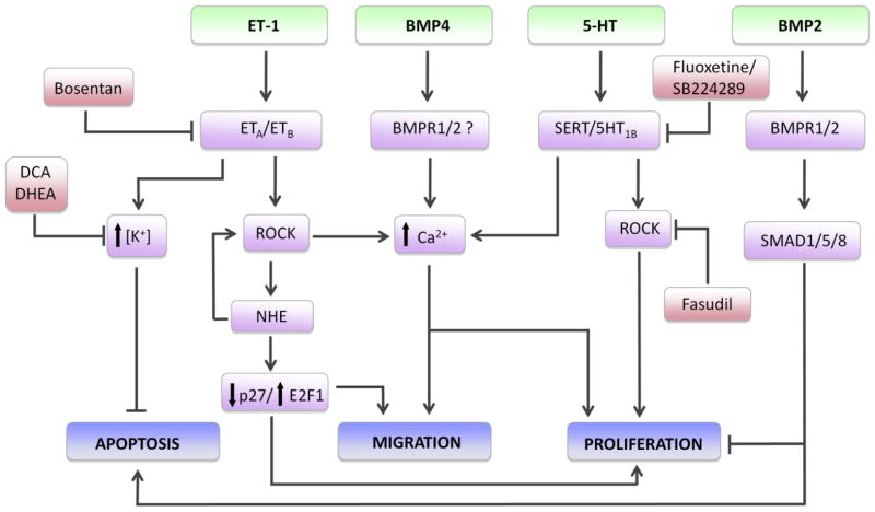
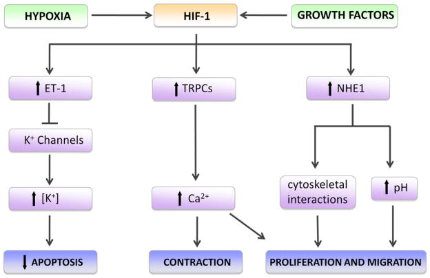
Pulmonary hypertension is a complex, progressive condition arising from a variety of genetic and pathogenic causes. Patients present with a spectrum of histologic and pathophysiological features, likely reflecting the diversity in underlying pathogenesis. It is widely recognized that structural alterations in the vascular wall contribute to all forms of pulmonary hypertension. Features characteristic of the remodeled vasculature in patients with pulmonary hypertension include increased stiffening of the elastic proximal pulmonary arteries, thickening of the intimal and/or medial layer of muscular arteries, development of vaso-occlusive lesions, and the appearance of cells expressing smooth muscle-specific markers in normally non-muscular small diameter vessels, resulting from proliferation and migration of pulmonary arterial smooth muscle cells and cellular transdifferentiation. The development of several animal models of pulmonary hypertension has provided the means to explore the mechanistic underpinnings of pulmonary vascular remodeling, although none of the experimental models currently used entirely replicates the pulmonary arterial hypertension observed in patients. Herein, we provide an overview of the histological abnormalities observed in humans with pulmonary hypertension and in preclinical models and discuss insights gained regarding several key signaling pathways contributing to the remodeling process. In particular, we will focus on the roles of ion homeostasis, endothelin-1, serotonin, bone morphogenetic proteins, Rho kinase, and hypoxia-inducible factor 1 in pulmonary arterial smooth muscle and endothelial cells, highlighting areas of cross-talk between these pathways and potentials for therapeutic targeting.
Figures
References
-
- Galie N, Hoeper MM, Humbert M, Torbicki A, Vachiery JL, Barbera JA, Beghetti M, Corris P, Gaine S, Gibbs JS, et al. Guidelines for the diagnosis and treatment of pulmonary hypertension: the Task Force for the Diagnosis and Treatment of Pulmonary Hypertension of the European Society of Cardiology (ESC) and the European Respiratory Society (ERS), endorsed by the International Society of Heart and Lung Transplantation (ISHLT) Eur Heart J. 2009;30:2493–2537. - PubMed
-
- Badesch DB, Champion HC, Sanchez MA, Hoeper MM, Loyd JE, Manes A, McGoon M, Naeije R, Olschewski H, Oudiz RJ, et al. Diagnosis and assessment of pulmonary arterial hypertension. J Am Coll Cardiol. 2009;54:S55–66. - PubMed
-
- Stenmark KR, Meyrick B, Galie N, Mooi WJ, McMurtry IF. Animal models of pulmonary arterial hypertension: the hope for etiological discovery and pharmacological cure. Am J Physiol Lung Cell Mol Physiol. 2009;297:L1013–1032. - PubMed
-
- Tuder RM. Pathology of pulmonary arterial hypertension. Semin Respir Crit Care Med. 2009;30:376–385. - PubMed
Publication types
MeSH terms
Substances
Grants and funding
LinkOut - more resources
Full Text Sources
Other Literature Sources
Medical
