. 2013 Feb 7;494(7435):116-20.
doi: 10.1038/nature11809. Epub 2013 Jan 13.

Microbiota restricts trafficking of bacteria to mesenteric lymph nodes by CX(3)CR1(hi) cells

Affiliations

Microbiota restricts trafficking of bacteria to mesenteric lymph nodes by CX(3)CR1(hi) cells

Gretchen E Diehl et al. Nature. .

Abstract

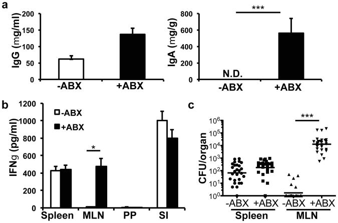
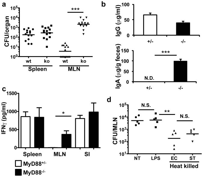
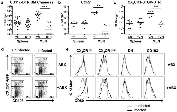
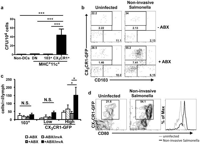
The intestinal microbiota has a critical role in immune system and metabolic homeostasis, but it must be tolerated by the host to avoid inflammatory responses that can damage the epithelial barrier separating the host from the luminal contents. Breakdown of this regulation and the resulting inappropriate immune response to commensals are thought to lead to the development of inflammatory bowel diseases such as Crohn's disease and ulcerative colitis. We proposed that the intestinal immune system is instructed by the microbiota to limit responses to luminal antigens. Here we demonstrate in mice that, at steady state, the microbiota inhibits the transport of both commensal and pathogenic bacteria from the lumen to a key immune inductive site, the mesenteric lymph nodes (MLNs). However, in the absence of Myd88 or under conditions of antibiotic-induced dysbiosis, non-invasive bacteria were trafficked to the MLNs in a CCR7-dependent manner, and induced both T-cell responses and IgA production. Trafficking was carried out by CX(3)CR1(hi) mononuclear phagocytes, an intestinal-cell population previously reported to be non-migratory. These findings define a central role for commensals in regulating the migration to the MLNs of CX(3)CR1(hi) mononuclear phagocytes endowed with the ability to capture luminal bacteria, thereby compartmentalizing the intestinal immune response to avoid inflammation.


Figures

Figure 1
Figure 1. Induction of immune response against non-invasive Salmonella after antibiotic treatment
Mice were left untreated or antibiotic treated for four weeks. (a) Mice were orally infected with non-invasive, non-pathogenic Salmonella (invA/aroA) and Salmonella-specific IgG in the blood and IgA in the feces were measured by ELISA. Bars represent the average from 5 mice from one of 3 independent experiments. ***P<0.001, unpaired t-test. Error bars represent S.E.M. (b) T cells from spleen (SP), mesenteric lymph node (MLN), and small intestine lamina propria (SI) of animals infected with non-invasive Salmonella were cultured with irradiated splenocytes and boiled Salmonella antigen and IFNγ was measured by ELISA. Bars represent three mice per treatment group and are representative of two independent experiments. *P<0.05, unpaired t-test. Error bars represent S.E.M. (c) Bacterial titers in the spleen and MLN were determined for mice infected with non-invasive Salmonella (invA). Data points represent organs from a single mouse; data were pooled from six experiments. ***P<0.0001, Mann-Whitney Test. Error bars represent the geometric mean.
Figure 2
Figure 2. MyD88-dependent signals limit non-invasive Salmonella entry in the MLN
(a) Myd88-/- or heterozygous littermates were infected with noninvasive Salmonella (invA) and bacterial titers were determined in the spleen and MLN. Data points represent organs from a single mouse. Data were pooled from three independent experiments. ***P<0.0001, Mann-Whitney Test. Error bars represent the geometric mean. (b) Myd88-/- or heterozygous littermates were infected with non-invasive, non-pathogenic Salmonella and blood and feces were analyzed for Salmonella-specific IgG and IgA. Bars represent data from three mice per genotype. Data are representative of three independent experiments. ***P<0.001, unpaired t-test. Error bars represent S.E.M. (c) IFNγ production by spleen, MLN and small intestinal lamina propria (SI) T cells from Salmonella-infected mice of the indicated genotype. Analysis was as in Figure 1b. Bars represent three animals per genotype. Data are representative of two independent experiments. *P<0.05, unpaired t-test. Error bars represent S.E.M. (d) Antibiotic-treated B6 mice were left untreated (NT) or gavaged twice with LPS or heat killed E. coli (EC) or Salmonella (ST). 24h later the mice were infected orally with non-invasive Salmonella. Bacterial titers in the MLN were determined two days later. Each point represents individual mice from one of two independent experiments. **P<0.002, one-way ANOVA. Error bars represent the geometric mean.
Figure 3
Figure 3. Colonization of MLN by non-invasive Salmonella requires CCR7-dependent trafficking of CX3CR1hicells
(a-c) Antibiotic-treated mice of the indicated genotype and littermate controls were orally infected with non-invasive Salmonella, and bacterial titers in spleen and MLN were determined. For (a) and (c), animals were treated with diphtheria toxin for two consecutive days before infection. For (a) ***P<0.0001, for (b) **P<0.005, for (c) ***P=0.0006, all Mann-Whitney Test. Error bars represent the geometric mean. (d) Analysis of dendritic cell subsets in MLN of untreated or antibiotic-treated Cx3cr1gfp/+ mice that were mock infected or infected with non-invasive Salmonella. MLN cells were isolated at 48h, gated on the MHCII+CD11c+ population, and analyzed for expression of CX3CR1 and CD103. Percentages are shown in each gate. Absolute numbers are shown in Figure S9a. (e) Expression of CD80 on intestinal myeloid cell subsets. Cells were gated on the indicated cell populations as shown in (d). For data in (a-c), points represents individual mice pooled from independent experiments. Panels in (d) represent individual mice from one of five independent experiments.
Figure 4
Figure 4. CD103-CX3CR1+cells migrate into afferent lymphatics of antibiotic-treated animals
(a) CX3CR1+ or CD103+ cells (gated on the MHCII+CD11c+ population, as in Figure 3d) from the MLN were isolated from antibiotic-treated, non-invasive Salmonella-infected mice. The numbers of bacteria per 104 cells were determined by plating on LB-strep plates. No bacteria were observed from cells isolated from uninfected or infected but antibiotic-untreated mice. Bars represent pooled data from 15 individual mice from 4 independent experiments. ***P<0.0001, one-way ANOVA with Bonferroni correction. Error bars represent S.E.M. (b) – (d) Untreated or antibiotic-treated Cx3cr1gfp/+ were left uninfected or were infected with non-invasive Salmonella. At 48h, cells in the intestinal lymph were isolated and analyzed by flow cytometry. (b) Cells were gated on the MHCII+CD11c+ population and analyzed for expression of CX3CR1-GFP and CD103. Data are representative of three independent experiments. (c) Quantitation of MHCII+CD11c+CX3CR1-GFP+ cells in the afferent lymph of the indicated mice. **P<0.01, one-way ANOVA with Bonferroni correction. Error bars represent S.E.M. (d) CD80 expression on CX3CR1-GFP+ cells from lymph of antibiotic-treated mice with and without Salmonella infection.

Comment in

References

    1. Macpherson AJ, Uhr T. Compartmentalization of the mucosal immune responses to commensal intestinal bacteria. Ann N Y Acad Sci. 2004;1029:36–43. - PubMed
    1. Hooper LV, Gordon JI. Commensal host-bacterial relationships in the gut. Science. 2001;292:1115–1118. - PubMed
    1. Hooper LV, Midtvedt T, Gordon JI. How host-microbial interactions shape the nutrient environment of the mammalian intestine. Annu Rev Nutr. 2002;22:283–307. - PubMed
    1. Hooper LV, et al. Molecular analysis of commensal host-microbial relationships in the intestine. Science. 2001;291:881–884. - PubMed
    1. Rakoff-Nahoum S, Paglino J, Eslami-Varzaneh F, Edberg S, Medzhitov R. Recognition of commensal microflora by toll-like receptors is required for intestinal homeostasis. Cell. 2004;118:229–241. - PubMed

Publication types

MeSH terms