. 2013 Feb 14;494(7436):243-6.
doi: 10.1038/nature11823. Epub 2013 Jan 16.

A direct and melanopsin-dependent fetal light response regulates mouse eye development

Affiliations

A direct and melanopsin-dependent fetal light response regulates mouse eye development

Sujata Rao et al. Nature. .

Abstract

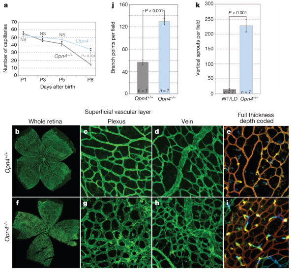
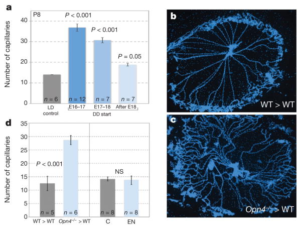
Vascular patterning is critical for organ function. In the eye, there is simultaneous regression of embryonic hyaloid vasculature (important to clear the optical path) and formation of the retinal vasculature (important for the high metabolic demands of retinal neurons). These events occur postnatally in the mouse. Here we have identified a light-response pathway that regulates both processes. We show that when mice are mutated in the gene (Opn4) for the atypical opsin melanopsin, or are dark-reared from late gestation, the hyaloid vessels are persistent at 8 days post-partum and the retinal vasculature overgrows. We provide evidence that these vascular anomalies are explained by a light-response pathway that suppresses retinal neuron number, limits hypoxia and, as a consequence, holds local expression of vascular endothelial growth factor (VEGFA) in check. We also show that the light response for this pathway occurs in late gestation at about embryonic day 16 and requires the photopigment in the fetus and not the mother. Measurements show that visceral cavity photon flux is probably sufficient to activate melanopsin-expressing retinal ganglion cells in the mouse fetus. These data thus show that light--the stimulus for function of the mature eye--is also critical in preparing the eye for vision by regulating retinal neuron number and initiating a series of events that ultimately pattern the ocular blood vessels.


Figures

Figure 1
Figure 1. | Hyaloid regression is regulated by light
a, Hyaloid vessel preparations at the indicated postnatal (P) days from pups reared under normal light conditions (LD) or under constant darkness (DD) from E16–17. Original magnification, ×50. b, As in a but a quantification of vessel number from P1 to P8. P values obtained by analysis of variance (ANOVA). c, P5 apoptotic index in hyaloid vascular cells (isolated apoptosis) or vessels undergoing a segmental pattern of apoptosis. P values obtained by Student's t-test. Sample size (n) as labelled. NS, not significant. Error bars are s.e.m.
Figure 2
Figure 2. |Hyaloid regression and retinal angiogenesis are regulated by melanopsin
a, Quantification of hyaloid vessels in Opn4+/+ and Opn4−/− mice over a P1 to P8 time course. P values obtained by ANOVA. b–i, Low (×100; b, f) and high (×200; c–e, g–i) magnification images of isolectin-labelled P8 retina from wild-type (b–e) and Opn4−/− (f–i) pups raised in normal lighting.e, i, Depth-coded z stack images for wild type (e) and Opn4−/− (i) indicate the appearance of vertical angiogenic sprouts. j, k, Graphs show quantification of branch points (j) and vertical sprouts (k) in animals of the indicated genotypes. WT, wild type. P values obtained by Student's t-test. Errors bars are s.e.m. Sample sizes (n) as labelled.
Figure 3
Figure 3. | Light and melanopsin-dependent regulation of VEGFA expression and hypoxia in the retina
a, Hyaloid vessel number from P1 to P8 in mice of labelled genotypes. P values obtained by ANOVA. b, Vitreous VEGFA immunoblot (IB) for wild-type mice at P1, P5 and P8 with quantification histogram. c, Immunoblot for P1 or P5 vitreous VEGFA in wild-type and Opn4−/− mice or in mice reared in LD or DD light conditions as labelled. d, ELISA quantification of VEGFA levels in the P5 vitreous of control/LD mouse pups (grey bar) from Opn4−/− mice (pale blue bar)or those raised in constant darkness from E16–17 (DD, blue bar). e, qPCR detection of Vegfa mRNA in P5 retina from control/LD (grey bar), Opn4−/− mice (light blue bar) and dark-reared mice (DD, dark blue bar). P values in b, d, e were obtained by Student's t-test. Sample sizes (n) as labelled. Error bars are s.e.m. f, g, Labelling of flat-mount P5 retinas from wild-type (f) and Opn4−/− (g) mice for blood vessels (isolectin, green) and for hypoxia (red). Retinal myeloid cells label faintly with isolectin. Original magnification, ×100. h, i, Quantification of the relative levels of hypoxyprobe labelling in the retinas of LD and DD mice (c) and wild-type versus Opn4−/− (d) retinas.
Figure 4
Figure 4. | Gestational light controls vascular development in the eye
a, Quantification of hyaloid vessels in mice raised in normal lighting (LD, grey bar) and those dark-reared from E16–17 (dark-blue bar), E17–18 (medium-blue bar), or after E18 (light-blue bar). b, c, P8 hyaloid vessel preparations from a wild-type embryo transferred into a wild-type pseudopregnant female (WT>WT) and an Opn4−/− embryo transferred into a wild-type pseudopregnant female (Opn4−/− > WT). Original magnification, ×50. d, Left panel: quantification of hyaloid vessels in P8 WT>WT (n = 5) and Opn4−/−>WT (n = 6) pups. Right panel: quantification of hyaloid vessels in normal control pups at P8 (C; n = 8)and P8 pups (n = 8) born to an enucleated female (EN). Sample sizes (n) as labelled. P values in a by ANOVA; P values in d were obtained by Student's t-test. Errors bars are s.e.m.

Comment in

References

    1. Ito M, Yoshioka M. Regression of the hyaloid vessels and pupillarymembrane of the mouse. Anat Embryol (Berl) 1999;200:403–411. - PubMed
    1. Fruttiger M. Development of the retinal vasculature. Angiogenesis. 2007;10:77–88. - PubMed
    1. Provencio I, Jiang G, De Grip WJ, Hayes WP, Rollag MD. Melanopsin: an opsin in melanophores, brain, and eye. Proc Natl Acad Sci USA. 1998;95:340–345. - PMC - PubMed
    1. Hattar S, Liao HW, Takao M, Berson DM, Yau KW. Melanopsin-containing retinal ganglion cells: architecture, projections, and intrinsic photosensitivity. Science. 2002;295:1065–1070. - PMC - PubMed
    1. Panda S, et al. Melanopsin (Opn4) requirement for normal light-induced circadian phase shifting. Science. 2002;298:2213–2216. - PubMed

Publication types

MeSH terms

Substances