. 2013 Feb 7;494(7435):60-4.
doi: 10.1038/nature11783. Epub 2013 Jan 13.

Structural basis for viral 5'-PPP-RNA recognition by human IFIT proteins

Affiliations

Structural basis for viral 5'-PPP-RNA recognition by human IFIT proteins

Yazan M Abbas et al. Nature. .

Abstract

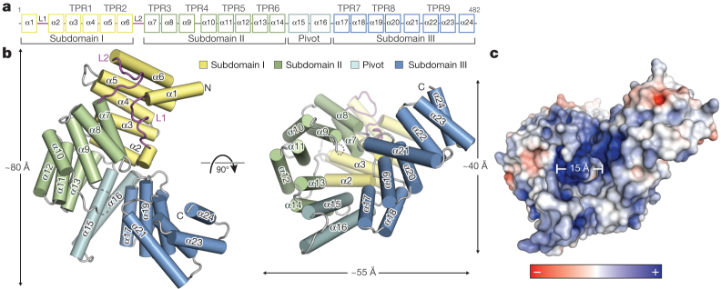
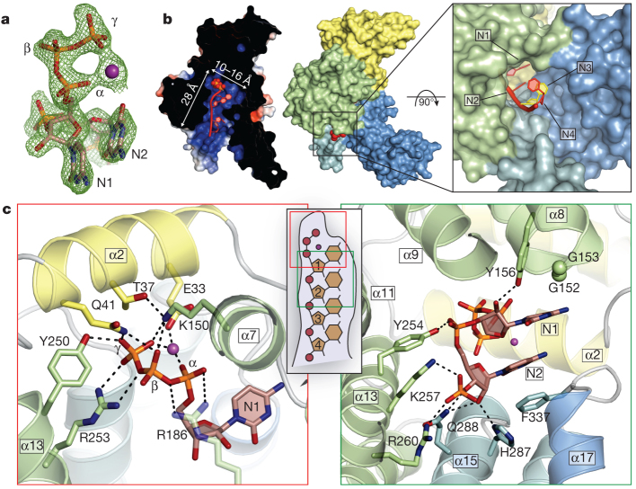
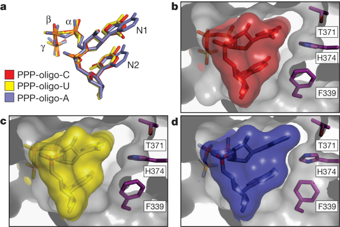
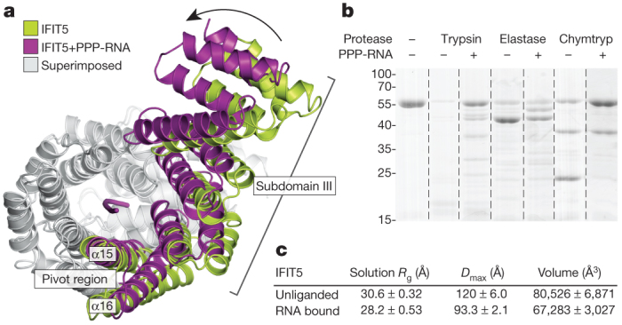
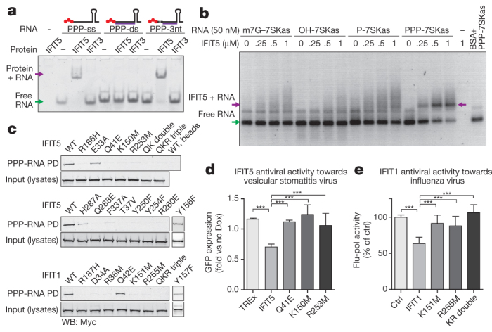
Interferon-induced proteins with tetratricopeptide repeats (IFITs) are innate immune effector molecules that are thought to confer antiviral defence through disruption of protein-protein interactions in the host translation-initiation machinery. However, it was recently discovered that IFITs can directly recognize viral RNA bearing a 5'-triphosphate group (PPP-RNA), which is a molecular signature that distinguishes it from host RNA. Here we report crystal structures of human IFIT5, its complex with PPP-RNAs, and an amino-terminal fragment of IFIT1. The structures reveal a new helical domain that houses a positively charged cavity designed to specifically engage only single-stranded PPP-RNA, thus distinguishing it from the canonical cytosolic sensor of double-stranded viral PPP-RNA, retinoic acid-inducible gene I (RIG-I, also known as DDX58). Mutational analysis, proteolysis and gel-shift assays reveal that PPP-RNA is bound in a non-sequence-specific manner and requires a 5'-overhang of approximately three nucleotides. Abrogation of PPP-RNA binding in IFIT1 and IFIT5 was found to cause a defect in the antiviral response by human embryonic kidney cells. These results demonstrate the mechanism by which IFIT proteins selectively recognize viral RNA, and lend insight into their downstream effector function.


Conflict of interest statement

The authors declare no competing financial interests.

Figures

Figure 1
Figure 1. Structural overview of human IFIT5.
a, Secondary structure, TPR motif and subdomain organization of IFIT5. b, Orthogonal views of IFIT5 with helices represented as cylinders. c, Surface representation of IFIT5 coloured by electrostatic potential (using APBS) from negative ( −5kTe−1; red) to positive (+5kTe−1; blue). PowerPoint slide
Figure 2
Figure 2. Structure of IFIT5 bound to PPP-RNA.
a, FoFc electron-density map of the triphosphate and first two nucleotides contoured at 3.5σ before inclusion of RNA into the model. The metal ion is indicated with a purple sphere. b, Left, cross-section of the complex coloured by surface electrostatic potential. The triphosphate is shown as spheres and RNA nucleotides are shown in red. Middle, surface representation of IFIT5 bound to PPP-RNA coloured by subdomain. Protruding RNA is shown as red spheres. Right, close-up view looking down the axis of the RNA-binding pocket. c, Close-up view of the residues making specific contacts with the triphosphate group (left) and the first two nucleotides, N1 and N2 (right). Helices are coloured according to the subdomain to which they belong. Hydrogen bond and salt-bridge interactions are indicated with black dashed lines. PowerPoint slide
Figure 3
Figure 3. The interaction between IFIT5 and PPP-RNA is non-sequence specific.
Close-up of the RNA-binding pocket in an orientation similar to that shown in Fig. 2b, right panel. a, Alignment of the first two nucleotides from the three IFIT5-RNA complexes. b, c, Surface and stick representation of the first two nucleotides within the IFIT5-oligo-C and IFIT5-oligo-U complexes. The protein surface is depicted as a transparent grey cutaway. d, IFIT5-oligo-A complex. Detailed binding between the protein and all nucleotides is depicted in Supplementary Fig. 12. PowerPoint slide
Figure 4
Figure 4. IFIT5 undergoes a conformational change upon binding PPP-RNA.
a, Comparison of IFIT5 bound to PPP-RNA (magenta) and the unbound form (green). Superimposed regions are coloured light grey. b, SDS–polyacrylamide gel electrophoresis of limited protease digestion of IFIT5 in the presence and absence of RNA taken from each experiment at the 15-min time point (see also Supplementary Fig. 15). Chymtryp, chymotrypsin. c, Summary of SAXS results. Measurements are the average from three concentrations, with the corresponding standard deviation. PowerPoint slide
Figure 5
Figure 5. Functional analyses of IFIT binding to PPP-RNA.
a, Mobility shift assay between IFIT5 or IFIT3 and ssRNA, dsRNA with blunt ends, or dsRNA with a three-nucleotide (3nt) overhang as indicated by the schematics above each set of lanes. PPP is indicated by red spheres, in vitro transcribed top strand is indicated by the black line, and synthetic complementary RNA is shown in purple. b, Agarose gel-shift assay between IFIT5 and the various RNAs indicated. 7SKas, 7SKantisense; BSA, bovine serum albumin; m7G; 7-methyl-guanosine cap. c, Biotinylated RNA pull-downs (PD) of wild-type (WT) and mutant IFIT1 and IFIT5 from HEK293 cell lysates. QK double indicates Q41E/K150M and QKR triple indicates Q41E/K150M/R253M. Y156F and Y157F were carried out separately, and the appropriate positive and negative controls are described in Supplementary Fig. 17. d, e, PPP-RNA binding is required for antiviral activities of IFIT5 and IFIT1. d, Replication of vesicular stomatitis virus expressing green fluorescent protein (GFP) in doxycycline (Dox)-inducible HEK Flp-In TREx cells expressing IFIT5 (and mutants). Average fold change (± s.d.) in doxycycline-treated versus untreated cells of ten measurements. e, Influenza virus in 293T cells transfected with IFIT1 (and mutants). Average percentage (± s.d.) of influenza polymerase (flu-pol) activity as compared to control (ctrl) of four independent experiments done in duplicate measurements. ***P < 0.001 (one-way analysis of variance, Tukey’s multiple comparison test). PowerPoint slide

References

    1. Barbalat R, Ewald SE, Mouchess ML, Barton GM. Nucleic acid recognition by the innate immune system. Annu. Rev. Immunol. 2011;29:185–214. doi: 10.1146/annurev-immunol-031210-101340. - DOI - PubMed
    1. Pichlmair A, et al. IFIT1 is an antiviral protein that recognizes 5′-triphosphate RNA. Nature Immunol. 2011;12:624–630. doi: 10.1038/ni.2048. - DOI - PubMed
    1. Fensterl V, Sen GC. The ISG56/IFIT1gene family. J. Interferon Cytokine Res. 2011;31:71–78. doi: 10.1089/jir.2010.0101. - DOI - PMC - PubMed
    1. Main ERG, Xiong Y, Cocco MJ, D’Andrea L, Regan L. Design of stable α-helical arrays from an idealized TPR motif. Structure. 2003;11:497–508. doi: 10.1016/S0969-2126(03)00076-5. - DOI - PubMed
    1. Guo J, Hui DJ, Merrick WC, Sen GC. A new pathway of translational regulation mediated by eukaryotic initiation factor 3. EMBO J. 2000;19:6891–6899. doi: 10.1093/emboj/19.24.6891. - DOI - PMC - PubMed

Publication types