. 2013 Feb;15(2):214-21.
doi: 10.1038/ncb2660. Epub 2013 Jan 20.

Direct lineage reprogramming of post-mitotic callosal neurons into corticofugal neurons in vivo

Affiliations

Direct lineage reprogramming of post-mitotic callosal neurons into corticofugal neurons in vivo

Caroline Rouaux et al. Nat Cell Biol. 2013 Feb.

Abstract

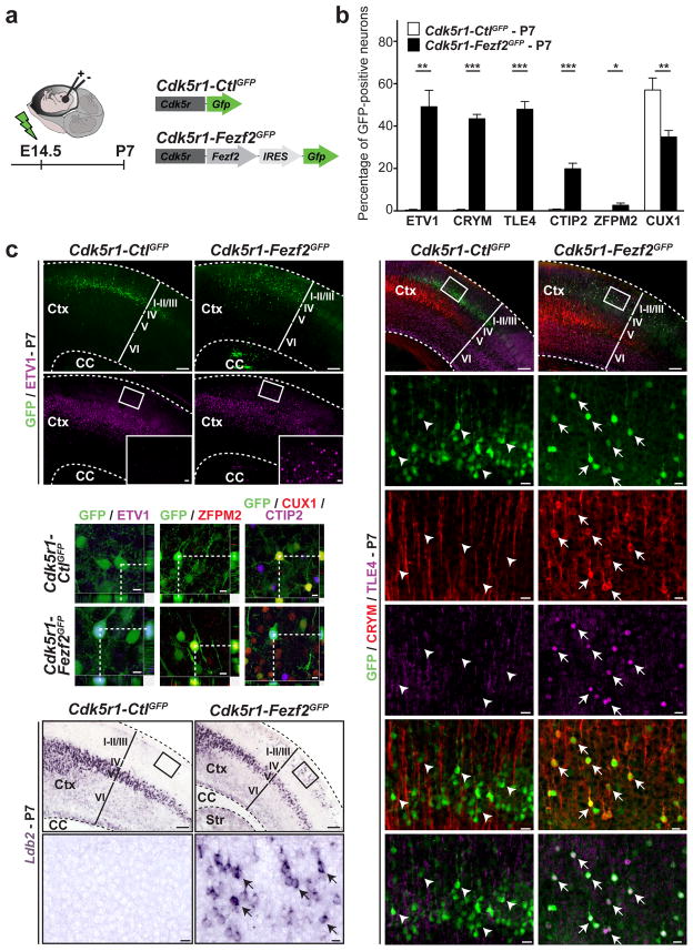
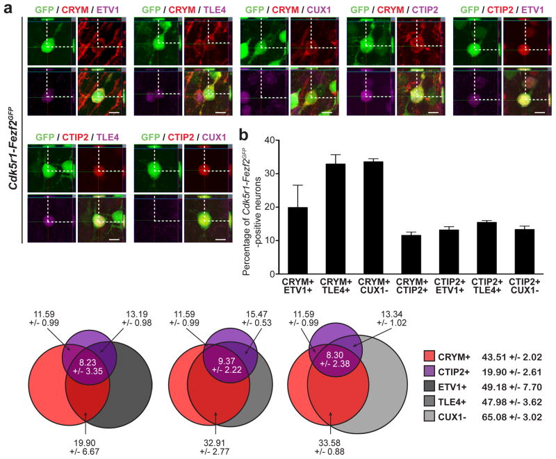
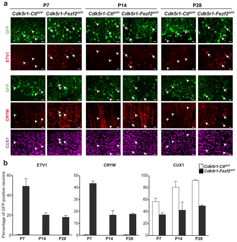
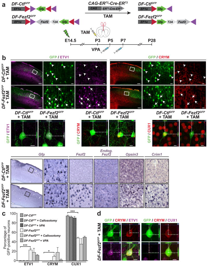
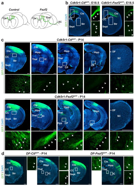
Once programmed to acquire a specific identity and function, cells rarely change in vivo. Neurons of the mammalian central nervous system (CNS) in particular are a classic example of a stable, terminally differentiated cell type. With the exception of the adult neurogenic niches, where a limited set of neuronal subtypes continue to be generated throughout life, CNS neurons are born only during embryonic and early postnatal development. Once generated, neurons become permanently post-mitotic and do not change their identity for the lifespan of the organism. Here, we have investigated whether excitatory neurons of the neocortex can be instructed to directly reprogram their identity post-mitotically from one subtype into another, in vivo. We show that embryonic and early postnatal callosal projection neurons of layer II/III can be post-mitotically lineage reprogrammed into layer-V/VI corticofugal projection neurons following expression of the transcription factor encoded by Fezf2. Reprogrammed callosal neurons acquire molecular properties of corticofugal projection neurons and change their axonal connectivity from interhemispheric, intracortical projections to corticofugal projections directed below the cortex. The data indicate that during a window of post-mitotic development neurons can change their identity, acquiring critical features of alternative neuronal lineages.


Figures

Figure 1
Figure 1. Expression of Fezf2 in migratory, postmitotic CPN induces them to acquire molecular features of CFuPN
a, Schematic representation of the experimental approach. Fezf2 or control vectors were electroporated at E14.5 and expression was restricted to CPN at the earliest stages of postmitotic development using the promoter of the Cdk5r gene. b, Quantification of the percentage of electroporated CPN expressing CFuPN-specific markers (ER81, CRYM, TLE4, CTIP2 and ZFPM2) and downregulating CPN-specific markers (CUX1). Results are expressed as the mean ± s.e.m. The paired, two-tailed t test was used for statistical analysis. * p<0.05; ** p<0.01; *** p<0.001. c, Immunocytochemistry and in situ hybridization for a panel of CFuPN and layer II/III CPN markers. Ctx, cortex; CC, corpus callosum; Str, striatum. Scale bars, 200 μm, 20 μm in high magnification panels and 10 μm in confocal images.
Figure 2
Figure 2. Fezf2 expression in postmitotic CPN induces a program of CFuPN-specific genes
a, Confocal analysis showing that CFuPN-specific genes are co-expressed in Cdk5r-Fezf2eGFP- electroporated CPN. Scale bars, 10 μm. b, Quantification of the percentage of Cdk5r-Fezf2eGFP-electroporated CPN expressing a combination of two CFuPN-specific markers. Results are expressed as the mean ± s.e.m. c, Proportional Venn diagrams representing an estimate of the percentage of Cdk5r-Fezf2eGFP-electroporated CPN co-expressing combinations of three CFuPN-specific markers.
Figure 3
Figure 3. Reprogrammed CPN maintain stable expression of CFuPN markers
a, Immunocytochemistry for ER81, CRYM and CUX1 in Cdk5r-Fezf2eGFP-electroporated CPN versus control (Cdk5r-CtleGFP) at P7, P14 and P28. Scale bars 20 μm. b, Quantification of the percentage of Cdk5r-Fezf2eGFP- and Cdk5r-CtleGFP-electroporated CPN maintaining ER81, CRYM and CUX1 expression at P7, P14 and P28. Results are expressed as the mean ± s.e.m.
Figure 4
Figure 4. Callosal projection neurons can undergo molecular reprogramming of subtype-specific genes in response to Fezf2 expression at postnatal day P3
a, Schematic of the constructs used to conditionally express Fezf2 at P3 and of experimental approach. CPN received Fezf2 (or control DNA) alone or in combination with either VPA treatment or callosotomy. b, Immunocytochemistry and in situ hybridization on coronal sections of cortex. c, Quantification of the percentage of electroporated CPN expressing CFuPN and CPN proteins. Results are expressed as the mean ± s.e.m. The paired, two-tailed t test was used for statistical analysis. * p<0.05; ** p<0.01; *** p<0.001. d, Confocal analysis showing that a subset of DF-Fezf2eGFP-electroporated CPN co-expressed CRYM and ER81 and are CRYM-positive and CU7-negative. Scale bars, 200 μm, 20 μm in high magnification panels and 10 μm in confocal images.
Figure 5
Figure 5. CPN can reprogram their axonal projections from callosal to corticofugal targets in response to Fezf2
a, Graphic representation of changes in axonal connectivity by layer II/III CPN in response to Fezf2 versus control. b, GFP-positive CPN axons crossing the corpus callosum at E18.5 in Cdk5r-Fezf2eGFP and Cdk5r-CtleGFP-electroporated CPN. Callosal projections are greatly reduced in Cdk5r-Fezf2eGFP- versus Cdk5r-CtleGFP-electroporated CPN. c, At P14, GFP-positive axons are restricted to the corpus callosum in Cdk5r-CtleGFP-electroporated CPN. By contrast, axons of Cdk5r-Fezf2eGFP-electroporated CPN are also present in the internal capsule and extend caudally to the thalamus and the cerebral peduncle. A small number of GFP-positive axons are present in the dorsal funiculus of the spinal cord in Cdk5r-Fezf2eGFP- but not in control-electroporated animals. d, GFP-positive axons at P14 of DF-Fezf2eGFP- or DF-CtleGFP-electroporated CPN that received TAM postmitotically at E17.5. Callosal axons are restricted to the corpus callosum in DF-CtleGFP CPN while axons are also present within the internal capsule in DF-Fezf2eGFP CPN. CC, corpus callosum; CP, cerebral peduncle; Ctx, cortex; DF, dorsal funiculus; Hipp, hippocampus; Hyp, hypothalamus; IC, internal capsule; Str, striatum; Sept, septum; Thal, thalamus. Scale bars, 200 μm and 20 μm in high magnification panels.

References

    1. Waddington CH. The strategy of the genes; a discussion of some aspects of theoretical biology. Allen & Unwin; 1957.
    1. Ming GL, Song H. Adult neurogenesis in the mammalian central nervous system. Annu Rev Neurosci. 2005;28:223–250. - PubMed
    1. Zhao C, Deng W, Gage FH. Mechanisms and functional implications of adult neurogenesis. Cell. 2008;132:645–660. - PubMed
    1. Selvaraj V, Plane JM, Williams AJ, Deng W. Switching cell fate: the remarkable rise of induced pluripotent stem cells and lineage reprogramming technologies. Trends Biotechnol. 2010;28:214–223. - PMC - PubMed
    1. Vierbuchen T, Wernig M. Direct lineage conversions: unnatural but useful? Nat Biotechnol. 2011;29:892–907. - PMC - PubMed

Publication types

MeSH terms