Review
. 2013 Feb;72(2):78-90.
doi: 10.1097/NEN.0b013e31827e5762.

Friedreich ataxia: neuropathology revised

Affiliations
Review

Friedreich ataxia: neuropathology revised

Arnulf H Koeppen et al. J Neuropathol Exp Neurol. 2013 Feb.

Abstract

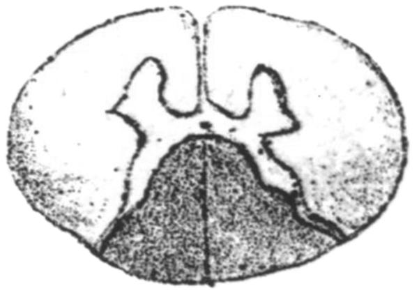
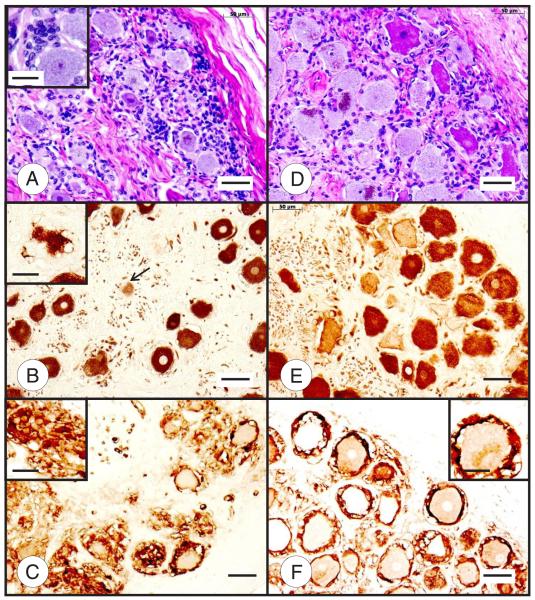
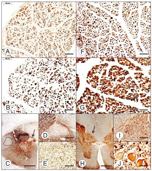
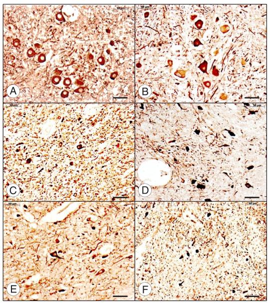
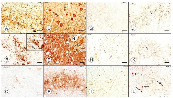
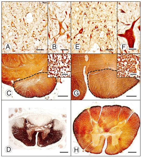
Friedreich ataxia is an autosomal recessive disorder that affects children and young adults. The mutation consists of a homozygous guanine-adenine-adenine trinucleotide repeat expansion that causes deficiency of frataxin, a small nuclear genome-encoded mitochondrial protein. Low frataxin levels lead to insufficient biosynthesis of iron-sulfur clusters that are required for mitochondrial electron transport and assembly of functional aconitase, and iron dysmetabolism of the entire cell. This review of the neuropathology of Friedreich ataxia stresses the critical role of hypoplasia and superimposed atrophy of dorsal root ganglia. Progressive destruction of dorsal root ganglia accounts for thinning of dorsal roots, degeneration of dorsal columns, transsynaptic atrophy of nerve cells in Clarke column and dorsal spinocerebellar fibers, atrophy of gracile and cuneate nuclei, and neuropathy of sensory nerves. The lesion of the dentate nucleus consists of progressive and selective atrophy of large glutamatergic neurons and grumose degeneration of corticonuclear synaptic terminals that contain γ-aminobutyric acid (GABA). Small GABA-ergic neurons and their projection fibers in the dentato-olivary tract survive. Atrophy of Betz cells and corticospinal tracts constitute a second intrinsic CNS lesion. In light of the selective vulnerability of organs and tissues to systemic frataxin deficiency, many questions about the pathogenesis of Friedreich ataxia remain.


Figures

FIGURE 1
FIGURE 1
Reproduction of Figure 1d of the spinal cord made by Friedreich in 1877 (1). Figure 1 was reproduced from Friedreich (1), under license by Copyrights Clearance Center on behalf of Springer Science and Business Media. Note the “upside-down” rendition in comparison with current practice. Friedreich used a mixture of carmine and hematoxylin for sections of the spinal cord but also discussed favorable results with gold chloride. He noticed the much more severe involvement of the dorsal columns than the anterolateral columns and suggested that the lesions were actually continuous across the gray matter of the dorsal horns. This drawing also seems to identify the lack of Clarke columns that are normally visible on low-power cross sections at this level.
FIGURE 2
FIGURE 2
Dorsal root ganglia (DRG) in Friedreich ataxia (FRDA) (A–C) and normal control (D–F). The DRG in FRDA (A) stands out by the smaller size of its neurons and subcapsular hypercellularity. The larger number of nuclei is caused by abundant satellite cells about neurons and in residual nodules ([A] insert). The heterogeneous staining intensity of normal DRG neurons with hematoxylin and eosin (D) and after immunohistochemistry (IHC) with anti–class III β-tubulin (E) is less evident in FRDA (A, B). The section of a DRG ganglion (B) also reveals the highly irregular outlines of several neurons caused by proliferation of satellite cells and ultimate total absorption of the neuron into residual nodules ([B] arrow and inset). The S100α immunostain reveals a single-cell layer of satellite cells around normal neurons in the normal DRG (F). In contrast, the much smaller neurons in FRDA display a highly irregular multilayered rim of these cells and replacement of neurons by nodules ([C] inset). Hematoxylin and eosin (A, D); IHC for class III β-tubulin (monoclonal antibody TUJ-1) (B, E); IHC for S100α (C, F). Scale bars = 50 μm; insets, 20 μm.
FIGURE 3
FIGURE 3
Dorsal spinal roots and dorsal nucleus of Clarke column of the spinal cord in Friedreich ataxia (FRDA) (A–E) and normal control (F–J). In comparison with the normal control (F), the dorsal root in FRDA (A) lacks large axons, but many thin axons remain. Myelinated fibers in the dorsal root of FRDA are abundant (B), but they are much smaller than in the normal (G). The dorsal nucleus of Clarke column is devoid of neurons (C); arrow in (C) indicates fields shown at higher magnification in (D) and (E). There is also long tract degeneration of the dorsal column, dorsal spinocerebellar tract, and corticospinal tract (interrupted line) (C). In the control case (H–J), the spinal cord (H) and the nucleus dorsalis are larger than those in FRDA. The normal nucleus dorsalis displays typical large round nerve cells (H); arrow in (H) indicates fields shown at higher magnification in (I) and (J). Immunohistochemistry (IHC) for phosphorylated neurofilament protein (A, F); IHC for myelin basic protein (B, G); IHC for of class III β-tubulin (C–E, H–J). Scale bars = (A, B, E–G, J) 50 μm; (C, H) 1 mm; (D, I) 200 μm.
FIGURE 4
FIGURE 4
Neuronal atrophy in gracile and lateral cuneate nuclei in Friedreich ataxia (FRDA). Normal gracile nucleus (A) and normal lateral cuneate nucleus (B). FRDA, gracile nuclei are shown in (C) and (E); FRDA, lateral cuneate nuclei are shown in (D) and (F). Panels (C, D) and (E, F) are from 2 different FRDA cases. Immunohistochemistry of nonphosphorylated neurofilament protein. Levels of the medulla oblongata and location of the nuclei were identified based on Olszewski and Baxter (63). Scale bar = 50 μm.
FIGURE 5
FIGURE 5
Sural nerve abnormalities in Friedreich ataxia (FRDA). (A, D) Double-label immunofluorescence of peripheral myelin protein P0 (Quantum Red) and axonal class III β-tubulin (Alexa 488, green) of the sural nerve in FRDA (A) shows persistence of axons, although axonal diameters are reduced and myelin is deficient compared with those of the normal (D). Larger myelinated fibers are entirely absent (A). (B, E) Few S100α-immunoreactive Schwann cells remain in the sural nerve of FRDA (B) in contrast to the normal control (E). (C, F) Electron microscopy of the nerve in FRDA confirms lack of thick myelin sheaths and an abundance of thin unmyelinated axons surrounded by tortuous Schwann cell processes ([C] arrow) in contrast to the normal control (F). Scale bars = (A, D) 25 μm; (B, E) 20 μm; (C, F) 5 μm. Dr Benjamin B. Gelman (Galveston, TX) provided the electron micrograph shown in (C).
FIGURE 6
FIGURE 6
The dentate nucleus (DN) in Friedreich ataxia (FRDA). Friedreich ataxia (A–C, G–I); normal control (D–F, J–L). All stains are immunohistochemical preparations: neuron-specific enolase (A, D); glutamic acid decarboxylase (GAD) (B, E); frataxin (C, F); vesicular glutamate transporter (VGluT1) (G, J); vesicular glutamate transporter 2 (VGluT2) (H, K); glycine transporter 2 (GlyT2) (I, L). Friedreich ataxia causes loss of large neurons (A), whereas small neurons persist ([A, D] arrows). Neuronal loss in FRDA causes an overall reduction of GABA (γ-aminobutyric acid)-ergic terminals in the DN (B) and grumose degeneration of the remaining nerve endings ([B] inset). Small neurons of the normal DN ([E] inset, arrow) and the DN in FRDA ([B] inset, arrow) display pale GAD reaction product. Numerous frataxin-positive axodendritic and axosomatic terminals outline large and small neurons of the normal DN (F). In FRDA, only traces of frataxin reaction product remain (C). Vesicular glutamate transporter 1 and VGluT2 reaction products reveal negative images of large neurons (N in [J] and [K], respectively). Vesicular glutamate transporter 1– and VGluT2-reactive terminals are less abundant in FRDA ([G] and [H], respectively), and negative images of neurons are absent. The normal DN contains GlyT2 reaction product that is as abundant as VGluT1 and VGluT2 (L). Arrows in (L) indicate the somata of 4 small glycinergic nerve cells in the normal DN. In FRDA, GlyT2 (I) is much less prominent than in the control. Scale bars = 50 μm; insets, 20 μm.
FIGURE 7
FIGURE 7
Motor cortex and corticospinal tract in Friedreich ataxia (FRDA). Friedreich ataxia (A–D); normal control (E–H); motor cortex immunostained for nonphosphorylated neurofilament protein (A, B, E, F); medullary pyramids immunostained for myelin basic protein (MBP) ([C], [G], and insets); upper lumbar spinal cord, immunostain for MBP (D, H). Most neurons in layer V of the motor cortex in FRDA (A) are comparable in size to those in the normal (E), but Betz cells are absent. The largest pyramidal neuron in layer V of FRDA is shown in (B). A Betz cell in the normal control is shown in (F). The limits of the medullary pyramids are outlined by interrupted lines. The pyramid in FRDA (C) is diminutive in comparison with the normal control (G), and the density of myelinated fibers is lower (insets in [C] and [G], respectively). Myelin basic protein immunostain of the upper lumbar cord shows the small cross-sectional area in FRDA (D) in comparison with the control specimen (H) and loss of myelinated fibers in dorsal columns, dorsal spinocerebellar tracts, and corticospinal tracts. Scale bars = (A, E) 50 μm; (B), (F) and insets in (C) and (G) 20 μm; (C, D, G, H) 1 mm.

References

    1. Friedreich N. Ueber Ataxie mit besonderer Berücksichtigung der hereditären Formen. Nachtrag. Virchows Arch Pathol Anat Physiol Klin Med. 1877;70:140–52.
    1. Koeppen AH, Morral JA, Davis AN, et al. The dorsal root ganglion in Friedreich’s ataxia. Acta Neuropathol. 2009;118:763–76. - PubMed
    1. Morral JA, Davis AN, Qian J, et al. Pathology and pathogenesis of sensory neuropathy in Friedreich’s ataxia. Acta Neuropathol (Berl) 2010;120:97–108. - PubMed
    1. Mott FW. Case of Friedreich’s disease, with autopsy and systematic microscopical examination of the nervous system. Arch Neurol Psychiat (Lond) 1907;3:180–200.
    1. Spiller WG. Friedreich’s ataxia. J Nerv Ment Dis. 1910;37:411–35.

Publication types

Substances