. 2013 Feb;72(2):119-29.
doi: 10.1097/NEN.0b013e3182801cde.

Alpha-synuclein pathology and axonal degeneration of the peripheral motor nerves innervating pharyngeal muscles in Parkinson disease

Affiliations

Alpha-synuclein pathology and axonal degeneration of the peripheral motor nerves innervating pharyngeal muscles in Parkinson disease

Liancai Mu et al. J Neuropathol Exp Neurol. 2013 Feb.

Abstract

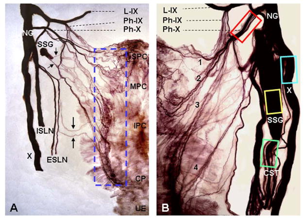
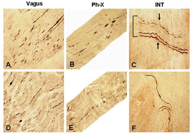
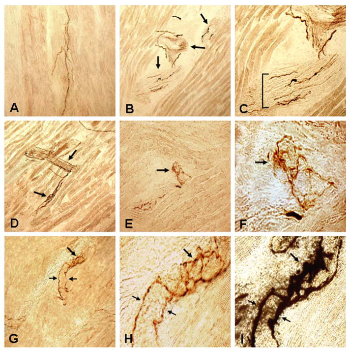
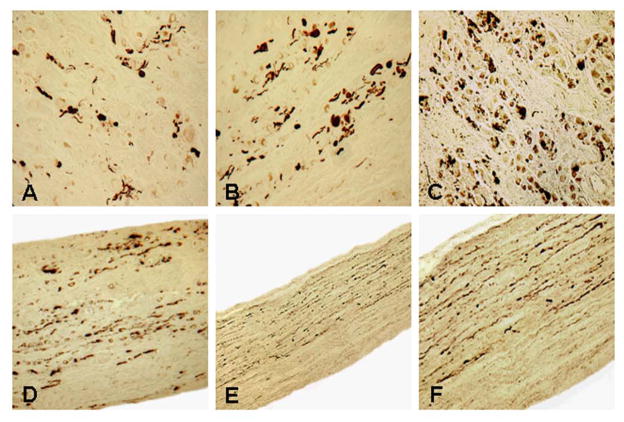
Parkinson disease (PD) is a neurodegenerative disease primarily characterized by cardinal motor manifestations and CNS pathology. Current drug therapies can often stabilize these cardinal motor symptoms, and attention has shifted to the other motor and nonmotor symptoms of PD that are resistant to drug therapy. Dysphagia in PD is perhaps the most important drug-resistant symptom because it leads to aspiration and pneumonia, the leading cause of death. Here, we present direct evidence for degeneration of the pharyngeal motor nerves in PD. We examined the cervical vagal nerve (cranial nerve X), pharyngeal branch of nerve X, and pharyngeal plexus innervating the pharyngeal muscles in 14 postmortem specimens, that is, from 10 patients with PD and 4 age-matched control subjects. Synucleinopathy in the pharyngeal nerves was detected using an immunohistochemical method for phosphorylated α-synuclein. Alpha-synuclein aggregates were revealed in nerve X and the pharyngeal branch of nerve X, and immunoreactive intramuscular nerve twigs and axon terminals within the neuromuscular junctions were identified in all of the PD patients but in none of the controls. These findings indicate that the motor nervous system of the pharynx is involved in the pathologic process of PD. Notably, PD patients who have had dysphagia had a higher density of α-synuclein aggregates in the pharyngeal nerves than those without dysphagia. These findings indicate that motor involvement of the pharynx in PD is one of the factors leading to oropharyngeal dysphagia commonly seen in PD patients.


Figures

FIGURE 1
FIGURE 1
Photographs of two human semipharynges processed with Sihler’s stain, wholemount nerve staining technique, showing the branching of cervical vagus (X) nerve, formation of the pharyngeal plexus, and tissue sampling sites for detecting neurodegenerative changes in the nerves and intramuscular nerve twigs. Pharyngeal plexus in humans provides motor innervation to pharyngeal constrictors (PCs) and cricopharyngeal (CP) sphincter. (A) Posterior view of a Sihler’s stained adult human semipharynx, showing the branching and distribution of the cervical X nerve and the pharyngeal plexus. Note that the X nerve gives off its pharyngeal branch (Ph-X) and superior laryngeal nerve (SLN). The SLN is then subdivided into internal (ISLN) and external (ESLN) branches which supply the laryngeal and pharyngeal mucosa (ISLN) and cricothyroid muscle (ESLN), respectively. The pharyngeal plexus is formed mainly by the Ph-X and less by the pharyngeal branch of the glossopharyngeal nerve (Ph-IX), ESLN (large arrows), and sympathetic nerve fibers (small arrows) from superior sympathetic ganglion (SSG). In this case, most of the SSG was removed in order to show nerve branching patterns. The enclosed region, which is the location of motor zone in the PCs and CP, indicates tissue sampling site of the PC and CP muscles for detecting neurodegenerative changes in the intramuscular nerve twigs and axon terminals. IPC, inferior pharyngeal constrictor; L-IX, lingual division of the IX nerve; MPC, middle pharyngeal constrictor; NG, nodose ganglion of X nerve; SPC, superior pharyngeal constrictor; UE, upper esophagus. x2. (B) Higher magnification image of a Sihler’s stained human semipharynx, showing anatomic relationship between different nerves and nerve branches and illustrating the nerve tissue sampling sites (enclosed regions) for histochemical and immunohistochemical studies. Note that the Ph-X gives off at least 4 primary branches (1, 2, 3, 4) to innervate the PC and CP muscles. x4. CST, cervical sympathetic trunk. Adapted and reproduced in part with permission from Mu and Sanders (35), Ann Otol Rhino Laryngol 2007;116:604–17.
FIGURE 2
FIGURE 2
Photomicrographs of the longitudinal sections of cervical X nerve trunk (A, D), pharyngeal branch of the X nerve (Ph-X) (B, E), and pharyngeal muscles (C, F) immunostained for phosphorylated α-synuclein (psyn) in PD subjects with dysphagia (A–C) and without dysphagia (D–F), showing Lewy neurites (LNs) in the nerves examined and immunoreactive intramuscular nerve twigs (INTs) in the muscles. (A) A stained section of a cervical X nerve trunk from a PD subject with dysphagia (PD 3, 78-year-old man with disease duration of 19 years, Hoehn & Yahr 4, and motor UPDRS 51; lesion severity: severe, +++). Note that there were numerous LNs (darkly stained threads and dots) in the X nerve. x200. (B) A stained section of the Ph-X from the same PD subject as in A. Note that the Ph-X in this case also contained frequent LNs (lesion severity: moderate, ++). x200. (C) A stained section of the inferior pharyngeal constrictor (IPC) from the same PD subject as in B. Note that an INT running across the muscle fibers was horizontally sectioned. Several axons on the superior and inferior margins of the INT were positively immunostained with anti-psyn immunohistochemistry (arrows), whereas the normal axons in the center of the INT remained unstained. x200. (D) A stained section of cervical X nerve trunk from a PD subject without dysphagia (PD 9, 80-year-old man with disease duration of 17 years, Hoehn & Yahr 5, and motor UPDRS 40; lesion severity: moderate, ++), showing frequent LNs in the X nerve. x200. (E) A stained section of the Ph-X from the same PD subject as in D. Note that there were several LNs in the Ph-X (lesion severity: mild, +). x200. (F) A stained section of cricopharyngeal (CP) sphincter from the same PD subject as in E, showing three immunoreactive axons in the muscle. x200.
FIGURE 3
FIGURE 3
Photomicrographs of the longitudinal sections of inferior pharyngeal constrictor (IPC) and cricopharyngeal (CP) sphincter immunostained with anti-phosphorylated α-synuclein immunohistochemistry in PD subjects, showing immunoreactive intramuscular nerve twigs (INTs) and axon terminals within neuromuscular junctions (NMJs). (A–C) Images of a stained section of IPC muscle from a PD subject with dysphagia (PD 8, 75-year-old man with disease duration of 30 years, Hoehn & Yahr 4, and motor UPDRS 66). (A) A stained muscle section showing immunoreactive axons (darkly stained threads). x100. (B) A stained muscle section containing three obliquely cut intramuscular nerve branches (arrows). Note that there were several α-synuclein immunoreactive axons (darkly stained threads) in each of the nerve branches. x100. (C) Magnification of B, showing the profiles of the nerve branches and α-synuclein positive and negative (unstained) axons within the intramuscular nerve branches. x200. (D–F) Images of stained sections of IPC muscle from a PD subject without dysphagia (PD 9, 80-year-old man with disease duration of 17 years, Hoehn & Yahr 5, and motor UPDRS 40), showing α-synuclein immunoreactive INTs and axon terminals within NMJs. (D) Low-power view of a stained muscle section, showing immunoreactive INTs (arrows) containing α-synuclein positive axons. x100. (E) Higher-power view of a stained muscle section, showing α-synuclein positive axon terminals within an NMJ (arrow). x200. (F) Closeup photograph of E, showing the profiles of the immunoreactive axon terminals within the NMJ (arrow). x640. (G–I) Photomicrographs of stained sections of CP muscle from a PD subject with dysphagia (PD 10, 79-year-old man with disease duration of 23 years, Hoehn & Yahr 2, and motor UPDRS 28), showing immunoreactive axon terminals. (G) Higher-power view of a stained PD CP, showing two α-synuclein positive preterminal axons (small arrows) and their terminals within the NMJ (large arrow). x200. (H) Closeup photograph of G, showing the organization of the axon terminals within the NMJ. Note that two immunoreactive preterminal axons (small arrows) innervating an NMJ give off terminals within the NMJ (large arrow). x640. (I) The muscle section used in H was restained with AChE-Ag. Note that the preterminal axons (small arrows) innervated an NMJ (large arrow) and that the immunoreactive axon terminals as shown in H were located within the AChE-Ag stained NMJ. x640.
FIGURE 4
FIGURE 4
Photomicrographs of cross sections of the cervical superior sympathetic ganglion (SSG) (A–C) and longitudinal sections of the cervical sympathetic nerve (D–F) from subjects with PD immunostained with anti-phosphorylated α-synuclein immunohistochemistry. (A) A stained section of SSG from a PD subject without dysphagia (PD 7, 79-year-old woman with disease duration of 11 years, Hoehn & Yahr 4, and motor UPDRS 47). The SSG was affected slightly as it contained some α-synuclein aggregates. x200. (B) A stained section of SSG from a PD subject without dysphagia (PD 5, 80-year-old man with disease duration of 11 years, Hoehn & Yahr 4, and motor UPDRS 53). The SSG was affected moderately as it exhibited abundant α-synuclein aggregates. x200. (C) A stained section of SSG from a PD subject with dysphagia (PD 10, 79-year-old man with disease duration of 23 years, Hoehn & Yahr 2, and motor UPDRS 28). The SSG was affected severely as it contained numerous α-synuclein aggregates. x200. (D) A stained longitudinal section of a cervical sympathetic trunk from the same PD subject as in B (PD 5). Note that there were abundant α-synuclein aggregates in the sympathetic nerve. x200. (E) Low-power view of a stained longitudinal section of a cervical sympathetic trunk from the same PD subject as in C (PD 10). Note that this sympathetic nerve contained numerous α-synuclein aggregates. Also note that α-synuclein immunoreactive axons were not randomly distributed throughout the nerve, instead they were concentrated in two-thirds of the nerve. x100. (F) Higher-power view of E. x200.

References

    1. Jellinger KA. Pathology of Parkinson’s disease. Changes other than the nigrostriatal pathway. Mol Chem Neuropathol. 1991;14:153–97. - PubMed
    1. Johnston BT, Li Q, Castell JA, et al. Swallowing and esophageal function in Parkinson’s disease. Am J Gastroenterol. 1995;90:1741–46. - PubMed
    1. Leopold NA, Kagel MC. Prepharyngeal dysphagia in Parkinson’s disease. Dysphagia. 1996;11:14–22. - PubMed
    1. Coates C, Bakheit AM. Dysphagia in Parkinson’s disease. Eur Neurol. 1997;38:49–52. - PubMed
    1. Fuh JL, Lee RC, Wang SJ, et al. Swallowing difficulty in Parkinson’s disease. Clin Neurol Neurosurg. 1997;99:106–12. - PubMed

Publication types

MeSH terms