

Case Reports
doi: 10.1007/s00384-012-1624-2.
Epub 2013 Jan 19.
Exceptional dissemination of perineal tuberculosis up to the right flank: a tribute to J.P. Nesselrod's study on the anatomy of pelvic lymphatics published in 1936 in the Annals of Surgery
Affiliations
- PMID: 23334691
- PMCID: PMC3646164
- DOI: 10.1007/s00384-012-1624-2
Item in Clipboard
Case Reports
Exceptional dissemination of perineal tuberculosis up to the right flank: a tribute to J.P. Nesselrod's study on the anatomy of pelvic lymphatics published in 1936 in the Annals of Surgery
Int J Colorectal Dis.
2013 May.
No abstract available
Figures
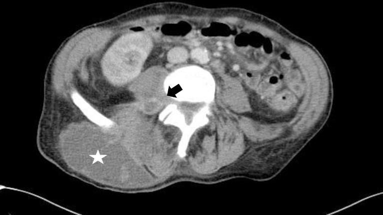
The patients' CT scan at admission, showing a large subcutaneous abscess (star) on the flank at the level of the fourth lumbar vertebral body, communicating with the iliopsoas muscle, the paraspinal muscles, and the epidural space (arrow)
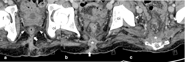
Coronary reconstruction of the patients' CT scan on the level of the femur (f) and os ischium (oi). Panel a shows a subcutaneous fistula (arrow) reaching the levator ani (triangles). In b, the subcutaneous abscess (star) is linked over a fistula (arrow) with the skin. In c, the presacral space is filled with abscess (star) whereas bilateral lymph nodes are visible (triangles)
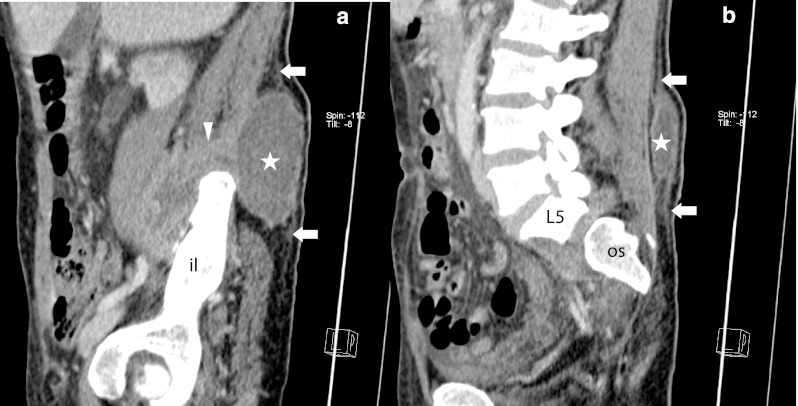
Sagittal reconstructions of the patient's CT scan at admission. On a, the subcutaneous abscess (star) and its breakthrough to the iliopsoas muscle (triangle) over the right crista iliaca (il os ilium) are shown. A thin subcutaneous spur (arrows) delineates potential routes of disease expansion (b) on the level of the lumbar vertebral column (L5 fifth lumbar vertebral body, OS os sacrum)
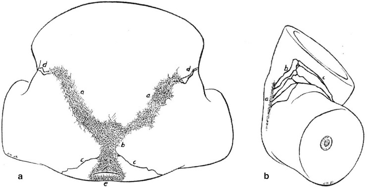
Original figure from Nesselrod's publication in 1936 [12] in Annals of Surgery showing the perineal and gluteal distribution of lymphatic vessels after injections of mercury in the skin, respectively. Panel a shows a dorsal view of the sacrococcygeal and superior gluteal region of a male white fetus at term (original description). Perianal (e), presacral (b), gluteal plexuses (a) and anastomotic vessels (d). Panel b shows a lateral view on the same specimen as in a, showing not only lymphatic vessels above the crista iliaca but also their link (c) to the inguinal zone
References
-
- Tai WC, Hu TH, Ch L, et al. Ano-perianal tuberculosis: 15 years of clinical experiences in Southern Taiwan. Colorectal Dis. 2010;12(7 Online):e120–e224. - PubMed
Publication types
MeSH terms
LinkOut - more resources
Full Text Sources
Other Literature Sources
Medical
