. 2013 Mar;242(3):254-68.
doi: 10.1002/dvdy.23923. Epub 2013 Feb 13.

PDGF receptor alpha+ mesoderm contributes to endothelial and hematopoietic cells in mice

Affiliations
Free PMC article

PDGF receptor alpha+ mesoderm contributes to endothelial and hematopoietic cells in mice

Guo Ding et al. Dev Dyn. 2013 Mar.
Free PMC article

Abstract

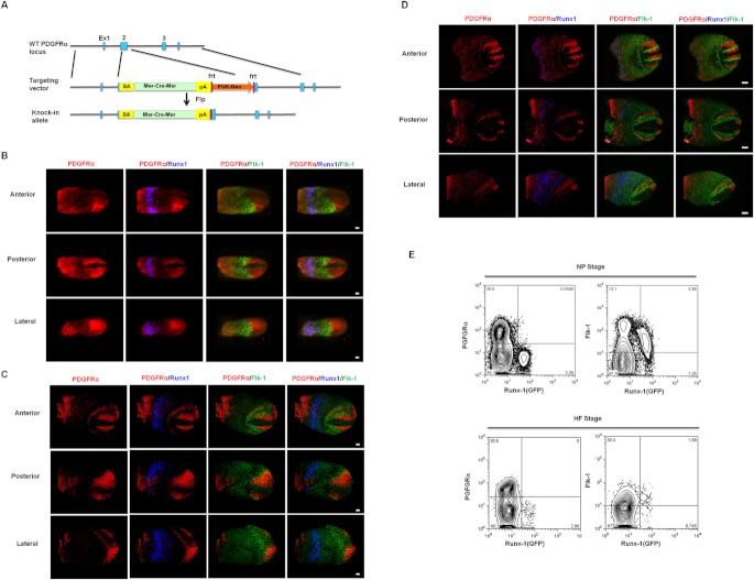
Background: Early mesoderm can be classified into Flk-1+ or PDGF receptor alpha (PDGFRα)+ population, grossly representing lateral and paraxial mesoderm, respectively. It has been demonstrated that all endothelial (EC) and hematopoietic (HPC) cells are derived from Flk-1+ cells. Although PDGFRα+ cells give rise to ECs/HPCs in in vitro ES differentiation, whether PDGFRα+ population can become hemato-endothelial lineages has not been proved in mouse embryos.

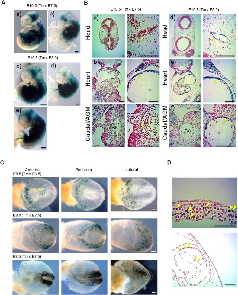
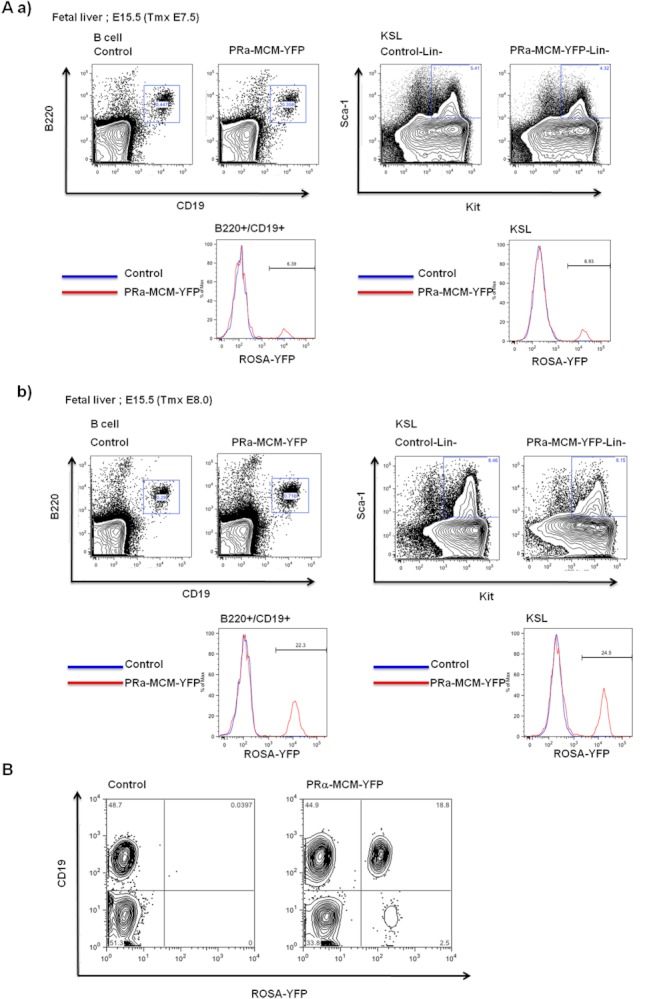
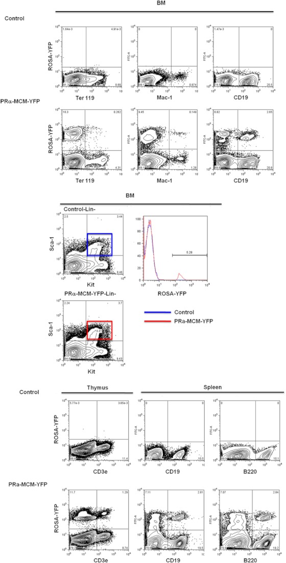
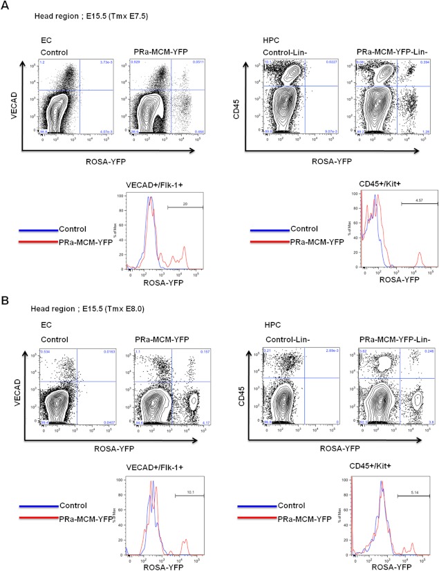
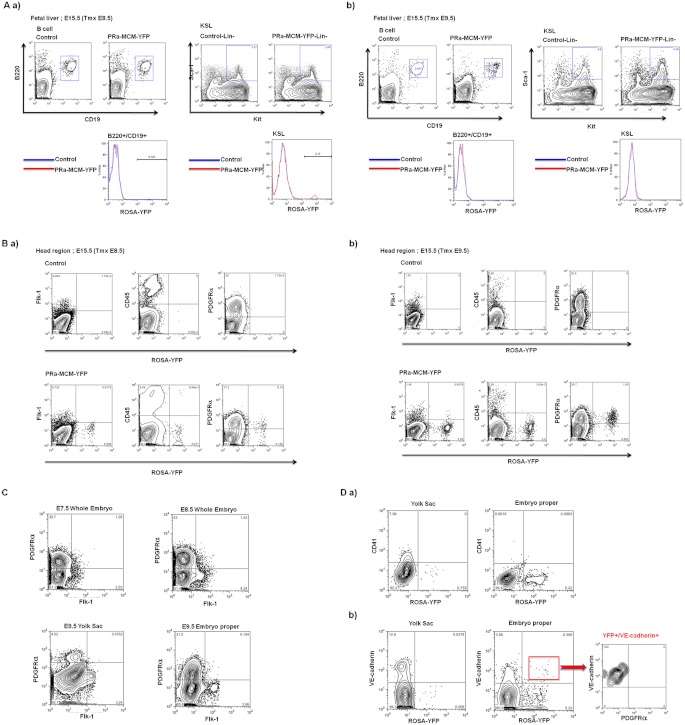
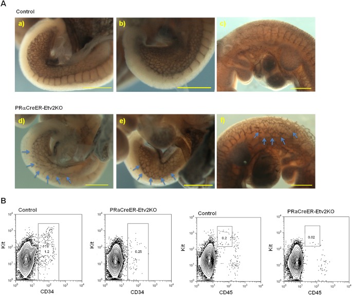
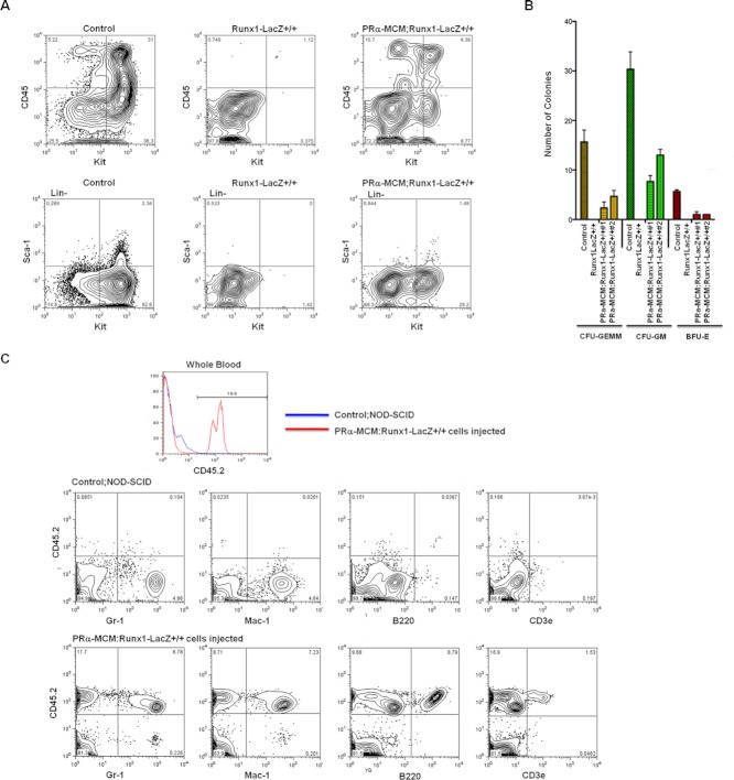
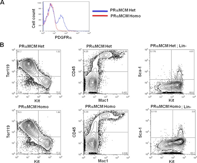
Results: Using PDGFRαMerCreMer mice, PDGFRα+ early mesoderm was shown to contribute to endothelial cells including hemogenic ECs, fetal liver B lymphocytes, and Lin-Kit+Sca-1+ (KSL) cells. Contribution of PDGFRα+ mesoderm into ECs and HPCs was limited until E8.5, indicating that PDGFRα+/Flk-1+ population that exists until E8.5 may be the source for hemato-endothelial lineages from PDGFRα+ population. The functional significance of PDGFRα+ mesoderm in vascular development and hematopoiesis was confirmed by genetic deletion of Etv2 or restoration of Runx1 in PDGFRα+ cells. Etv2 deletion and Runx1 restoration in PDGFRα+ cells resulted in abnormal vascular remodeling and rescue of fetal liver CD45+ and Lin-Kit+Sca-1+ (KSL) cells, respectively.

Conclusions: Endothelial and hematopoietic cells can be derived from PDGFRα+ early mesoderm in mice. PDGFRα+ mesoderm is functionally significant in vascular development and hematopoiesis from phenotype analysis of genetically modified embryos.


Figures

Fig. 1
Fig. 1
A: Generation of PDGFRα-MerCreMer (PRα-MCM) knock-in mice. Tmx-inducible MerCreMer was knocked into the PDGFRα locus using homology arms corresponding to 5′ side, 79,307–85,194; 3′ side, 85,253–89,284 from RP23–55P22. Correct integration was confirmed by Southern blotting. PCR genotyping can be performed by primers, PRα WT Rev1; ggagaacaaggacgcatgtgtgg, PRα5′F2; gcttccctcttatctatctgactg, MCM Rev2; aaggtggacctgatcatggagattc (WT, 260 bp; Targeted, 700 bp). SA, splice acceptor; pA, polyadenylation signal. B: Immunostaining of PDGFRα, Flk-1, and Runx1 in NP stage (E7.5) mouse embryos. NP-stage wild type embryos were stained by PDGFRα, Flk-1, and Runx1 antibodies. There was a narrow overlap of PDGFRα and Flk-1 domains. Note, however, that PDGFRα and Runx1 expression was seen in distinct mesoderm subsets. Scale bar=100 μm. C: Immunostaining of PDGFRα, Flk-1, and Runx1 in HF stage (E8.0) mouse embryos. In HF-stage embryos, similar staining was observed as in NP-stage samples. Note also in this stage that there is no overlap between PDGFRα and Runx1+ populations. Scale bar=100 μm. D: Immunostaining of PDGFRα, Flk-1, and Runx1 in E8.5 somite stage embryos. As seen in E7.5 or E8.0 embryos, there is some overlap between PDGFRα and Flk-1 areas, however no overlap between PDGFRα and Runx1. In this stage, PDGFRα or Flk-1 antibody stains form somite structure or aortas, respectively. Scale bar=100 μm. E: FACS analysis of NP- and HF-stage embryos. NP- (left) or HF- (right) stage embryos were stained by indicated antibodies to examine the relationship between Runx1, PDGFRα, and Flk-1. In both stages, almost no Runx1+/PDGFRα+ cells were detected, while more that 70% of Runx1-GFP+ cells were Flk-1+.
Fig. 2
Fig. 2
A: Whole mount LacZ staining of PRα-MCM-LacZ embryos. To activate MerCreMer, Tmx was injected into pregnant females at E7.5 (neural plate stage, top panels) or E8.0 (head fold stage, middle panels), when PDGFRα starts to be clearly detected in mesoderm with initiation of vasculogenesis and hematopoiesis. Embryos were LacZ stained at E10.5. LacZ-stained cells were distributed mainly in the intraembryonic area including the head, heart, and umbilical cord (arrows in lower panel). Note that only a few labeled cells were seen in the yolk sac except the umbilical cord (b, d). Scale bar=1 mm. B: Histological analysis of LacZ-stained PRα-MCM-LacZ embryos with Tmx injection at E7.5 (a–c) or E8.0 (d–f) into pregnant females. Embryos were LacZ stained and sectioned at E10.5. LacZ-positive cells were distributed in cranial mesenchyme, atrial and ventricular cardiomyocytes. Higher magnification is shown on the right side of a–f. Note the presence of labeled ECs in cranial vessels (a, d) and ventral wall of the descending aorta (arrows on right in c, f). Presence of LacZ+ ECs and HPCs in the aorta-gonad-mesonephron region is consistent with the finding that fetal liver HSCs exist as E7.5–8.5 labeled cells in the fetal liver (see Figs. 3A and 6A,a). Scale bar=100 μm. C, D: Short-term tracing of PDGFRα+ cells from E7.5 to E8.0–8.5. C: PRα-MCM-LacZ embryos were analyzed at E8.0 and E8.5 after Tmx injection into pregnant females at E6.5 or E7.5. Note that labeled cells are present in the lateral mesoderm area that overlaps Flk-1 staining (see Fig. 1C) at E8.0 (“Posterior” panels). Existence of some labeled cells in this lateral area where Flk-1+ cells exist at E8.0 supports that part of the labeled cells might become Flk-1+. At E8.5,labeled cells become more concentrated around the paraxial zone (bottom panels). Tmx injection at E6.5 labeled YS HPCs that are different from those labeled by E7.5 injection (top panels). Scale=100 μm. D: Sections of LacZ-stained E8.5 embryos after E6.5 injection demonstrate the labeling in YS blood islands (arrowheads) and cardiac tissue (arrows). Note that part of YS HPCs (top panel) and cardiac tissue (bottom panel) contains LacZ+ cells. Scale=100 μm.
Fig. 3
Fig. 3
A: PDGFRα+ cells labeled at E7.5 or E8.0 contribute to fetal liver HPCs. a: Pregnant females with PRα-MCM-YFP embryos were Tmx injected at E7.5. Contribution of YFP+ cells to fetal liver HPCs was analyzed at E15.5. To evaluate the contribution of labeled cells to definitive HPCs, B cell and KSL populations were gated. YFP+ cells constituted 6–7% of B or KSL population gated. (YFP expression from ROSA locus is the indicator of Cre recombination by PRα-MCM transgene.) b: PRα-MCM-YFP embryos were analyzed at E15.5 after Tmx injection at E8.0 into pregnant females. To evaluate the contribution of labeled cells to definitive HPCs, B cell and KSL populations were gated. YFP+ cells constituted 22 or 25% of B and KSL population, respectively. B: PDGFRα+ cells from embryo proper develop into B cells in explant culture. After Tmx injection into pregnant females at E7.5, caudal part of E8.25 PRαMCM-YFP embryos was explanted and cultured on OP9 feeder cells with IL-7 (20 ng/ml) and Flt-3 ligand (10 ng/ml). After 2 days, explants were dissociated and further cultured in the same condition for another 2 weeks. Floating cells were harvested and stained by anti-CD19 antibody to demonstrate the B cell generation. Note that significant proportion of CD19+ B cells exist as YFP+ in cultured cells from the PRαMCM-YFP embryo (right panel).
Fig. 4
Fig. 4
E7.5 PDGFRα+ cells contribute to adult hematopoietic tissue. Bone marrow, thymus, and spleen cells from PRαMCM-YFP adult mice (Tmx injected at E7.5 during pregnancy) were analyzed for the contribution of YFP+-labeled cells. BM contains YFP+ cells in erythroid, macrophage/monocyte, and B cells as well as KSL population. Around 5% of YFP+ cells in KSL population reflects the similar proportion of YFP+ cells in fetal liver KSL population. YFP-labeled cells (E7.5 Tmx) also contribute to B and T cells in spleen and thymus.
Fig. 5
Fig. 5
PDGFRα+ cells labeled at E7.5 or E8.0 contribute to cranial ECs. A: Head regions from PRα-MCM-YFP embryos were analyzed at E15.5 after Tmx injection into pregnant females at E7.5. By FACS analysis, ∼20% of VE-cadherin+/Flk-1+ cells or ∼4.5% of CD45+/Kit+ cells were YFP+, indicating that E7.5 PDGFRα+ mesoderm contributes to cranial ECs and HPCs. B: Pregnant females with PRα-MCM-YFP embryos were Tmx injected at E8.0. Contribution of YFP+ cells to cranial ECs analyzed at E15.5. By FACS analysis, ∼10% of VE-cadherin+/Flk-1+ cells were labeled as YFP+, indicating that a significant proportion of cranial ECs are derived from E8.0 PDGFRα+ mesoderm. Note also that ∼5% of CD45+/Kit+ cells were YFP+, indicating the contribution of E8.0 PDGFRα+ mesoderm into HPCs.
Fig. 6
Fig. 6
A: PDGFRα+ cells labeled at E8.5 contribute to fetal liver HPCs, but with lower efficiency. By E9.5 labeling, almost no PDGFRα+ cells contribute to fetal liver HPCs. a: After E8.5 Tmx injection, fetal liver HPCs were analyzed. YFP+-labeled cells contributed to ∼2% of KSL cells but are almost negligible in B cell population, indicating that the contribution of PDGFRα+ mesoderm significantly declines around E8.5. b: In fetal liver HPCs, no YFP+ cells were observed after E9.5 Tmx injection into pregnant females. B: Contribution of YFP+ cells to cranial ECs/HPCs was analyzed in E15.5 PRα-MCM-YFP embryos after Tmx injection at E8.5 or E9.5 into pregnant females. a: In cranial samples, almost no E8.5-labeled YFP+ cells contribute to either Flk-1+ (VE-cadherin+) ECs or CD45+ HPCs, indicating that only early stage PDGFRα+ mesoderm can give rise to cranial ECs and HPCs. Note that the majority of YFP+ cells existed as PDGFRα+ population. b: In E15.5 cranial samples, YFP+ cells were present not in Flk-1+ (VE-cadherin+) or CD45+ population, but in the PDGFRα+ population after E9.5 Tmx injection. C: PDGFRα+/Flk-1+ cells that exist from E7.5–8.5 decreased profoundly at E9.5. Wild type embryos were stained by anti-PDGFRα and Flk-1 antibodies and analyzed by FACS. Note that the proportion of cells co-expressing PDGFRα and Flk-1 has significantly declined at E9.5 from earlier embryos. D: FACS analysis of the PRα-MCM-YFP embryos demonstrating that VE-cadherin+ ECs were labeled within 24 hr after E7.5 Tmx injection. To trace the short-term fate of PDGFRα+ cells, Tmx was injected into pregnant females at E7.5. YS (a), and embryo proper (b) from PRα-MCM-YFP embryos were analyzed at E8.0 after 12 hr. YFP+ cells were present mainly in the embryo proper (b), but very few in the YS (a). We could not detect any labeled CD41+ primitive HPCs either in yolk sac or embryo proper. Note, however, that part of VE-cadherin+ cells were labeled in the embryo proper (red box), suggesting that VE-cadherin+/PDGFRα-ECs can differentiate from PDGFRα+ mesoderm within 12 hr (b, right panel).
Fig. 7
Fig. 7
A: Etv2 inactivation in early PDGFRα+ mesoderm causes embryonic vascular changes. In PRαBAC-CreER-Etv2KO mice, Etv2 was inactivated in PDGFRα+ mesoderm by Tmx injection at E8.0. PRαBAC-CreER line was used because multi-copy insertions of the BAC construct will provide more efficient Cre activity than PDGFRαMCM one copy knock-in. Control (a–c) and PRαBAC-CreEREtv2KO (d–f) embryos were exposed to Tmx by injection at E8.0 and analyzed at E9.5 (a, b, d, e) or E10.5 (c, f) by PECAM staining. Note the sparse vitelline vascular network (d,e, arrows) or intersomatic vessels (f, arrows) observed in the PRαBAC-CreEREtv2KO embryos compared with the control. Scale bar=500 μm. B: Hematopoietic defects accompanying EC loss in PRαBAC-CreEREtv2KO embryos. Caudal half of E10.5 control and PRαBAC-CreEREtv2KO embryos were analyzed for CD45+ HPCs. Around 50% PRαBAC-CreEREtv2KO embryos showed a reduced number of CD34+ cells and CD45+ HPCs. Reduction in CD45low/Kithigh population in PRαBAC-CreEREtv2KO embryos suggests the loss of hematopoietic progenitors.
Fig. 8
Fig. 8
A: Restoration of Runx1 expression in PDGFRα+ mesoderm at E7.5 partly rescued. CD45+ or KSL cells in E12.5 fetal liver in Runx1 null background. At E7.5, Tmx was injected into pregnant females having embryos with PRα-MCM transgene over homozygous Runx1-LacZ allele (functionally Runx1 null but Runx1 restorable after Cre recombination). E12.5 fetal liver cells from control, Runx1-LacZ+/+ (functionally Runx1KO), and PRα-MCM;Runx1-LacZ+/+ (functionally Runx1 KO but Runx1 restored after Cre recombination in PDGFRα+ cells) embryos were analyzed by FACS. Note that CD45+ cells were restored substantially in KO embryos carrying PRα-MCM transgene (top panels). A small number of KSL cells were present in PRα-MCM; Runx1-LacZ+/+ embryos but totally absent from Runx1KO embryos (bottom panels). These results suggest that E7.5 PDGFRα+ cells can contribute to HPCs, including hematopoietic stem cells in the fetal liver. B: Colony-forming unit assay in PRα-MCM-mediated Runx1 restored fetal liver. Fetal liver cells (10,000 cells/35 mm) were plated in methylcellulose medium from Runx1-LacZ+/-(Control), PRα-MCM;Runx1-LacZ+/+ (PRα-MCM;Runx1KO), and Runx1-LacZ+/+ (Runx1KO) embryos. In PRα-MCM;Runx1-LacZ+/+ embryos, Runx1 expression was restored in E7.5 PDGFRα+ cells by Tmx injection. Note that colony formation was partly recovered in PRα-MCM;Runx1-LacZ+/+ samples compared to Runx1-LacZ+/ +. C: Reconstitution of multilineage HPCs by fetal liver cells with Runx1 rescued in PDGFRα+ mesoderm. About 5×10⁵ fetal liver cells from E12.5 PRα-MCM;Runx1-LacZ+/+ (Tmx E7.5) embryos were injected into sublethally irradiated Scid recipient mice. Two months later, contribution of donor-derived cells was assessed by peripheral blood analysis. CD45.2 donor cells were present in the recipient blood and contributed to multiple lineages, including B and T cells, indicating the possible restoration of HSCs from Runx1-restored E7.5 PDGFRα+ mesoderm.
Fig. 9
Fig. 9
PDGFRα is dispensable for fetal liver HPCs development. PRα-MCM heterozygous mice were used to generate PDGFRα-deficient embryos. Fetal liver HPCs from PRα-MCM heterozygous or homozygous embryos were analyzed at E12.5. A: Parts of embryos were stained by anti-PDGFRα antibody confirming that the PRα-MCM homozygous embryo lacked the population expressing PDGFRα B: Fetal liver HPCs from PRα-MCM heterozygous or homozygous embryos were analyzed. Homozygous PRα-MCM embryos were assumed to be functionally deficient for PDGFRα, which was confirmed by antibody staining. Ter119+, CD45+, and KSL populations were similar between control heterozygous and PDGFRα deficient embryos, indicating that PDGFRα is dispensable for the fetal liver HPCs development at least until E12.5. Numbers of colony-forming units using heterozygous and homozygous cells were also indistinguishable (data not shown).

References

    1. Bertrand JY, Giroux S, Golub R, Klaine M, Jalil A, Boucontet L, Godin I, Cumano A. Characterization of purified intraembryonic hematopoietic stem cells as a tool to define their site of origin. Proc Natl Acad Sci USA. 2005;102:134–139. - PMC - PubMed
    1. Corbel C, Salaun J, Belo-Diabangouaya P, Dieterlen-Lievre F. Hematopoietic potential of the pre-fusion allantois. Dev Biol. 2007;301:478–488. - PubMed
    1. Cumano A, Dieterlen-Lievre F, Godin I. Lymphoid potential, probed before circulation in mouse, is restricted to caudal intraembryonic splanchnopleura. Cell. 1996;86:907–916. - PubMed
    1. Davis RP, Ng ES, Costa M, Mossman AK, Sourris K, Elefanty AG, Stanley EG. Targeting a GFP reporter gene to the MIXL1 locus of human embryonic stem cells identifies human primitive streak-like cells and enables isolation of primitive hematopoietic precursors. Blood. 2008;111:1876–1884. - PubMed
    1. Drake CJ, Fleming PA. Vasculogenesis in the day 6.5 to 9.5 mouse embryo. Blood. 2000;95:1671–1679. - PubMed

Publication types

MeSH terms

Substances