. 2013 Mar;193(3):865-76.
doi: 10.1534/genetics.112.149088. Epub 2013 Jan 18.

A novel interaction between aging and ER overload in a protein conformational dementia

Affiliations

A novel interaction between aging and ER overload in a protein conformational dementia

Angela Schipanski et al. Genetics. 2013 Mar.

Abstract

Intraneuronal deposition of aggregated proteins in tauopathies, Parkinson disease, or familial encephalopathy with neuroserpin inclusion bodies (FENIB) leads to impaired protein homeostasis (proteostasis). FENIB represents a conformational dementia, caused by intraneuronal polymerization of mutant variants of the serine protease inhibitor neuroserpin. In contrast to the aggregation process, the kinetic relationship between neuronal proteostasis and aggregation are poorly understood. To address aggregate formation dynamics, we studied FENIB in Caenorhabditis elegans and mice. Point mutations causing FENIB also result in aggregation of the neuroserpin homolog SRP-2 most likely within the ER lumen in worms, recapitulating morphological and biochemical features of the human disease. Intriguingly, we identified conserved protein quality control pathways to modulate protein aggregation both in worms and mice. Specifically, downregulation of the unfolded protein response (UPR) pathways in the worm favors mutant SRP-2 accumulation, while mice overexpressing a polymerizing mutant of neuroserpin undergo transient induction of the UPR in young but not in aged mice. Thus, we find that perturbations of proteostasis through impairment of the heat shock response or altered UPR signaling enhance neuroserpin accumulation in vivo. Moreover, accumulation of neuroserpin polymers in mice is associated with an age-related induction of the UPR suggesting a novel interaction between aging and ER overload. These data suggest that targets aimed at increasing UPR capacity in neurons are valuable tools for therapeutic intervention.


Figures
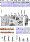
Figure 1
Figure 1
Accumulation of mutant neuroserpin in the ER correlates with neurodegeneration in FENIB mice. (A) Immunohistochemical staining for neuroserpin in CA1 and CA2 hippocampal regions of aged (8, 12, 20, 34, 45, 64, and >80 weeks) mice (n = 4). Neuroserpin accumulates progressively in Tg(NSS49P) but not in Tg(NS) over time. (B) Quantification of sections shown in A. S49P neuroserpin aggregation reaches a temporary plateau from weeks 34 to 45 with an exponential rise at a later timepoint. (C) Electron micrographs from cortical neurons of >70-week-old Tg(NSS49P) mice (left). Inclusion bodies (IBs) are found in the cytoplasm (Nuc, nucleus). IBs are positive in immunogold labeling for neuroserpin (IB, middle). In preembedding DAB-labeled tissue, neuroserpin positive IBs are found within rough ER (ER, right arrows). (D) Brain homogenates of 45-week-old Tg(NSS49P) and Tg(NS) mice, analyzed by nondenaturating PAGE show mutant neuroserpin as a polymer ladder, wild-type neuroserpin as monomers. (E and G) Immunohistochemical stainings for NeuN and caspase-3 in the hippocampus of mice. (F and H) Quantification of sections shown in E and G. (F) NeuN positive neurons show substantial loss of neurons only in aged (45 and 80 weeks) Tg(NSS49P) mice but not in Tg(NS) mice when compared to younger littermates (12 weeks). (H) Caspase-3 positive neurons show enhanced apoptosis in Tg(NSS49P) mice. Bars, (A, E, and G) 50 µm; (C, left to right) 5 µm, 500 nm, and 1 µm. Statistical significance *P ≤ 0.05; NS P ≥ 0.05.
Figure 2
Figure 2
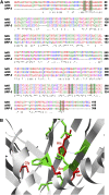
Comparison of neuroserpin and SRP-2. (A) Alignment of human (hNS) and mouse (mNS) neuroserpin with SRP-2. Sites causing FENIB are shadowed in red and amino acids forming a hydrophobic pocket in green (*, identical amino acids; : and ., amino acids with similar characteristics). (B) Structure of mouse neuroserpin showing close proximity of amino acids with disease-causing mutations (in red).
Figure 3
Figure 3
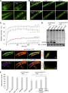
SRP-2H302R forms immobile protein aggregates in muscle cells leading to movement defects of transgenic worms. (A) In contrast to SRP-2 (wild-type), SRP-2H302R forms protein aggregates when expressed under its own and muscle-specific promoter. (B) Fluorescent images of representative FRAP experiments (B) and their graphic demonstration (C) show efficient fluorescence recovery of SRP-2 but not SRP-2H302R. SRP-2 (light green) shows slightly less fluorescence recovery than Q0::YFP (as control lacking inclusions, blue), whereas FRAP of SRP-2H302R (purple) is comparable to Q40::YFP (as control expressing 40 repeats of fluorescently tagged polyQ proteins with considerable inclusions, red). The traces are averages of five or more replicates (n = 7). (D, upper panel) 7.5 % native PAGE of extracts from indicated strains. SRP-2H302R (two independent integrated lines, hhls76, hhls77, and one extrachromosomal line hhEx39) forms high molecular weight aggregates (lanes 4 to 6) whereas SRP-2 (two independent integrated lines, hhls74, hhls75, and one extrachromosomal line, hhEx39, lanes 1 to 3) runs as a single band. Lower panel: SDS-Page to quantify tubulin for equal loading. (E) Confocal projections of transgenic worms showing the distribution of YFP–SRP-2 (green) and rhodamine-phalloidin that stained myofilaments (red). SRP-2 colocalizes with F-actin protein as shown by scatterplot, while SRP-2H302R deposits as discrete fluorescent foci that do not disrupt the myofilament structure. (F) Body bends of worms (lines as described in D) per minute were measured. (n = 30) Bars, (A, B, and E) 50 µm. Statistical significance *P ≤ 0.05; NS P ≥ 0.05.
Figure 4
Figure 4
Genetic deletion of HSF-1 and UPR pathways increase the amount of SRP-2H302R aggregates. (A) Fluorescent image of SRP-2H302R shows perinuclear localization consistent with ER localization (DAPI staining; right picture is a close-up of the merged image). (B) Representative fluorescent images of mutants lacking hsf-1, ire-1, atf-6, and pek-1. (C) Quantification of aggregates of SRP-2H302R worms described in B (n = 15). (D) Native and denaturing PAGE of extracts from indicated strains. Top native gel shows polymeric forms of SRP-2H302R; bottom denaturing gel shows the total level of SRP-2H302R probed with α-YFP antibodies. Tubulin was used as loading control. Integrated transgenic line hhIs76 was used for B–D. (E) Representative fluorescent images of mutant worms lacking either pek-1 or ire-1 after inactivation of hsf-1 for 48 hr by RNAi. The empty RNAi feeding vector pPD129.36 served as negative control. (F) Quantification of SRP-2H302R aggregates described in E (n = 25). Bars, (A) 10 μm, (B and E) 50 μm. Statistical significance *P ≤ 0.05; **P ≤ 0.01 (two-tailed Student’s t-test).
Figure 5
Figure 5
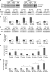
Transient and exhaustible induction of the UPR in Tg(NSS49P) mice negatively correlates with amounts of polymeric neuroserpin. (A) Reverse transcriptase–PCR from Tg(NSS49P) and Tg(NS) aged 12, 20, and 34 weeks and tunicamycin-treated (T+) or untreated (T) cells, to evaluate the levels of XBP1 (representative gel from three independent experiments). PstI digest distinguishes between processed and unprocessed XBP1. (B and D) Mutant Tg(NSS49P) and Tg(NS) mice (n = 3) were analyzed at 12, 20, 34, 45, 64, and >80 weeks by Western blot for presence of cleaved ATF6 (B) and phosphorylated eIF2α (D), with actin and eIF2α as loading controls. (C and E) Densitometric analysis of Western blots from B and D. Wild-type levels were set to one. (C) Tg(NSS49P) mice show increase of cleaved ATF6 with a maximum between 34 and 45 weeks. (E) Expression of phosphorylated eIF2α peaked in Tg(NSS49P) mice at 20 weeks. (F and G) Tg(NSS49P) and Tg(NS) mice (each n = 3) aged 8, 12, 20, 34, 45, 64, and >80 weeks were assessed for total and polymeric neuroserpin by ELISA. (F) Tg(NS) mice show no polymeric neuroserpin at any of the investigated timepoints. Polymeric neuroserpin was readily detectable in Tg(NSS49P) at 8 weeks of age. Surprisingly, levels of polymeric neuroserpin drop at 34 weeks. With disease progression, levels of polymeric neuroserpin increased exponentially. (G) Tg(NS) mice show constant low levels of total neuroserpin, whereas in Tg(NSS49P), neuroserpin plateaus between weeks 12 and 34; at later timepoints total neuroserpin increased exponentially. Statistical significance *P ≤ 0.05; NS P ≥ 0.05 (two-tailed Student’s t-test).
Figure 6
Figure 6
Hypothetical model how transient UPR induction and aggregation-prone proteins are interconnected in conformational dementia. In our proposed model, transient induction of the UPR in young mice limits polymer formation. With increasing age, these mechanisms cease resulting in an exponential increase of protein aggregates. Increase in aggregates (present as polymers and potentially in equilibrium with oligomers) cosegregates with neurodegeneration and clinical disease develops once the functional reserve capacity of the CNS is exhausted.

References

    1. Balch W. E., Morimoto R. I., Dillin A., Kelly J. W., 2008. Adapting proteostasis for disease intervention. Science 319: 916–919. - PubMed
    1. Borges V. M., Lee T. W., Christie D. L., Birch N. P., 2010. Neuroserpin regulates the density of dendritic protrusions and dendritic spine shape in cultured hippocampal neurons. J. Neurosci. Res. 88: 2610–2617. - PubMed
    1. Brandt R., Gergou A., Wacker I., Fath T., Hutter H., 2009. A Caenorhabditis elegans model of tau hyperphosphorylation: induction of developmental defects by transgenic overexpression of Alzheimer’s disease-like modified tau. Neurobiol. Aging 30: 22–33. - PubMed
    1. Brenner S., 1974. The genetics of Caenorhabditis elegans. Genetics 77: 71–94. - PMC - PubMed
    1. Brignull H. R., Moore F. E., Tang S. J., Morimoto R. I., 2006. Polyglutamine proteins at the pathogenic threshold display neuron-specific aggregation in a pan-neuronal Caenorhabditis elegans model. J. Neurosci. 26: 7597–7606. - PMC - PubMed

Publication types

MeSH terms