. 2013:19:16-24.
Epub 2013 Jan 7.

Lateral mobility of L-type calcium channels in synaptic terminals of retinal bipolar cells

Affiliations

Lateral mobility of L-type calcium channels in synaptic terminals of retinal bipolar cells

Wallace B Thoreson et al. Mol Vis. 2013.

Abstract

Purpose: Efficient and precise release of glutamate from retinal bipolar cells is ensured by the positioning of L-type Ca(2+) channels close to release sites at the base of the synaptic ribbon. We investigated whether Ca(2+) channels at bipolar cell ribbon synapses are fixed in position or capable of moving in the membrane.

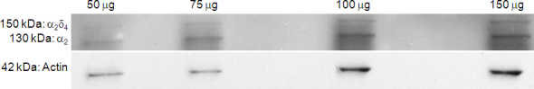
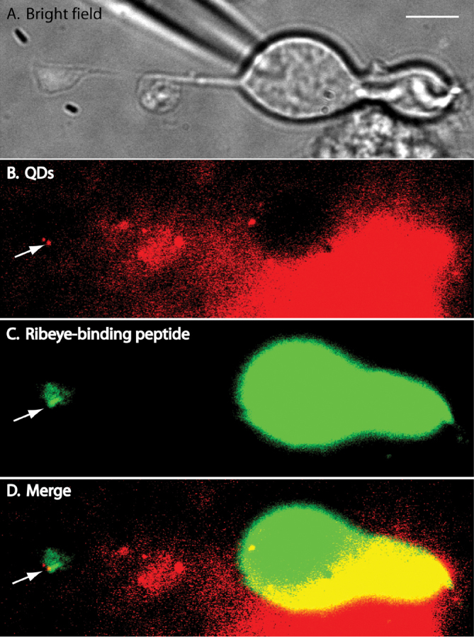
Methods: We tracked the movements of individual L-type Ca(2+) channels in bipolar cell terminals after labeling channels with quantum dots (QDs) attached to α(2)δ(4) accessory Ca(2+) channel subunits via intermediary antibodies.

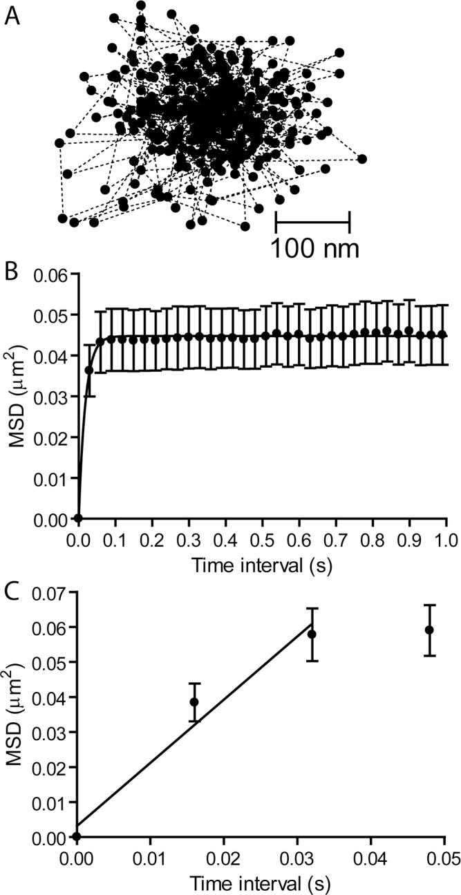
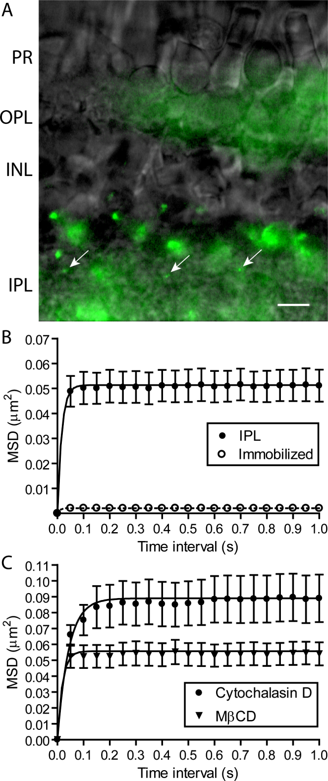
Results: We found that individual Ca(2+) channels moved within a confined domain of 0.13-0.15 μm(2) in bipolar cell terminals, similar to ultrastructural estimates of the surface area of the active zone beneath the ribbon. Disruption of actin expanded the confinement domain indicating that cytoskeletal interactions help to confine channels at the synapse, but the relatively large diffusion coefficients of 0.3-0.45 μm(2)/s suggest that channels are not directly anchored to actin. Unlike photoreceptor synapses, removing membrane cholesterol did not change domain size, indicating that lipid rafts are not required to confine Ca(2+) channels at bipolar cell ribbon synapses.

Conclusions: The ability of Ca(2+) channels to move within the presynaptic active zone suggests that regulating channel mobility may affect release from bipolar cell terminals.


Figures

Figure 1
Figure 1
Western blot analysis of the anti-α2δ4 antibody. Protein lysates from Ambystoma tigrinum retinas were resolved with western blot analysis. In this blot, we ran increasing quantities of protein lysate in each lane to optimize the visualization of the presumptive protein bands. Blots were first probed with the anti-α2δ4 antibody, which revealed two distinct bands at 150 kDa and 130 kDa, indicating the presence of the α2δ4 and α2 proteins, respectively. We then stripped the blot and probed it with an anti-β-actin antibody to assess the successive increase in total protein loaded into each lane.
Figure 2
Figure 2
Isolated bipolar cell with Ca2+ channels in the synaptic terminal labeled with quantum dots (QDs). A: A bright-field image of an isolated bipolar cell. B: A fluorescence image of QDs attached to the synaptic terminal of an isolated bipolar cell. The QD adjacent to the arrow was located in the focal plane; the dimmer QD above that one was located in a different focal plane. For this experiment, we used QDs whose emission peaked at 655 nm. The image was averaged from 100 frames acquired at 30 ms/frame. C: To label ribbons, we introduced a fluorescent peptide into the bipolar cell through a whole cell patch pipette. The HiLyte 488-conjugated peptide binds selectively to the CtBP domain of the ribbon protein, RIBEYE. D: Merged image showing that the bright QD overlapped with a region of bright HiLyte 488 fluorescence in the bipolar cell terminal (arrow).
Figure 3
Figure 3
Ca2+ channels move within confined domains on the terminals of isolated retinal bipolar cells. A: A plot of the trajectory of a single QD showing its position at 400 time points measured every 30 ms. B: A plot of mean squared displacement (MSD) versus time interval for individual Ca2+ channels in bipolar cell terminals (n=18). Confinement areas were calculated by fitting the data with Equation 3 (L=0.409±0.00479 μm). C: A diffusion coefficient of D=0.45±0.24 μm2/s (n=9) was determined from data acquired at 16 ms intervals using Equation 2. The straight line shows the fit to the MSD versus time interval relationship from the origin through the first two time points.
Figure 4
Figure 4
The confinement domains for Ca2+ channel movements on bipolar cell terminals in the inner plexiform layer (IPL) were expanded by disrupting actin but not by depleting membrane cholesterol. A: Three quantum dots (QDs) labeling individual Ca2+ channels in the IPL (arrows). Diffuse autofluorescence is also visible in the inner and outer plexiform layers. Scale bar=10 μm. B: MSD versus time interval for movements of individual Ca2+ channels in the IPL (n=34; filled circles). Confinement areas were calculated by fitting the data with Equation 3 (L=0.393±0.00237 μm). QDs immobilized in vacuum grease showed a small amount of jitter (L=0.0164±0.00447 μm, n=9; open circles). C: Actin disruption with cytochalasin D (20 μM) significantly expanded Ca2+ channel confinement domains relative to control (L=0.517±0.00680 μm, n=35, p<0.05; filled circles) but cholesterol depletion with MβCD (10 mM) did not (L=0.409±0.00252 μm, n=16; triangles).

References

    1. Heidelberger R, Matthews G. Calcium influx and calcium current in single synaptic terminals of goldfish retinal bipolar neurons. J Physiol. 1992;447:235–56. - PMC - PubMed
    1. Tachibana M, Okada T, Arimura T, Kobayashi K, Piccolino M. Dihydropyridine-sensitive calcium current mediates neurotransmitter release from bipolar cells of the goldfish retina. J Neurosci. 1993;13:2898–909. - PMC - PubMed
    1. von Gersdorff H, Sakaba T, Berglund K, Tachibana M. Submillisecond kinetics of glutamate release from a sensory synapse. Neuron. 1998;21:1177–88. - PubMed
    1. Coggins M, Zenisek D. Evidence that exocytosis is driven by calcium entry through multiple calcium channels in goldfish retinal bipolar cells. J Neurophysiol. 2009;101:2601–19. - PMC - PubMed
    1. Jarsky T, Tian M, Singer JH. Nanodomain control of exocytosis is responsible for the signaling capability of a retinal ribbon synapse. J Neurosci. 2010;30:11885–95. - PMC - PubMed

Publication types

MeSH terms

Substances

LinkOut - more resources