. 2013;11(1):e1001467.
doi: 10.1371/journal.pbio.1001467. Epub 2013 Jan 15.

Integration of canonical and noncanonical Wnt signaling pathways patterns the neuroectoderm along the anterior-posterior axis of sea urchin embryos

Affiliations

Integration of canonical and noncanonical Wnt signaling pathways patterns the neuroectoderm along the anterior-posterior axis of sea urchin embryos

Ryan C Range et al. PLoS Biol. 2013.

Abstract

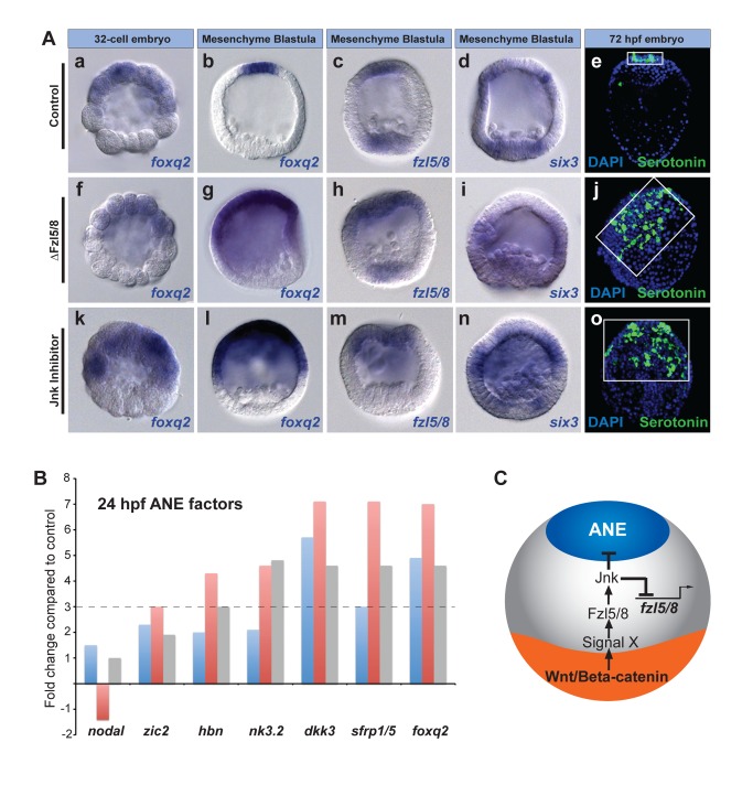
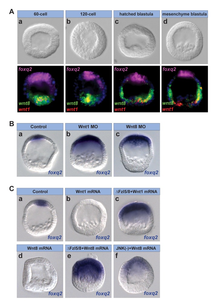
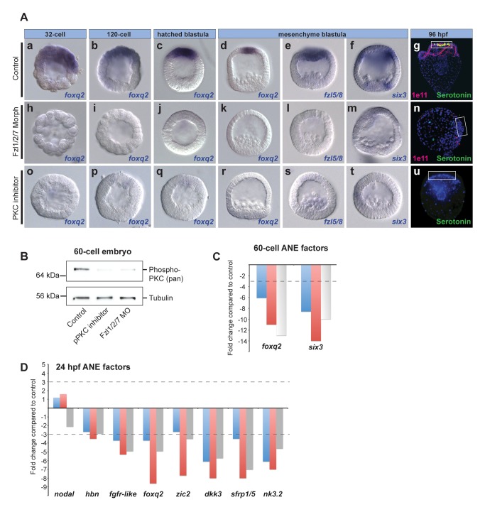
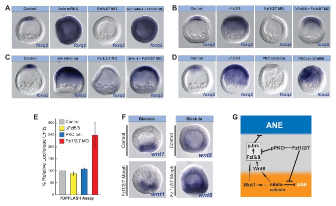
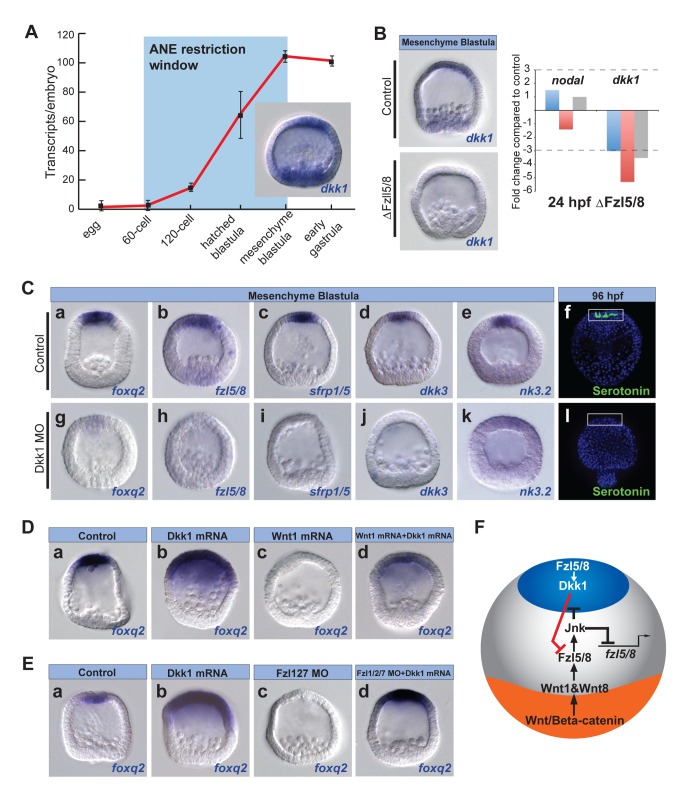
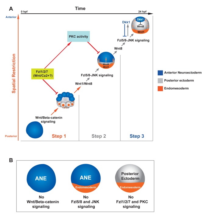
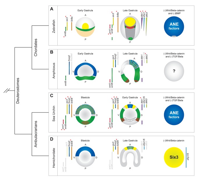
Patterning the neuroectoderm along the anterior-posterior (AP) axis is a critical event in the early development of deuterostome embryos. However, the mechanisms that regulate the specification and patterning of the neuroectoderm are incompletely understood. Remarkably, the anterior neuroectoderm (ANE) of the deuterostome sea urchin embryo expresses many of the same transcription factors and secreted modulators of Wnt signaling, as does the early vertebrate ANE (forebrain/eye field). Moreover, as is the case in vertebrate embryos, confining the ANE to the anterior end of the embryo requires a Wnt/β-catenin-dependent signaling mechanism. Here we use morpholino- or dominant negative-mediated interference to demonstrate that the early sea urchin embryo integrates information not only from Wnt/β-catenin but also from Wnt/Fzl5/8-JNK and Fzl1/2/7-PKC pathways to provide precise spatiotemporal control of neuroectoderm patterning along its AP axis. Together, through the Wnt1 and Wnt8 ligands, they orchestrate a progressive posterior-to-anterior wave of re-specification that restricts the initial, ubiquitous, maternally specified, ANE regulatory state to the most anterior blastomeres. There, the Wnt receptor antagonist, Dkk1, protects this state through a negative feedback mechanism. Because these different Wnt pathways converge on the same cell fate specification process, our data suggest they may function as integrated components of an interactive Wnt signaling network. Our findings provide strong support for the idea that the sea urchin ANE regulatory state and the mechanisms that position and define its borders represent an ancient regulatory patterning system that was present in the common echinoderm/vertebrate ancestor.


Conflict of interest statement

The authors have declared that no competing interests exist.

Figures

Figure 1
Figure 1. Preventing Wnt/β-catenin signaling allows activation of presumptive ANE specification throughout the embryo.
(A) Diagram showing the regulatory factors shared between the sea urchin ANE and the vertebrate forebrain/eye field. Both territories are restricted by mechanisms dependent on posterior Wnt signaling. (B, D, F, H) foxq2 expression in glycerol control (B, D) or Axin mRNA-injected embryos (F, H) at the 32-cell stage (B, F) and late blastula stage (D, H). six3 expression in glycerol control (C) and TCF-Eng mRNA-injected early blastulae (G). (E) A normal 3.5-d pluteus stage embryo. (I) A 3.5-d embryo misexpressing Axin mRNA. White box outlines the ANE. Serotonergic neurons (green), DAPI (nuclei, blue). (J) Schematic showing the stages of ANE restriction in 32-cell stage (6 hpf) (a), early blastula stage (7–15 hpf) (b), and mesenchyme blastula stage (24 hpf) embryos (c). ANE (blue), nβ-catenin (orange nuclei at 32-cell stage), endomesoderm territory (orange at blastula stages), posterior ectoderm (gray at blastula stages), and unknown restriction mechanism activated by posterior nβ-catenin (orange arrows).
Figure 2
Figure 2. Fzl5/8 signaling and JNK activity are required for ANE restriction.
(A) foxq2, fzl5/8, and six3 expression in 32-cell and late blastula-stage control embryos (Aa–d), ΔFzl5/8 mRNA-injected embryos (Af–i), and embryos treated with JNK inhibitor (Ak–n). (Ae, j, o) Serotonergic neurons in control, ΔFzl5/8 mRNA-injected, and JNK inhibitor-treated embryos, respectively. White boxes outline the ANE. Serotonergic neurons (green), DAPI (nuclei, blue). (B) qPCR measurements from three different cultures of embryos showing that ANE regulatory genes are up-regulated at the late blastula stage (24 hpf) in the absence of functional Fzl5/8 signaling. The y-axis shows the fold change in gene expression in ΔFzl5/8-containing embryos relative to controls. The dotted line marks a 3-fold change in expression. nodal expression is used as an internal control because it is unaffected in the absence of Fzl5/8 signaling (Croce et al., 2006) . (C) Diagram showing a model for ANE restriction consistent with the data presented in this figure.
Figure 3
Figure 3. Wnt1 and Wnt8 signaling are necessary for Fzl5/8-JNK-mediated ANE restriction.
(A) Three-color in situ hybridization for wnt1 (red), wnt8 (green), and foxq2 (magenta) transcripts during ANE restriction. wnt1 mRNA appears yellow when overlapping with wnt8 mRNA. (B) The foxq2 expression domain is not restricted in embryos injected with a Wnt1 (b) or Wnt8 (c) morpholino. (C) foxq2 expression is completely eliminated in embryos injected with either Wnt1 (b) or Wnt8 (d) mRNA. The Wnt1- and Wnt8-mediated inhibition of foxq2 expression requires functional Fzl5/8 (c, e); the Wnt8-mediated inhibition of foxq2 expression requires JNK activity (f). MO, morpholino.
Figure 4
Figure 4. Fzl1/2/7 signaling and PKC activity are necessary for ANE specification.
(A) foxq2 expression at stages from 32-cell to hatched blastula, as well as fzl5/8 and six3 expression at mesenchyme blastula stage in control embryos (Aa–f), Fzl1/2/7 morpholino-injected embryos (Ah–m), and embryos treated with PKC inhibitor (Ao–t). (Ag, n, u) Neurons in control, Fzl1/2/7 morpholino-injected, and PKC inhibitor-treated 96-hpf pluteus larvae. White boxes outline the ANE. Serotonergic neurons (green), DAPI (nuclei, blue), synaptotagminB/1e11 (pan-neural, magenta). (B) Western blot showing that phosphorylation of PKC is blocked to similar extents with a PKC inhibitor and Fzl1/2/7 MO. (C, D) qPCR measurements from three different cultures of embryos showing that the early ANE regulatory genes are down-regulated in the absence of Fzl1/2/7 signaling at the 60-cell stage (C) and late blastula stages (D). The y-axis shows the fold change in gene expression level in Fzl1/2/7 morpholino-containing embryos relative to controls. The dotted line marks a 3-fold change in expression. nodal expression is used as an internal control.
Figure 5
Figure 5. Fzl1/2/7 signaling and PKC activity antagonize the ANE restriction mechanism.
(A–D) Injected molecules are indicated above each panel. Inhibition of Fzl1/2/7 function (Fzl1/2/7 MO) does not interfere with foxq2 expression in the absence of nβ-catenin (Axin mRNA) (A) or functional Fzl5/8 (ΔFzl5/8 mRNA) (B) or when JNK was inhibited (C). (D) Blocking PKC activity in the absence of ΔFzl5/8 signaling rescues foxq2 expression. (E) TopFlash assays on three different cultures of embryos showing that the activity of Fzl1/2/7, but not those of Fzl5/8 or PKC, suppress nβ-catenin activity. (F) wnt1 and wnt8 are expressed in the endomesoderm of Fzl1/2/7 morphants. (G) Diagram showing a model for ANE restriction based on the data presented in this and the preceding figures. MO, morpholino.
Figure 6
Figure 6. Dkk1 activity defines the ANE territory by antagonizing Wnt signaling.
(A) qPCR measurements from three different batches of embryos showing the number of Dkk1 transcripts/embryo at the indicated stages during early development. Values were normalized to z12 transcripts (see Materials and Methods). (B) In situ hybridization (left) and qPCR (right) show that dkk1 expression requires Fzl5/8 signaling (ΔFzl5/8 mRNA). (C) Six ANE markers show that ANE specification requires Dkk1. (D, E) Injected molecules are indicated at the top of panels. (D) Overexpression of Dkk1 prevents foxq2 restriction to the anterior pole and suppression of foxq2 expression by ectopic Wnt1. (E) Overexpression of Dkk1 rescues foxq2 expression in embryos co-injected with Fzl1/2/7 morpholino. (F) Diagram showing a model for ANE restriction based on the data presented in this and previous figures. MO, morpholino.
Figure 7
Figure 7. Three-step model for the balance of Wnt signaling interactions during ANE restriction.
(A) The y-axis monitors the progress of spatial restriction of the ANE (blue), and the x-axis indicates the developmental timing of ANE restriction. Each image represents the position of the ANE in time and space. (Step 1) Initially, the maternal regulatory state in the absence of Wnt signaling supports ANE specification throughout the embryo. Then, nβ-catenin signaling in the posterior half of the embryo activates an unknown negative regulatory activity that blocks the accumulation of ANE factors, either by blocking their transcription directly or the activity of their ubiquitously expressed maternal activators. (Step 2) As development progresses, posterior nβ-catenin activates production of at least two Wnt ligands, Wnt1 and Wnt8, that are necessary to initiate the ANE restriction mechanism in posterior ectoderm beginning at the 60-cell stage. These secreted ligands signal through the Wnt receptor, Fzl5/8, activating the JNK pathway. The Wnt/JNK pathway progressively down-regulates expression of ANE factors during early blastula stages in all but the most anterior ectoderm. During Step 1 and possibly Step 2, Fzl1/2/7 signaling attenuates the nβ-catenin- and Fzl5/8-JNK-mediated down-regulation of ANE factors, preventing complete shutdown of ANE specification. PKC activity also antagonizes Fzl5/8-JNK-mediated ANE restriction, downstream of Fzl1/2/7 signaling. (Step 3) Expression of Wnt8, Wnt1, and/or another ligand X is activated in more anterior blastomeres. These ligands continue Fzl5/8-mediated ANE restriction until the late blastula-stage embryo activates production of the secreted Wnt antagonist, Dkk1, via Fzl5/8. Through negative feedback, Dkk1 limits Fzl5/8 activity, thereby defining the borders of the ANE. Orange arrows indicate the Wnt/β-catenin-mediated mechanism, gray arrows indicate the Wnt/JNK mediated mechanism, and red indicates Fzl1/2/7 and PKC interactions. (B) Diagram showing the state of the ANE in embryos lacking either Wnt/β-catenin, Fzl5/8 and pJNK, or Fzl1/2/7 and pPKC signaling.
Figure 8
Figure 8. Conservation of expression patterns of orthologs of sea urchin genes that function in the development of ANE.
The territorial expression of orthologs in each embryo is shown to the left and right of each diagram. In cases where there is no information concerning the expression pattern of a particular ortholog, it is represented in light gray. Red asterisks designate that functional studies show these factors are involved in AP patterning of the presumptive neuroectoderm. The images of embryos are colored to indicate the expression patterns of foxq2, six3, wnt1, and wnt8. (A) ANE factors are initially expressed throughout the presumptive zebrafish neuroectoderm at late blastula/early gastrula stages (left-hand diagram). These ANE factors are progressively down-regulated in posterior regions of the presumptive neuroectoderm (dark gray) during gastrulation by a mechanism involving Fzl8a, Wnt8, Wnt1, and Dkk1, until they are confined to the anterior pole (middle diagram). Factors such as sFrp1 and Dkk3 subsequently pattern the forebrain. The right-hand diagram shows that, in the absence of Wnt/β-catenin and BMP signaling, ANE factors are expressed throughout most of the embryo. Embryos are oriented with their dorsal sides facing up from the page. Data taken from ,,,–. (B) In amphioxus embryos, foxq2 is expressed throughout the anterior half, and wnt8 throughout the vegetal plate, of early gastrula embryos (left-hand diagram). By late gastrula, foxq2 and the putative ANE factors dkk1, six3, and dkk3 are expressed in the anterior-most ectoderm. wnt8 and wnt1 are expressed posterior to these putative ANE factors, consistent with a role in the restriction of foxq2, six3, and dkk3 expression to the anterior pole (middle diagram). Data taken from –. (C) Sea urchin embryo ANE factors are initially expressed throughout the presumptive ectoderm, and wnt1 and wnt8 are both expressed in the posterior half of blastula stage embryos (left-hand diagram). Then, ANE factors are progressively down-regulated from posterior ectoderm by a Wnt1, Wnt8, Fzl5/8, and Dkk1-dependent mechanism (middle diagram). In the absence of Wnt/β-catenin and TGF-β signaling, the entire sea urchin embryo expresses ANE factors (right-hand diagram). Data taken from this study and ,. (D) In blastula stage hemichordate embryos, foxq2 is expressed broadly in the anterior half of the embryo (data show that six3 and fzl5/8 also are expressed broadly by early gastrula stages) (left-hand diagram). By late blastula stages, putative ANE factors foxq2, six3, and sfrp1/5 are restricted to the anterior-most ectoderm, and functional data show that sfrp1/5 restriction involves Fzl5/8 (middle diagram). In the absence of Wnt/β-catenin signaling the entire hemichordate embryo expresses putative ANE factors six3 and sfrp1/5 (right-hand diagram). Data taken from ,,,.

References

    1. Adamska M, Degnan SM, Green KM, Adamski M, Craigie A, et al. (2007) Wnt and TGF-beta expression in the sponge Amphimedon queenslandica and the origin of metazoan embryonic patterning. PloS One 2: e1031 doi:10.1371/journal.pone.0001031. - DOI - PMC - PubMed
    1. Darras S, Gerhart J, Terasaki M, Kirschner M, Lowe CJ (2011) beta-catenin specifies the endomesoderm and defines the posterior organizer of the hemichordate Saccoglossus kowalevskii. Development 138: 959–970. - PMC - PubMed
    1. Momose T, Derelle R, Houliston E (2008) A maternally localised Wnt ligand required for axial patterning in the cnidarian Clytia hemisphaerica. Development 135: 2105–2113. - PubMed
    1. Logan CY, Miller JR, Ferkowicz MJ, McClay DR (1999) Nuclear beta-catenin is required to specify vegetal cell fates in the sea urchin embryo. Development 126: 345–357. - PubMed
    1. Wikramanayake AH, Hong M, Lee PN, Pang K, Byrum CA, et al. (2003) An ancient role for nuclear beta-catenin in the evolution of axial polarity and germ layer segregation. Nature 426: 446–450. - PubMed

Publication types

MeSH terms