. 2013 Jan 14:6:119.
doi: 10.3389/fncir.2012.00119. eCollection 2012.

Frequency discrimination and stimulus deviance in the inferior colliculus and cochlear nucleus

Affiliations

Frequency discrimination and stimulus deviance in the inferior colliculus and cochlear nucleus

Yaneri A Ayala et al. Front Neural Circuits. .

Abstract

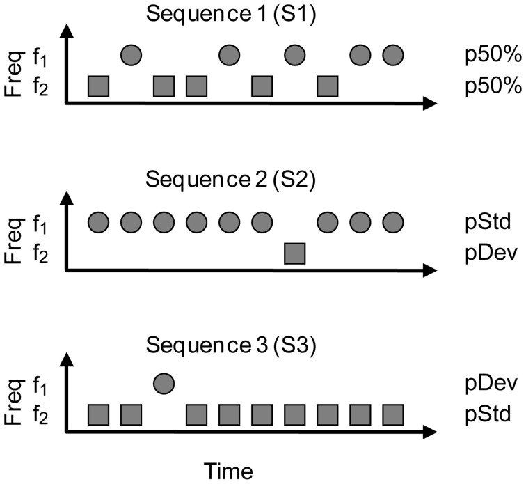
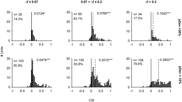
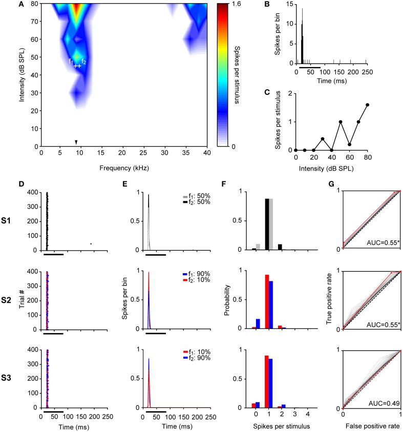
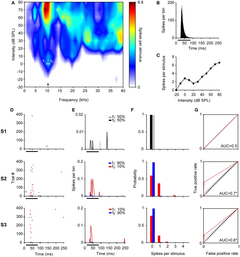
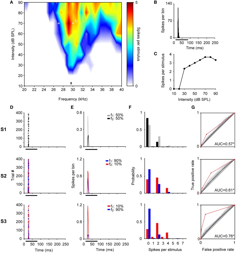
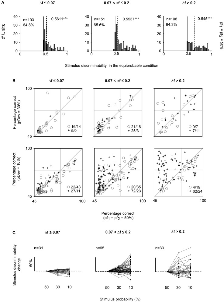
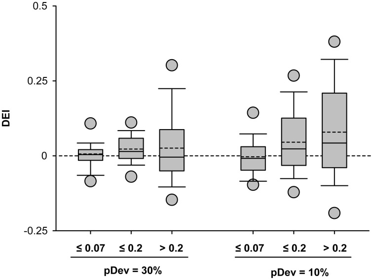
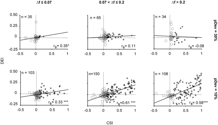
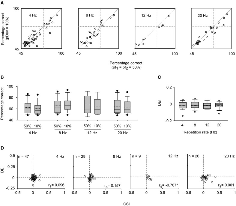
Auditory neurons that exhibit stimulus-specific adaptation (SSA) decrease their response to common tones while retaining responsiveness to rare ones. We recorded single-unit responses from the inferior colliculus (IC) where SSA is known to occur and we explored for the first time SSA in the cochlear nucleus (CN) of rats. We assessed an important functional outcome of SSA, the extent to which frequency discriminability depends on sensory context. For this purpose, pure tones were presented in an oddball sequence as standard (high probability of occurrence) or deviant (low probability of occurrence) stimuli. To study frequency discriminability under different probability contexts, we varied the probability of occurrence and the frequency separation between tones. The neuronal sensitivity was estimated in terms of spike-count probability using signal detection theory. We reproduced the finding that many neurons in the IC exhibited SSA, but we did not observe significant SSA in our CN sample. We concluded that strong SSA is not a ubiquitous phenomenon in the CN. As predicted, frequency discriminability was enhanced in IC when stimuli were presented in an oddball context, and this enhancement was correlated with the degree of SSA shown by the neurons. In contrast, frequency discrimination by CN neurons was independent of stimulus context. Our results demonstrated that SSA is not widespread along the entire auditory pathway, and suggest that SSA increases frequency discriminability of single neurons beyond that expected from their tuning curves.

Keywords: ROC analysis; SSA; change detection; deviant sensitivity; mismatch negativity; non-lemniscal pathway.


Figures

Figure 1
Figure 1
The oddball stimulation paradigm. Two frequencies (f1, f2) were presented pseudo-randomly with different probabilities of occurrence. In Sequence 1 (S1), f1, and f2 occurred with equal probability (p50%), which served as a control condition. This condition is useful to see the neuron's tuning to the frequencies chosen. For the oddball condition, the probability of the frequencies was modified such that one frequency (f1, circles) was the standard tone, occurring with high probability, and the other (f2, squares) was the deviant tone, with low probability of occurrence (Sequence 2, S2). The probabilities of f1 and f2 were reversed in Sequence 3 (S3) in order to have each frequency presented as deviant and standard. We tested two probabilities for the deviant tone (pDev), 30 and 10%, so the corresponding probabilities for the standard (pStd) were 70 and 90%, respectively.
Figure 2
Figure 2
Distribution of stimulus-specific adaptation indices of IC neurons. Histograms of the common SSA index (CSI) displayed according to the frequency separation intervals (columns: Δf ≤ 0.07, 0.07 < Δf ≤ 0.2, Δf > 0.2) and the probability of the deviant tone (rows: pDev = 30 and 10%). The CSI was calculated from the responses recorded in S2 and S3. A CSI = 0 indicates an equal neuronal response when the tones were presented as deviant or standard, while positive and negative values represent higher firing rates when the tones were deviant or standard, respectively. For each stimulus condition the CSI values were tested against zero (solid line). The numbers next to the dashed line indicate the value of the median and the statistical significance (Signed Rank Test; *p < 0.05, ***p < 0.001). The distributions moved toward positive values when Δf was larger and pDev smaller. The percentages indicate the amount of neurons with CSI > 0.1.
Figure 3
Figure 3
Example of a non-adapting neuron in the IC. (A) Narrow FRA in color code for response magnitude. The tested frequencies (f1: 8.7 kHz, f2: 9.6 kHz, white crosses) were chosen around the BF (8.8 kHz) (arrowhead), at 45 dB SPL. (B) PSTH of the accumulated response to all the frequencies (0.5 – 40 kHz) and intensities (0 – 80 dB SPL) presented (1 ms bins). (C) Rate-level function at BF. (D–G) The responses of the neuron for each pair of stimuli for each of the three sequences (S1, S2, S3) are shown as dot raster plots (D), PSTH (3 ms bins) (E), spike probability distributions (F), and ROC curves (G). In the dot raster each dot represents the occurrence of a spike. The black bar under the PSTH and dot raster plots indicates the duration of the stimulus (75 ms). The probability of each frequency for each sequence is indicated on the upper left of the (E) panels. In the ROC curves (G) the dashed line corresponds to random guessing or no stimulus discrimination (AUC = 0.5), indicating complete overlap of the spike probability distributions. The red line represents the ROC curve calculated using the recorded data, the curves plotted in gray were obtained with the permutation method of the original spike count distributions, and the black line is represents the mean ROC curve of permutations. A total of 10,000 permutations were calculated, but for visual clarity only 100 curves are displayed. For each ROC curve, the area under the ROC curve (AUC) is shown corresponding to the original AUC value minus the mean AUC from permutations, as well as, the significance value for AUC > 0.5 (Permutation test; *p < 0.05). The repetition rate was 4 Hz and the frequency separation was 0.141 octaves. This neuron did not show SSA (CSI = 0.04, Bootstrapping; p > 0.05), displaying a very similar response to f1 and f2 across the three sequences regardless the probability of each tone.
Figure 4
Figure 4
Example of an adapting neuron in the IC. (A) FRA of a broadly tuned neuron with a BF of 10 kHz (arrowhead). The frequencies tested are indicated by the white crosses around the BF (f1: 9.5 kHz, f2: 9.6 kHz), at 0 dB SPL. (B–G) Same format as in Figure 3. This neuron showed strong SSA (CSI = 0.88, Bootstrapping; p < 0.05) reducing its firing to the high probability tone in S2 and S3 while still responding to the low probability one across most stimulus presentations. This differential firing is reflected in an AUC larger than 0.5 (Permutation test; *p < 0.05) in oddball sequences (S2 and S3).
Figure 5
Figure 5
Example of a partially-adapting neuron in the IC. (A) FRA from a neuron with a BF of 29.9 kHz (arrowhead). The frequencies tested were f1: 27.1 kHz and f2: 30 kHz, at 70 dB SPL. (B–G) Same format as in Figure 3. This neuron displayed a significant level of SSA (CSI = 0.5, Bootstrapping; p < 0.05), responding to both tones across the 400 stimulus trials. Although, the neuron displayed significant discriminability in the equiprobable condition (S1, AUC = 0.57) (Permutation test; *p < 0.05), this was improved under the oddball sequences (S2, AUC = 0.61; S3, AUC = 0.76).
Figure 6
Figure 6
Neurometric performance under equiprobable and oddball conditions of IC neurons. (A) Distributions of the AUC values for the equiprobable condition (AUC50%) indicating the median (dashed line) significantly differs from 0.5 (Signed Rank Test; ***p < 0.001). The percentage of neurons whose AUC50% was higher than 0.5 is indicated for each panel (Permutation test; p < 0.05). (B) Scatter plots showing the neurometric performance for frequency discrimination expressed as percentage correct under the oddball condition (rows: pDev = 30, 10%) versus the equiprobable condition (pf1 = pf2 = 50%), for each frequency contrast interval (columns: Δf ≤ 0.07, 0.07 < Δf ≤ 0.2, Δf > 0.2). Separately are represented the neurons with CSI ≤ 0.1 (gray circles) and CSI > 0.1 (dark crosses). For the oddball condition, the percentage correct corresponds to the mean AUC value obtained from S2 and S3. The number of neurons above and below the diagonal line (equal performance in both conditions) is indicated by the inset on the bottom right of each panel. (C) Sensitivity curves of individual neurons expressed as percentage of change elicited when pDev = 10% and 30% regarding the pf1 = pf2 = 50% condition.
Figure 7
Figure 7
Stimulus discriminability enhancement of IC neurons across different stimulus conditions. Box plots of the discriminability enhancement under the oddball condition (DEI) showing the mean (dashed line) and the median (solid line) values, as well as, the 5th and 95th outliers. All the mean values were positive (except for the 10%f ≤ 0.07 condition), reflecting a better stimulus discrimination when one of the frequencies is presented as a deviant tone, that is, with low probability of ocurrence (30 or 10%). The DEIs were only affected by the frequency separation [F(2, 489) = 5.72, p < 0.01] (Two-Way ANOVA, deviant probability × frequency separation).
Figure 8
Figure 8
The stimulus discriminability reflects the degree of stimulus-specific adaptation exhibited by IC neurons. Correlation of the stimulus discriminability enhancement (DEI) and the SSA index (CSI). In gray circles are represented the neurons with CSI ≤ 0.1 and in dark crosses those with CSI > 0.1. The linear correlation was reflected by the Spearman's correlation coefficient (rs), whose strength varied according to the frequency contrast (columns) and probability of the deviant tone (rows). *p < 0.05, ***p < 0.001.
Figure 9
Figure 9
Bandwidth of frequency response areas and SSA level of IC neurons. (A) Box plots of the bandwidth (BW) values grouped into CSI ranges for the three Δf intervals with the mean (dashed line) and median values (solid line) indicated, as well as, the 5th and 95th outliers. There is an increment in the bandwidths as neurons have higher CSI [F(1, 688) = 37.5, p = 0] and as the level increased [F(1, 688) = 77.4, p = 0] (Analysis of covariance of BW with level, Δf and CSI as factors). (B) Box plots of BW values grouped into DEI ranges. The BW increases for neurons that displayed higher discriminability improvement under the oddball condition [F(1, 688) = 26.7, p = 0]. Same format as panel (A). (Analysis of covariance of BW with level, Δf and DEI as factors).
Figure 10
Figure 10
Histological identification of recording sites of IC and CN neurons. (A) Example of recording sites marked with an electrolytic lesion (arrowheads) in the lateral cortex of the IC at 1.9 mm lateral, according to Paxinos and Watson (2007). Two different tracts are indicated by Tr #1 and Tr #2. (B,C) Recording sites (arrowheads) and tracts (Tr) located in DCN (at 11.52 mm from bregma) and VCN (at 11.04 mm from bregma), respectively. The slices were Nissl stained and cut at 40 μm in a sagital (A) and coronal plane (B,C). Scale bars of 500 μm. D, dorsal; C, caudal; M, medial.
Figure 11
Figure 11
Example of a CN neuron. The format for all panels is the same as in Figures 3–5. (A) The BF was 23.4 kHz (indicated by the arrowhead) and the tested frequencies (f1 = 23.4 kHz and f2 = 25.9 kHz, white crosses) differ by 0.144 octaves. The PSTH of the accumulated response to all the frequencies (0.5 – 40 KHz) and intensities (0 – 80 dB SPL) presented (1 ms bins), as well as, the rate-level function at BF are shown in (B) and (C), respectively. The neuron exhibited a low CSI = 0.1 (Bootstrapping; p > 0.05) with a robust response across trials (D,E) and its frequency discriminability was not sensitive to the oddball condition (F,G). The AUC values in all conditions were slightly higher than 0.5 (Permutation test; *p < 0.05).
Figure 12
Figure 12
CN neurons do not exhibit SSA. (A) Spike count to deviant (dev) and to standard (std) stimuli elicited at different repetition rates represented in a color code. This color code is the same for (B–E) panels. (4 Hz, n = 47; 8 Hz, n = 29; 12 Hz, n = 9; 20 Hz, n = 26) (B) Scattergraph of the Frequency-Specific SSA indices (SI) for f1 and f2 presented at different repetitions rates. This index reflects the normalized spikes counts elicited when each frequency was the deviant tone regarding the response evoked when it was the standard one. (C) Scattergraph of the median first spike latency (FSL) for f1 and f2 when they were the deviant (FSLdev) or the standard (FSLstd) stimulus. In the right column, are displayed the box plots of the median FSL for the population of neurons to deviant (red) and to standard (blue) stimulus across repetition rates. The n for this panel is the double of the number of neurons tested, since two frequencies were tested as deviant and as standard stimulus for each neuron. (D,E) Box plots of the CommonSSA index (CSI) when increasing the rate of stimulation and scattergraph of CSI when varying the frequency separation factor (Δf), respectively.
Figure 13
Figure 13
Stimulus sensitivity in CN neurons. (A) Neurometric boxes displaying the percentage of correct identifications in the equiprobable and oddball condition (pDev = 10%) for different repetitions rates (4, 8, 12, and 20 Hz). (B) Box plots indicating that no improvement was elicited under the oddball condition regarding the equiprobable one (Signed Rank Test; p > 0.05). (C) Box plots that indicate that the discrimination enhancement indexes (DEI) remained at zero and did not change across repetition rate (Kruskal–Wallis Test; p > 0.05). (D) No positive correlation between the CSI and DEI was found (Spearman's correlation coefficient, rs; *p < 0.05).

References

    1. Anderson L. A., Christianson G. B., Linden J. F. (2009). Stimulus-specific adaptation occurs in the auditory thalamus. J. Neurosci. 29, 7359–7363 10.1523/JNEUROSCI.0793-09.2009 - DOI - PMC - PubMed
    1. Anderson L. A., Izquierdo M. A., Antunes F. M., Malmierca M. S. (2009). A monosynaptic pathway from dorsal cochlear nucleus to auditory cortex in rat. Neuroreport 20, 462–466 10.1097/WNR.0b013e328326f5ab - DOI - PubMed
    1. Anderson L. A., Malmierca M. S. (2013). The effect of auditory cortical deactivation on stimulus-specific adaptation in the inferior colliculus of the rat. Eur. J. Neurosci. 37, 52–62 10.1111/ejn.12018 - DOI - PubMed
    1. Antunes F. M., Malmierca M. S. (2011). Effect of auditory cortex deactivation on stimulus-specific adaptation in the medial geniculate body. J. Neurosci. 31, 17306–17316 10.1523/JNEUROSCI.1915-11.2011 - DOI - PMC - PubMed
    1. Antunes F. M., Nelken I., Covey E., Malmierca M. S. (2010). Stimulus-specific adaptation in the auditory thalamus of the anesthetized rat. PLoS ONE 5:e14071 10.1371/journal.pone.0014071 - DOI - PMC - PubMed