. 2012 Sep;7(9):885-899.
doi: 10.2217/fvl.12.81.

HSV, axonal transport and Alzheimer's disease: in vitro and in vivo evidence for causal relationships

Affiliations

HSV, axonal transport and Alzheimer's disease: in vitro and in vivo evidence for causal relationships

Elaine L Bearer. Future Virol. 2012 Sep.

Abstract

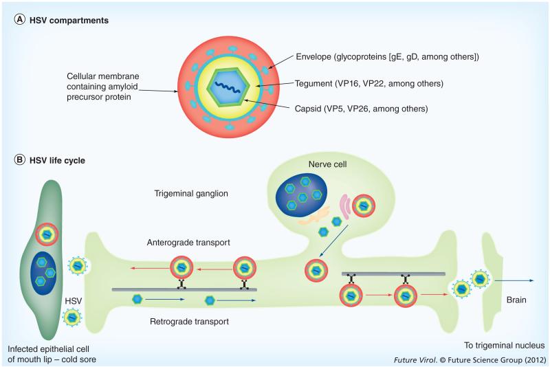
HSV, a neurotropic virus, travels within neuronal processes by fast axonal transport. During neuronal infection HSV travels retrograde from the sensory nerve terminus to the neuronal cell body, where it replicates or enters latency. During replication HSV travels anterograde from the cell body to the nerve terminus. Postmortem studies find a high frequency of HSV DNA in the trigeminal ganglia as well as the brain. Studies correlating HSV with Alzheimer's disease (AD) have been controversial. Here we review clinical evidence supporting such a link. Furthermore, the author describes experimental data showing physical interactions between nascent HSV particles and host transport machinery implicated in AD. The author concludes that the complexity of this relationship has been insufficiently explored, although the relative ease and nontoxicity of a potential anti-HSV treatment for AD demands further study.


Figures

Figure 1
Figure 1. HSV structure and life cycle
(A) HSV compartments: this schematic depicts the four concentric compartments of the infective particle encased in a fifth compartment provided by a cellular membrane system found encircling cytoplasmic viral particles during productive infections and containing amyloid precursor protein. The capsid is approximately 135 nm in diameter and the whole assemblage may be as large as 350–400 nm. (B) HSV life cycle: here we outline the process the virus follows to infect the trigeminal ganglion and progress into the trigeminal nucleus in the brain. Note that the trigeminal ganglion cells project two processes: one to the lip, the other to the brain. Virus traveling in a single neuron can move within these processes to go either to the lip or into the brain. The route from the lip to the brain can be >10 cm in length.
Figure 2
Figure 2. HSV-1 interacts with cellular amyloid precursor protein during egress
(A) VP26-GFP HSV-1 (green) particles colocalize with APP (red) during egress by immunofluorescence after synchronous infection with VP26-GFP HSV-1 (green) and fixation at 7 h postinfection. Examples of two infected cells also stained by immunofluorescence for gE (blue). Lower right inset shows a higher magnification of the boxed region. In this inset, one VP26-GFP-labeled particle displays all three labels (gE, APP and VP26), another has only APP (white arrowhead) or only gE (pink arrowhead). Such single labels demonstrate that colocalization is not due to bleed-through from other fluorescent channels. The inset graph shows intensity profiles along the line drawn across the right-hand cell. Superimposed peaks in the graph for the different colors are indicated by vertical arrows. (B) The first frame of a video sequence showing the initial positions of HSV capsids (VP26-GFP, green) and cellular APP-labeled with red fluorescent protein (APP-monomeric red fluorescent protein, red) in an infected cell at 7–9 h postinfection from a 3-s time-lapse 900-s (15 min) video. Double-labeled particles appear yellow (arrows). Many capsids (64%) colocalize with APP compartments in this frame. Capsids travel with APP vesicles, and sometimes join and separate from them. Positions of the N and of the cell boundaries are delineated by white lines. (C) The tracks of selected VP26-GFP HSV particles that move with the APP-monomeric red fluorescent protein label. Each trace has been assigned a different pseudocolor. Beginnings and ends of movements are indicated by dots and arrowheads, respectively. (D) An example of a double-labeled HSV-APP particle that moves away from the nucleus (arrow). In the last panel eight frames are superimposed to demonstrate the particle’s trajectory. (E) Immunogold-labeled electron micrograph of an HSV-1 particle inside an infected cell cytoplasm probed with anti-C-APP followed by protein-A labeled with 10 nm gold. Gold particles decorate both cellular and viral membranes surrounding capsids. Scale bar = 100 nm. APP: Amyloid precursor protein; GFP: Green fluorescent protein; N: Nuclei. Modified with permission from [7]. Supporting videos for these images can be found on the PLoS One journal website linked to the online publication.
Figure 3
Figure 3. Schematics showing intracellular interactions between cellular amyloid precursor protein and HSV particles at an early stage (top) and a later stage (bottom) of productive infection in epithelial cells
Intracellular dynamics at early stages during viral replication (top panel): (A) in the perinuclear region, viral particles move around and within the large membranous compartments near the nucleus, and also frequently colocalize with the viral envelope glycoproteins, gE and gD, and with cellular transmembrane organelle proteins in this area of the cell, such as: LAMP2, a lysosomal membrane protein; TGN46, associated with the trans-Golgi network; and APP, whose physiologic function is unknown. In the mid-cytoplasmic area, the LAMP2 compartments are separate from this apparent Golgi network, and rarely colocalize with any viral or Golgi components at the periphery. (B) In the outer cytoplasm neither LAMP2 nor TGN46-positive organelles colocalize with viral particles, while APP particles remain associated both with viral capsids and with viral envelope glycoproteins, gE and gD, probably on their way to the cell surface. (C) Capsids entering smaller post-Golgi compartments that stain uniquely for APP undergo fast transport towards the cell surface. APP: Amyloid precursor protein; GFP: Green fluorescent protein. Adapted from [7], with permission as modified from [61]. (D) After leaving the nucleus, viral particles are also found without detectible APP or the other cellular membrane proteins studied here. These particles could be inside some other unlabeled membrane system, or be free in the cytoplasm. These solo particles will be transported only rarely. (E) Capsids, possibly with tegument, may ride on the cytoplasmic surface of APP-labeled membrane systems, attach and detach from these membranes, or bud into them. Any particular capsid may employ all of these mechanisms during transit in the cytoplasm. In each case, we hypothesize that microtubule motors, such as one or more of the kinesin family, are recruited to the capsid-containing membrane-bound particles, possibly via APP or another cellular motor receptor. Dimensions are not to scale. Intracellular dynamics at later stages (bottom panel): as the microtubule network breaks down and reorganizes during productive infection, capsids must travel further to reach the cellular membrane synthesis and packaging centers in the Golgi apparatus, which drifts out during productive infections. In this case capsids travel directly on microtubules, possibly recruiting retrograde machinery as for infective entry, and yet reach the cell surface due to the disorganization of the microtubule network. Upon arrival at the cortex, where the Golgi has drifted, viral envelopment takes place. Dimensions are not to scale. APP: Amyloid precursor protein; GFP: Green fluorescent protein. Adapted from [7], with permission as modified from [61].

References

    1. Roizman B, Knipe D. Herpes simplex viruses and their replication. In: Knipe D, Howley P, editors. Fields Virology. Lippincott Williams & Wilkins; PA, USA: 2001. pp. 2399–2461.
    1. Su YH, Moxley MJ, Ng AK, et al. Stability and circularization of herpes simplex virus type 1 genomes in quiescently infected PC12 cultures. J. Gen. Virol. 2002;83(Pt 12):2943–2950. - PubMed
    1. Mellerick DM, Fraser NW. Physical state of the latent herpes simplex virus genome in a mouse model system: evidence suggesting an episomal state. Virology. 1987;158(2):265–275. - PubMed
    1. Farooq AV, Shukla D. Corneal latency and transmission of herpes simplex virus-1. Future Virol. 2011;6(1):101–108. - PMC - PubMed
    1. Bearer EL. Perspectives on herpes-APP interactions. Aging Cell. 2004;3(2):81–84. - PMC - PubMed

LinkOut - more resources