. 2013;8(1):e53404.
doi: 10.1371/journal.pone.0053404. Epub 2013 Jan 15.

The yeast transcription factor Crz1 is activated by light in a Ca2+/calcineurin-dependent and PKA-independent manner

Affiliations

The yeast transcription factor Crz1 is activated by light in a Ca2+/calcineurin-dependent and PKA-independent manner

Kristofer Bodvard et al. PLoS One. 2013.

Abstract

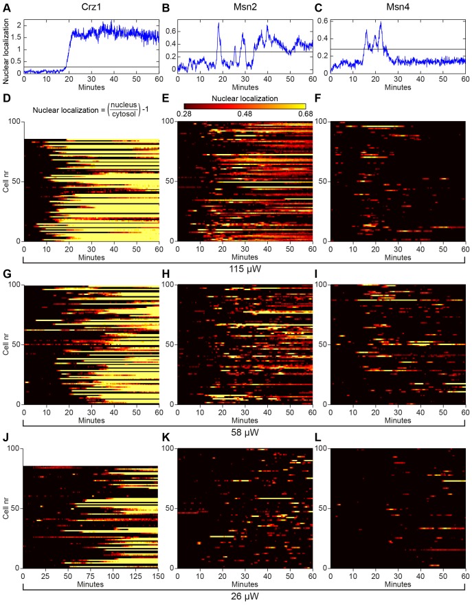
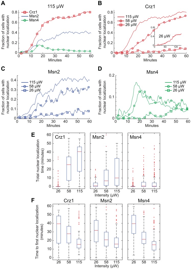
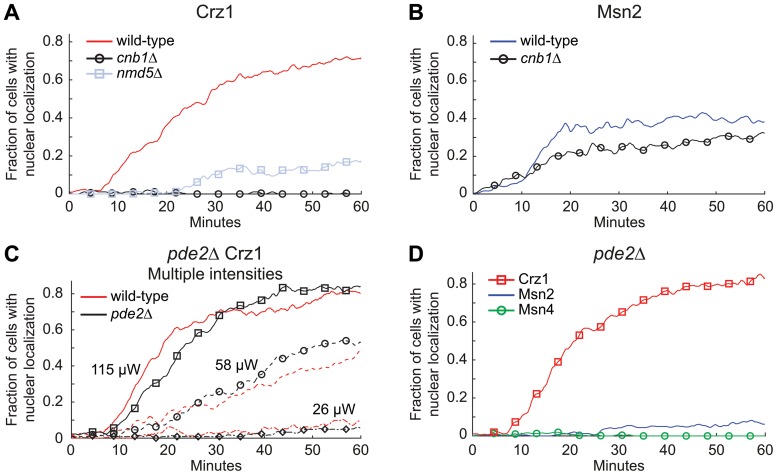
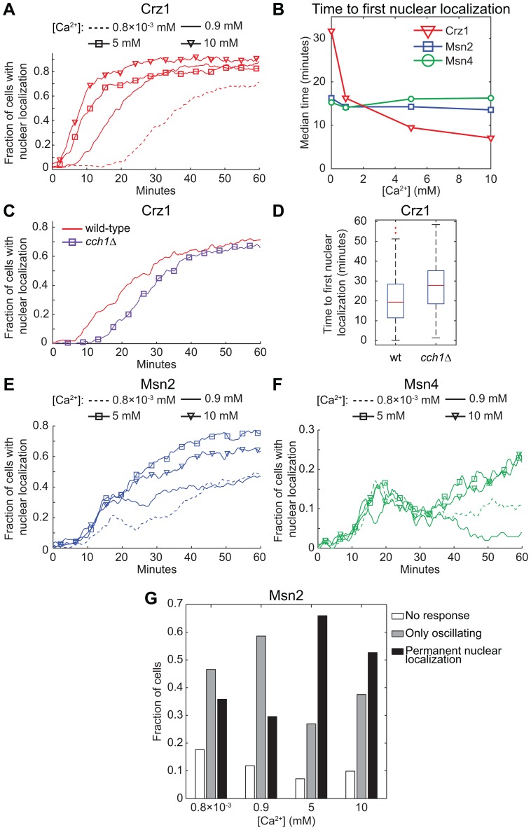
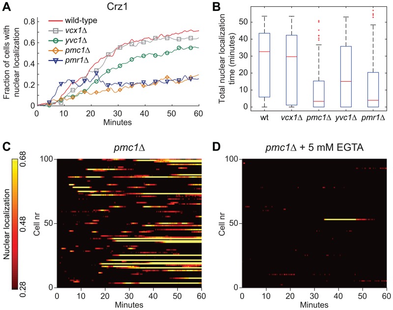
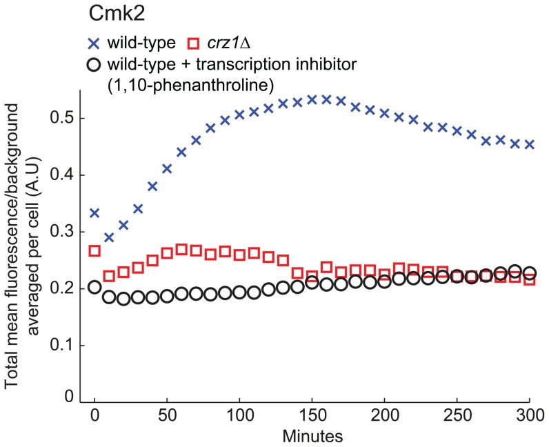
Light in the visible range can be stressful to non-photosynthetic organisms. The yeast Saccharomyces cerevisiae has earlier been reported to respond to blue light via activation of the stress-regulated transcription factor Msn2p. Environmental changes also induce activation of calcineurin, a Ca(2+)/calmodulin dependent phosphatase, which in turn controls gene transcription by dephosphorylating the transcription factor Crz1p. We investigated the connection between cellular stress caused by blue light and Ca(2+) signalling in yeast by monitoring the nuclear localization dynamics of Crz1p, Msn2p and Msn4p. The three proteins exhibit distinctly different stress responses in relation to light exposure. Msn2p, and to a lesser degree Msn4p, oscillate rapidly between the nucleus and the cytoplasm in an apparently stochastic fashion. Crz1p, in contrast, displays a rapid and permanent nuclear localization induced by illumination, which triggers Crz1p-dependent transcription of its target gene CMK2. Moreover, increased extracellular Ca(2+) levels stimulates the light-induced responses of all three transcription factors, e.g. Crz1p localizes much quicker to the nucleus and a larger fraction of cells exhibits permanent Msn2p nuclear localization at higher Ca(2+) concentration. Studies in mutants lacking Ca(2+) transporters indicate that influx of extracellular Ca(2+) is crucial for the initial stages of light-induced Crz1p nuclear localization, while mobilization of intracellular Ca(2+) stores appears necessary for a sustained response. Importantly, we found that Crz1p nuclear localization is dependent on calcineurin and the carrier protein Nmd5p, while not being affected by increased protein kinase A activity (PKA), which strongly inhibits light-induced nuclear localization of Msn2/4p. We conclude that the two central signalling pathways, cAMP-PKA-Msn2/4 and Ca(2+)-calcineurin-Crz1, are both activated by blue light illumination.


Conflict of interest statement

Competing Interests: The authors have declared that no competing interests exist.

Figures

Figure 1
Figure 1. Nucleocytoplasmic localization responses of Crz1p, Msn2p and Msn4p during continuous exposure to blue light.
(A)–(C) Nuclear localization trajectories in a single cell and (D)–(L) temporal nucleocytoplasmic localization profiles for populations of 80–100 cells extracted from fluorescence microscopy images for Crz1-GFP (left column), Msn2-GFP (middle column) and Msn4-GFP (right column) at three different light intensities (rows). (A)–(C) The horizontal line represents the threshold used for determining nuclear localization. (D)–(L) Nuclear localization (colour scale) visualizes the relative fluorescence intensity between the nucleus and the cytoplasm in individual cells ((Inucleus/Icytosol)−1). Crz1p permanently entered the nucleus in a comparatively uniform fashion throughout the whole cell population and exhibited occasionally only one or two oscillations in some cells prior to permanent nuclear localization (A, D, G, and J). Note the different time scale in (J). Msn2p and Msn4p exhibited nucleocytoplasmic oscillations, where the oscillatory response of Msn2p was much more pronounced. A subpopulation of cells showed permanent Msn2p nuclear localization after a period of oscillations (mainly seen in B and E).
Figure 2
Figure 2. Quantitative analyses of the effects of illumination on nuclear localization of Crz1p, Msn2p, and Msn4p.
(A)–(D) Fraction of cells with transcription factors localized to the nucleus at different light intensities. Inset (B): Crz1-GFP exposed to 26 µW during 150 minutes. (E) The total time in the nucleus for the transcription factors Crz1p, Msn2p and Msn4p, during 60 minutes of continuous light exposure at different intensities. There is no statistically significant difference between the responses at the two highest light intensities for Msn4p (p = 0.2145). (F) Time to first nuclear localization of Crz1p, Msn2p and Msn4p at different light intensities. The changes in time to the first nuclear localization as a function of light intensity are statistically significant (Msn2p 58 µW vs. 115 µW p = 0.011, others p<0.001), except for Crz1p 26 µW vs. 58 µW (p = 0.109). Note that Crz1p has only entered the nucleus in ∼25% of the cells at 26 µW during the first 60 minutes considered here, giving rise to a relatively fast median value of 39 minutes. Statistical tests were carried out using the Mann–Whitney U-test.
Figure 3
Figure 3. Transcription factor nuclear localization dependence on calcineurin and protein kinase A activity.
(A) Fraction of cells with Crz1-GFP localized to the nucleus. In both cnb1Δ and nmd5Δ strains Crz1p nuclear localization was drastically reduced. (B) Fraction of cells with Msn2-GFP localized to the nucleus in a cnb1Δ strain. (C) Fraction of cells with Crz1-GFP nuclear localization in a pde2Δ strain at different light intensities (115 µW, 58 µW and 26 µW). (D) Fraction of cells with transcription factor nuclear localization in pde2Δ mutants (115 µW). Corresponding wild-type/reference experiments are shown for each sample set.
Figure 4
Figure 4. Effect of the extracellular [Ca2+] and cellular influx on Crz1p, Msn2p and Msn4p localization in response to light.
Fraction of cells with the transcription factors (A) Crz1-GFP, (E) Msn2-GFP and (F) Msn4-GFP localized to the nucleus as a function of time during continuous exposure to blue light (450–490 nm, 115 µW). (C)–(D) A delayed nuclear localization response was found in cells lacking the plasma membrane calcium channel CCH1. The difference is statistically significant (p<0.0001). (A)–(B) and (E)–(F) Cells were allowed to adapt to the specified extracellular Ca2+ concentration by growing them first overnight, and then in a fresh culture that was allowed to reach OD600 = 0.4–0.5 at the start of the light exposure experiment. Msn2p nuclear localization increased at 5 mM and 10 mM compared to standard conditions (0.9 mM Ca2+). Nuclear localization of Msn4p increased after 30 minutes of light exposure (5 mM and 10 mM). The curves have been smoothed with an averaging filter for visualization purposes. (B) Median values of the time to first nuclear localization as a function of extracellular Ca2+ concentration. (G) Msn2-GFP nuclear localization trajectory classification. Cells were manually classified as either: No response (Msn2p located only in the cytoplasm), Only oscillating (Msn2p oscillates continuously, never exhibited sustained nuclear localization), or Permanent nuclear localization (Msn2p resided in the nucleus for a longer period of time towards the end of the experiment). The latter class increased drastically at 5 mM Ca2+. [Ca2+]: 0.8×10⁻³ mM (0.4 mg/l from calcium pantothenate, without CaCl2), 0.9 mM (standard medium), 5 mM and 10 mM.
Figure 5
Figure 5. Internal Ca2+ pumps are important for the wild-type light-induced response of Crz1p.
(A) Fraction of cells with Crz1-GFP localized to the nucleus as a function of time during continuous exposure to blue light (450–490 nm, 115 µW). (B) Total nuclear localization time of Crz1p during 60 minutes of light exposure. (C)–(D) Nucleocytoplasmic localization profiles of Crz1p extracted from fluorescence microscopy images of pmc1Δ cells. (D) EGTA was added to the medium to a final concentration of 5 mM just prior to exposure to blue light.
Figure 6
Figure 6. Illumination increases Cmk2-GFP fluorescence in a manner dependent on Crz1p and de novo transcription.
Increase in Cmk2-GFP average fluorescence (population average per cell, Ncell,wt = 107, Ncell,crz1Δ = 99, Ncell,transcription inhibitor = 89) after 40 minutes of exposure to blue light (115 µW, 450–490 nm). To verify that the fluorescence increase in wild-type cells was due to de novo transcription of CMK2-GFP the transcription inhibitor 1,10-phenanthroline (100 µg/ml) was added just prior to illumination.
Figure 7
Figure 7. Schematic overview of the two signalling pathways responsive to illumination.
This study has identified that both intra- and extracellular Ca2+-depots are important for the wild-type Crz1p response during illumination. Importantly, the Crz1p nuclear localization is calcineurin dependent and PKA independent.

References

    1. Berry DB, Gasch AP (2008) Stress-activated genomic expression changes serve a preparative role for impending stress in yeast. Molecular Biology of the Cell 19: 4580–4587. - PMC - PubMed
    1. Cai L, Dalal CK, Elowitz MB (2008) Frequency-modulated nuclear localization bursts coordinate gene regulation. Nature 455: 485–490. - PMC - PubMed
    1. Bodvard K, Wrangborg D, Tapani S, Logg K, Sliwa P, et al. (2011) Continuous light exposure causes cumulative stress that affects the localization oscillation dynamics of the transcription factor Msn2p. BBA-Mol Cell Res 1813: 358–366. - PubMed
    1. Garmendia-Torres C, Goldbeter A, Jacquet M (2007) Nucleocytoplasmic oscillations of the yeast transcription factor Msn2: Evidence for periodic PKA activation. Current Biology 17: 1044–1049. - PubMed
    1. Hao N, O’Shea EK (2011) Signal-dependent dynamics of transcription factor translocation controls gene expression. Nat Struct Mol Biol 19: 31–39. - PMC - PubMed

Publication types

MeSH terms