Randomized Controlled Trial
. 2013;8(1):e53716.
doi: 10.1371/journal.pone.0053716. Epub 2013 Jan 15.

Hyperbaric oxygen induces late neuroplasticity in post stroke patients--randomized, prospective trial

Affiliations
Randomized Controlled Trial

Hyperbaric oxygen induces late neuroplasticity in post stroke patients--randomized, prospective trial

Shai Efrati et al. PLoS One. 2013.

Abstract

Background: Recovery after stroke correlates with non-active (stunned) brain regions, which may persist for years. The current study aimed to evaluate whether increasing the level of dissolved oxygen by Hyperbaric Oxygen Therapy (HBOT) could activate neuroplasticity in patients with chronic neurologic deficiencies due to stroke.

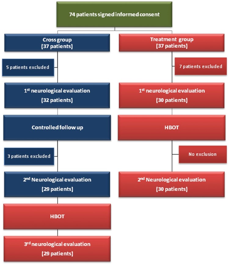
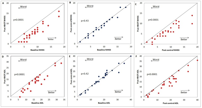
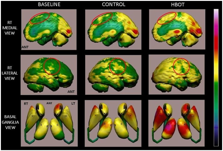
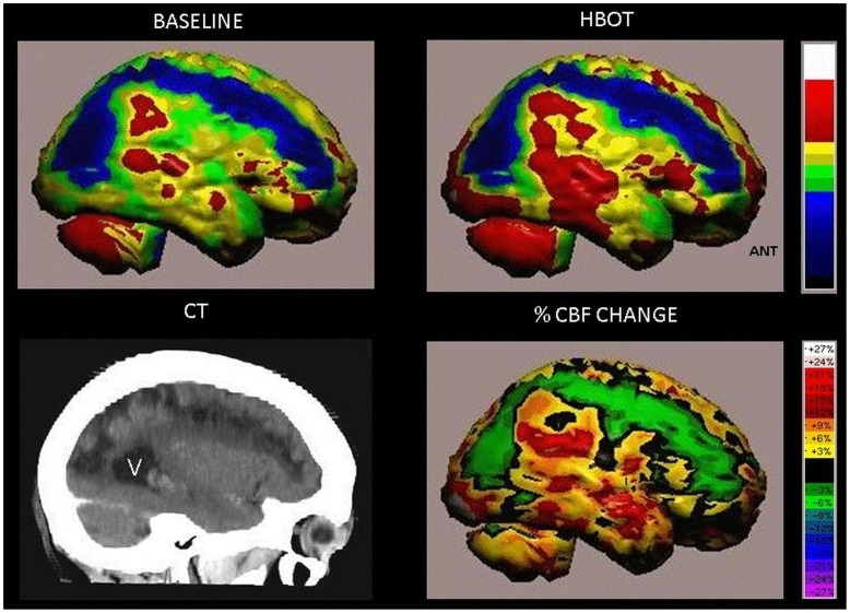
Methods and findings: A prospective, randomized, controlled trial including 74 patients (15 were excluded). All participants suffered a stroke 6-36 months prior to inclusion and had at least one motor dysfunction. After inclusion, patients were randomly assigned to "treated" or "cross" groups. Brain activity was assessed by SPECT imaging; neurologic functions were evaluated by NIHSS, ADL, and life quality. Patients in the treated group were evaluated twice: at baseline and after 40 HBOT sessions. Patients in the cross group were evaluated three times: at baseline, after a 2-month control period of no treatment, and after subsequent 2-months of 40 HBOT sessions. HBOT protocol: Two months of 40 sessions (5 days/week), 90 minutes each, 100% oxygen at 2 ATA. We found that the neurological functions and life quality of all patients in both groups were significantly improved following the HBOT sessions while no improvement was found during the control period of the patients in the cross group. Results of SPECT imaging were well correlated with clinical improvement. Elevated brain activity was detected mostly in regions of live cells (as confirmed by CT) with low activity (based on SPECT) - regions of noticeable discrepancy between anatomy and physiology.

Conclusions: The results indicate that HBOT can lead to significant neurological improvements in post stroke patients even at chronic late stages. The observed clinical improvements imply that neuroplasticity can still be activated long after damage onset in regions where there is a brain SPECT/CT (anatomy/physiology) mismatch.

Trial registration: ClinicalTrials.gov NCT00715897.


Conflict of interest statement

Competing Interests: The authors herby declare that, although Prof. Eshel Ben-Jacob is a PLOS ONE Editorial Board member, this does not alter the authors' adherence to all the PLOS ONE policies on sharing data and materials.

Figures

Figure 1
Figure 1. Flowchart of the patients in the study.
Figure 2
Figure 2. The results of the neurological evaluation. Each point represents a patient.
(A–C) NIHSS score: (A) Scores of the treated group patients before and after the HBOT period (B) Scores of the cross group before and after the control (no treatment) period. (C) Scores of the cross group after the HBOT period. (D–F) The same as (A)-(C) for the activities of daily living (ADL) scores. We note that the lines indicate the diagonal. Abbreviation: NIHSS =  National Institutes of Health Stroke Scale; ADL =  Activities of Daily Living.
Figure 3
Figure 3. Scatter plot analysis of the changes in the combined neurological evaluations.
The scatter plot shows changes in the NIHSS and ADL scores in terms of the scaled relative differences as is defined in the methods section (averaged difference (j–SIj>) is not divided by STD(SFj−SIj)). The color code is – changes during the treatment periods for the HBOT treated group (red diamonds), changes during for the HBOT-treated cross group (red circles) and changes during the control (non-treatment) period of the HBOT-treated cross group (blue circles).
Figure 4
Figure 4. Volume rendered Brain SPECT perfusion maps of Example 1.
The results are of a patient in the cross group, suffering from left hemiparesis due to ischemic stroke that occurred 1 year prior to inclusion in the study. Baseline and control volume rendered brain perfusion views show diffuse hypoperfusion in the right hemisphere involving the fronto-parietal region and right postero-medial frontal (right motor cortex), right medial parietal and posterior-parietal (sensory cortex and associative motor cortex) (red circles). The HBOT SPECT scan done at the end of HBOT treatments shows disappearance of the perfusion deficits that were still demonstrated at the end of the control period. In addition, a significant global cortical and subcortical (basal ganglia and thalamic nuclei) perfusion improvement is seen.
Figure 5
Figure 5. Volume rendered Brain SPECT perfusion maps of Example 2.
The results are of a patient in the treated group, suffering from right hemiparesis due to ischemic stroke that occurred 14 months prior to her inclusion in the study. Comparison of pre- and post-hyperbaric treatment SPECT scans. These SPECT images demonstrate significant improvement of perfusion deficits in the left hemisphere involving the medial and posterolateral frontal area (motor cortex, red circles) and lateral inferior frontal region (Broca's area, blue circles) in comparison to the baseline SPECT. HBOT SPECT findings correlate positively with the patient's improved motor and verbal functions.
Figure 6
Figure 6. Volume rendered Brain SPECT perfusion maps of Example 3.
The results are of a patient in the treated group suffering from left hemiparesis due to ischemic stroke that occurred 26 months prior to inclusion in the study. The brain perfusion maps (upper two images) show the infracted brain (deep blue color) involving the right antero-postero-lateral frontal, right superior-parietal and right parieto-occipital regions. Curved sagittal view in CT MIP reconstruction of the brain shows the anatomical stroke area (left lower image, V = posterior horn of right ventricle). The peri-infarct region show improved perfusion as demonstrated by HBOT image (right upper image). Quantitation of the cerebral blood flow (CBF) change (delta between baseline and HBOT) is demonstrated in the right lower image.

References

    1. Teasell R (2009) Forward. An international perspective of stroke rehabilitation. Top Stroke Rehabil 16: v. - PubMed
    1. Prvu Bettger JA, Stineman MG (2007) Effectiveness of multidisciplinary rehabilitation services in postacute care: state-of-the-science. A review. Arch Phys Med Rehabil 88: 1526–1534. - PMC - PubMed
    1. Anderson DC, Bottini AG, Jagiella WM, Westphal B, Ford S, et al. (1991) A pilot study of hyperbaric oxygen in the treatment of human stroke. Stroke 22: 1137–1142. - PubMed
    1. Nighoghossian N, Trouillas P, Adeleine P, Salord F (1995) Hyperbaric oxygen in the treatment of acute ischemic stroke. A double-blind pilot study. Stroke; a journal of cerebral circulation 26: 1369–1372. - PubMed
    1. Rusyniak DE, Kirk MA, May JD, Kao LW, Brizendine EJ, et al. (2003) Hyperbaric oxygen therapy in acute ischemic stroke: results of the Hyperbaric Oxygen in Acute Ischemic Stroke Trial Pilot Study. Stroke; a journal of cerebral circulation 34: 571–574. - PubMed

Publication types

Associated data