. 2013 Apr;125(1):63-73.
doi: 10.1111/jnc.12153. Epub 2013 Feb 17.

Aging is associated with altered inflammatory, arachidonic acid cascade, and synaptic markers, influenced by epigenetic modifications, in the human frontal cortex

Affiliations

Aging is associated with altered inflammatory, arachidonic acid cascade, and synaptic markers, influenced by epigenetic modifications, in the human frontal cortex

Vasken L Keleshian et al. J Neurochem. 2013 Apr.

Retraction in

  • Retraction.
    [No authors listed] [No authors listed] J Neurochem. 2017 Mar;140(6):980. doi: 10.1111/jnc.13948. Epub 2017 Feb 3. J Neurochem. 2017. PMID: 28261875 Free PMC article.

Abstract

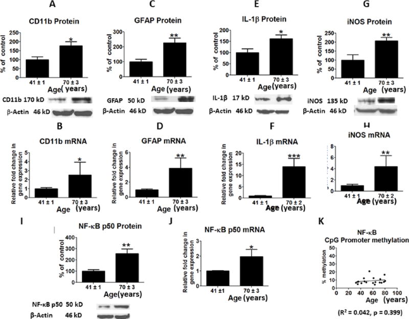
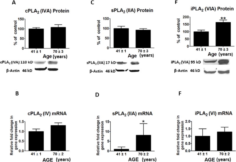
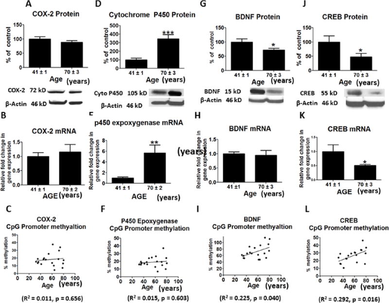
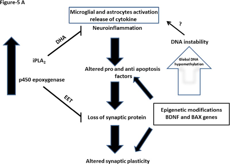
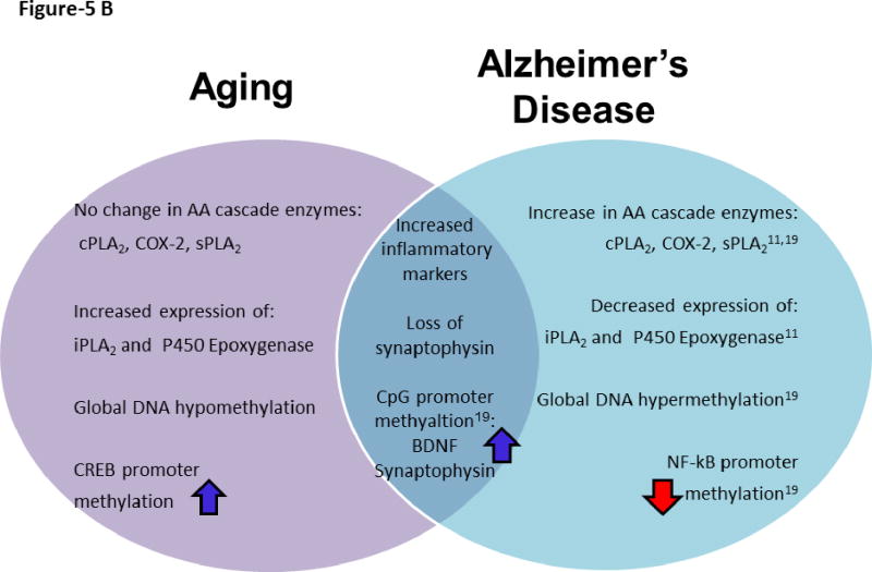
Aging is a risk factor for Alzheimer's disease (AD) and is associated with cognitive decline. However, underlying molecular mechanisms of brain aging are not clear. Recent studies suggest epigenetic influences on gene expression in AD, as DNA methylation levels influence protein and mRNA expression in postmortem AD brain. We hypothesized that some of these changes occur with normal aging. To test this hypothesis, we measured markers of the arachidonic acid (AA) cascade, neuroinflammation, pro- and anti-apoptosis factors, and gene specific epigenetic modifications in postmortem frontal cortex from nine middle-aged [41 ± 1 (SEM) years] and 10 aged subjects (70 ± 3 years). The aged compared with middle-aged brain showed elevated levels of neuroinflammatory and AA cascade markers, altered pro and anti-apoptosis factors and loss of synaptophysin. Some of these changes correlated with promoter hypermethylation of brain derived neurotrophic factor (BDNF), cyclic AMP responsive element binding protein (CREB), and synaptophysin and hypomethylation of BCL-2 associated X protein (BAX). These molecular alterations in aging are different from or more subtle than changes associated with AD pathology. The degree to which they are related to changes in cognition or behavior during normal aging remains to be evaluated.


Conflict of interest statement

Conflicts of interests: None

Figures

Figure 1
Figure 1
Mean protein levels of neuroinflammatory markers (with representative immunoblots) are shown in Figure: A, Cd11b; C, GFAP; E, IL-1beta; G, iNOS and I, NF-kBp50. Bar graphs are ratios of optical densities of individual protein bands to β-actin, expressed as percent of control. Mean mRNA levels of neuroinflammatory markers are shown in Figure B, Cd11b; D, GFAP; F, IL-1beta; H, iNOS and J, NF-kBp50. mRNA levels in postmortem frontal cortex from the middle aged (n = 9) and aged subjects (n = 10), measured using quantitative RT-PCR. mRNA levels of Cd11b;, GFAP, IL-1beta, iNOS and NF-kBp50 in aged group normalized to the endogenous control (β-globulin) and relative to the control (calibrator), using the ΔΔCT method. CpG promoter methylation of NF-kBp50 is shown in Figure 1K. Values are Mean ± SEM and t-tests are used to compare old vs. middle age groups. * p < 0.05, ** p < 0.01, *** p < 0.001 as compared to middle aged group.
Figure 2
Figure 2
Mean protein levels (with representative immunoblots) of arachidonic acid cascade markers A, cPLA2IVA; C, sPLA2 IIA and E, iPLA2 VIA. Bar graphs are ratios of optical densities of individual protein bands to β-actin, expressed as percent of control. Mean mRNA levels of arachidonic acid cascade markers are shown in Figure: B, cPLA2IVA; D, sPLA2 IIA and F, iPLA2VIA. mRNA levels in postmortem frontal cortex from the middle aged (n = 9) and aged group subjects (n = 10), measured using quantitative RT-PCR. mRNA levels of cPLA2IVA, sPLA2 IIA and iPLA2VIA in aged group normalized to the endogenous control (β-globulin) and relative to the control (calibrator), using the ΔΔCT method. Values are Mean ± SEM and t-tests are used to compare old vs. middle age groups. * p < 0.05, ** p < 0.01, *** p < 0.001 as compared to middle aged group.
Figure 3
Figure 3
Mean protein levels (with representative immunoblots) of A, COX-2; D, Cytochrome p450 epoxygenase; G, BDNF and J, CREB. Bar graphs are ratios of optical densities of individual protein bands to β-actin, expressed as percent of control. Mean mRNA levels of B, COX-2; E, Cytochrome p450 epoxygenase; H, BDNF and K, CREB in postmortem frontal cortex from the middle aged (n = 9) and aged group subjects (n = 10), measured using quantitative RT-PCR. -mRNA levels of COX-2, Cytochrome p450 epoxygenase, BDNF and CREB in aged group normalized to the endogenous control (β-globulin) and relative to the control (calibrator), using the ΔΔCT method. Mean levels of CpG promoter methylation at C, COX-2; F, Cytochrome p450 epoxygenase; I, BDNF and L, CREB. Values are Mean ± SEM and t-tests are used to compare old vs. middle age groups. * p < 0.05, ** p < 0.01, *** p < 0.001 as compared to aged group.
Figure 4
Figure 4
Mean protein levels (with representative immunoblots) of Figure : A, BCL-2; D, BAX; I, synaptophysin in postmortem frontal cortex from the middle aged (n = 9) and aged group subjects (n = 10). Bar graphs are ratios of optical densities of individual protein bands to β-actin, expressed as percent of control. Mean mRNA levels of B, BCL-2; E, BAX; G, BAX/BCL-2 mRNA ratio; J, synaptophysin in postmortem frontal cortex from the middle aged (n = 9) and aged group subjects (n = 10), measured using quantitative RT-PCR. mRNA levels of BCL-2, BAX and synaptophysin in aged group normalized to the endogenous control (β-globulin) and relative to the control (calibrator), using the ΔΔCT method. Mean levels of CpG promoter methylation shown in Figure: C, BCL-2; F, BAX; H, BAX/BCL-2 and K, synaptophysin. Figure L represents levels of global DNA methylation in postmortem frontal cortex from the middle aged (n = 9) and aged group subjects (n = 10). Values are Mean ± SEM and t-tests are used to compare old vs. middle age groups. * p < 0.05, ** p < 0.01, *** p < 0.001 as compared to aged group.
Figure 5
Figure 5
Figure 5A. Brain aging involves balance between protective and progressive factors. Increased expression of iPLA2 and cytochrome p450 expoxygenase and their products are neuroprotective. At the same time, increased neuroinflammatory markers and altered pro and anti-apoptotic factors could contribute to loss of synaptic proteins. This may lead to altered synaptic plasticity. These molecular alterations are influenced by gene specific epigenetic modifications. Figure 5B. Schematic representation of distinct and common molecular features of aging and Alzheimer’s disease.
Figure 5
Figure 5
Figure 5A. Brain aging involves balance between protective and progressive factors. Increased expression of iPLA2 and cytochrome p450 expoxygenase and their products are neuroprotective. At the same time, increased neuroinflammatory markers and altered pro and anti-apoptotic factors could contribute to loss of synaptic proteins. This may lead to altered synaptic plasticity. These molecular alterations are influenced by gene specific epigenetic modifications. Figure 5B. Schematic representation of distinct and common molecular features of aging and Alzheimer’s disease.

References

    1. Abdelmohsen K, Hutchison ER, Lee EK, et al. miR-375 inhibits differentiation of neurites by lowering HuD levels. Mol Cell Biol. 2010;30:4197–4210. - PMC - PubMed
    1. Abdolmaleky HM, Cheng KH, Russo A, Smith CL, Faraone SV, Wilcox M, Shafa R, Glatt SJ, Nguyen G, Ponte JF, Thiagalingam S, Tsuang MT. Hypermethylation of the reelin (RELN) promoter in the brain of schizophrenic patients: A preliminary report. American Journal of Medical Genetics Part B: Neuropsychiatric Genetics. 2005;134B:60–66. - PubMed
    1. Agrawal A, Tay J, Yang GE, Agrawal S, Gupta S. Age-associated epigenetic modifications in human DNA increase its immunogenicity. Aging (Albany NY) 2010;2:93–100. - PMC - PubMed
    1. Akiba S, Mizunaga S, Kume K, Hayama M, Sato T. Involvement of group VI Ca2+-independent phospholipase A2 in protein kinase C-dependent arachidonic acid liberation in zymosan-stimulated macrophage-like P388D1 cells. J Biol Chem. 1999;274:19906–19912. - PubMed
    1. Albrecht S, Bourdeau M, Bennett D, Mufson EJ, Bhattacharjee M, LeBlanc AC. Activation of Caspase-6 in Aging and Mild Cognitive Impairment. The American Journal of Pathology. 2007;170:1200–1209. - PMC - PubMed

Publication types