. 2013 Jan 21;200(2):173-86.
doi: 10.1083/jcb.201203128.

A noncatalytic function of the ligation complex during nonhomologous end joining

Affiliations

A noncatalytic function of the ligation complex during nonhomologous end joining

Jessica Cottarel et al. J Cell Biol. .

Abstract

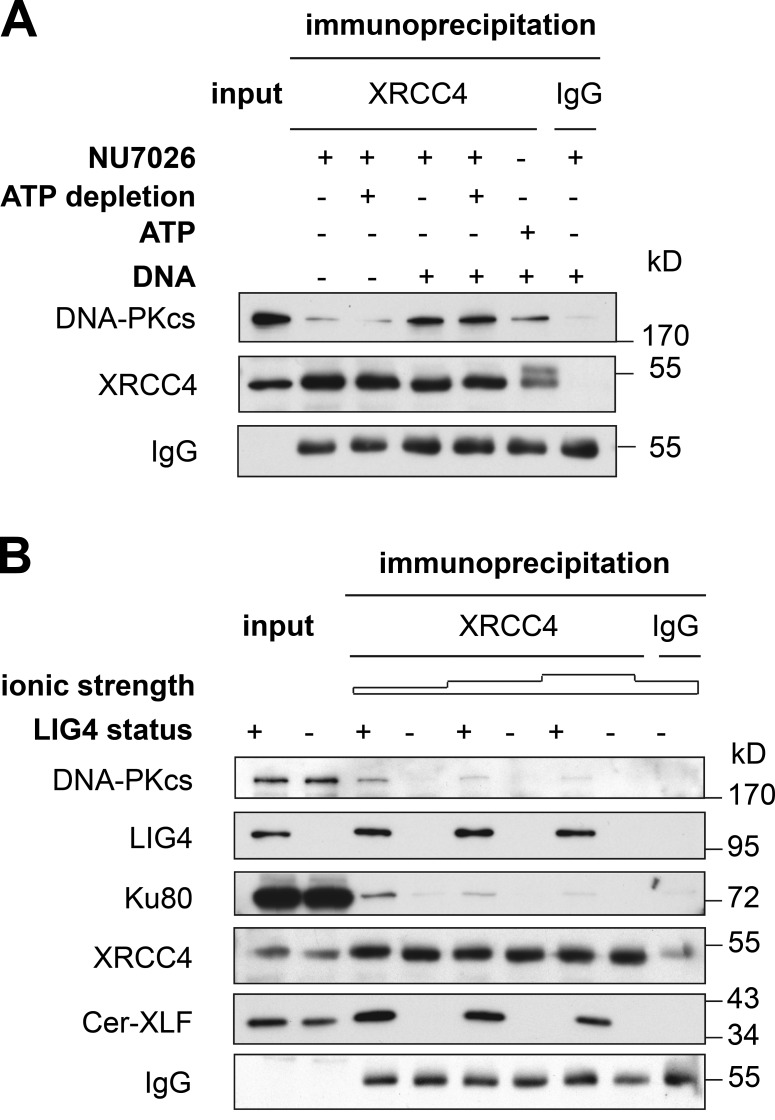
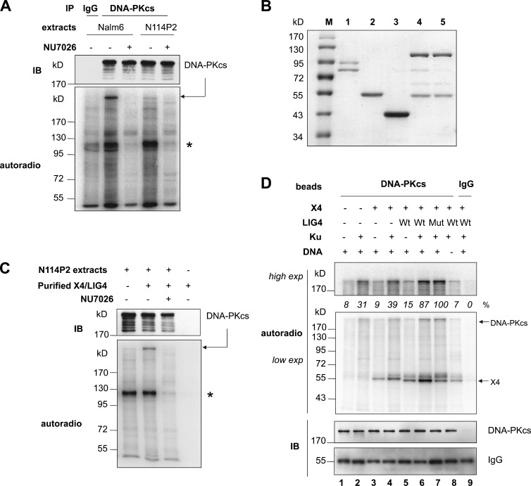
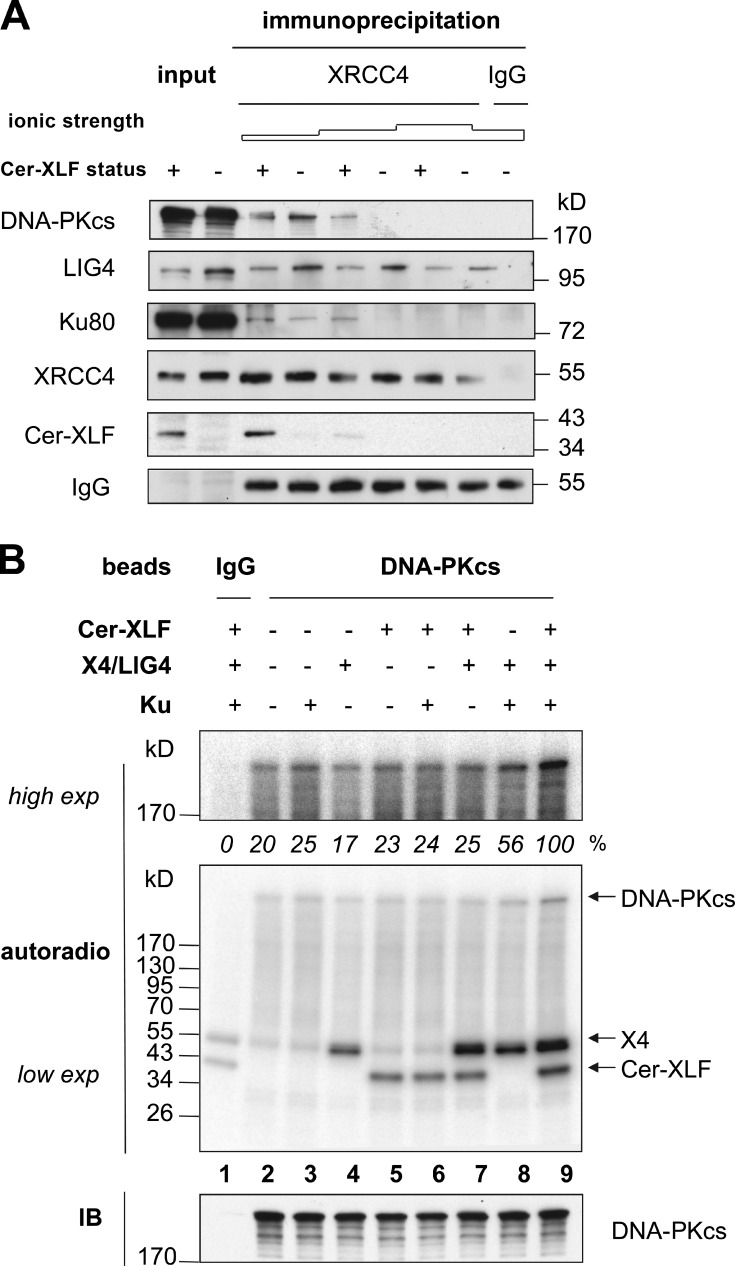
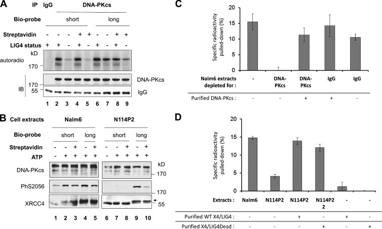
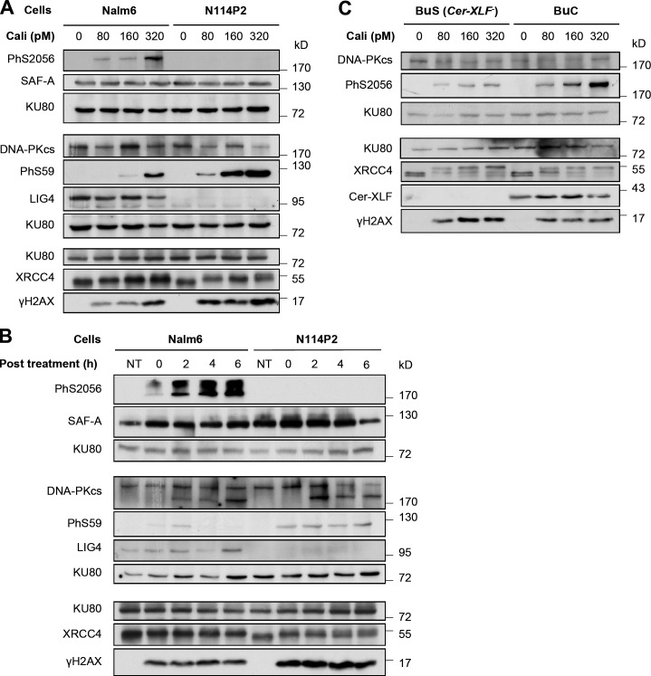
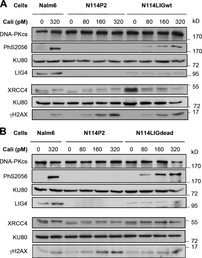
Nonhomologous end joining is the primary deoxyribonucleic acid (DNA) double-strand break repair pathway in multicellular eukaryotes. To initiate repair, Ku binds DNA ends and recruits the DNA-dependent protein kinase (DNA-PK) catalytic subunit (DNA-PKcs) forming the holoenzyme. Early end synapsis is associated with kinase autophosphorylation. The XRCC4 (X4)-DNA Ligase IV (LIG4) complex (X4LIG4) executes the final ligation promoted by Cernunnos (Cer)-X4-like factor (XLF). In this paper, using a cell-free system that recapitulates end synapsis and DNA-PKcs autophosphorylation, we found a defect in both activities in human cell extracts lacking LIG4. LIG4 also stimulated the DNA-PKcs autophosphorylation in a reconstitution assay with purified components. We additionally uncovered a kinase autophosphorylation defect in LIG4-defective cells that was corrected by ectopic expression of catalytically dead LIG4. Finally, our data support a contribution of Cer-XLF to this unexpected early role of the ligation complex in end joining. We propose that productive end joining occurs by early formation of a supramolecular entity containing both DNA-PK and X4LIG4-Cer-XLF complexes on DNA ends.


Figures

Figure 1.
Figure 1.
LIG4 is required for DNA-dependent coimmunoprecipitation of X4, Cer-XLF, and DNA-PK. (A) Nalm6 extracts were incubated for 15 min under in vitro NHEJ conditions and with components as indicated. ATP depletion refers to addition of the ATP-consuming system (glucose + hexokinase) as previously described (Calsou et al., 2003). Then, the samples were mixed with control IgG or anti-X4 magnetic beads. Immunoprecipitation proceeded for 4 h at 4°C on a wheel followed by three washes for 5 min in 0.5× PBS adjusted to a 100-mM final NaCl concentration, denaturation, and separation on 10% SDS-PAGE gel. The input lane corresponds to 50% of the volume of extracts used in the immunoprecipitation. (B) NHEJ-type extracts from Nalm6 control or N114P2 LIG4-null pre-B cells (LIG4 status + or −, respectively) were incubated with ds DNA and NU7026 as defined in A. Incubation was followed by immunoprecipitation with control IgG or anti-X4 antibodies and washes with increasing salt concentration (100, 150, or 200 mM NaCl). Reaction samples were heat denatured, separated on SDS-PAGE gels, and electrotransferred onto membrane that was blotted with antibodies as indicated. The input lanes correspond to 50% of the volume of extracts used in the immunoprecipitation.
Figure 2.
Figure 2.
LIG4 protein, but not activity, is required for DNA-PKcs autophosphorylation activated by ds DNA ends in cell extracts. (A) Nalm6 control and N114P2 LIG4-null extracts were incubated under reaction conditions with γ-[32P]ATP and addition of components as specified. Incubation was followed by immunoprecipitation (IP) with control IgG2a or anti–DNA-PKcs antibodies. Reaction samples were heat denatured, separated on SDS-PAGE gels, and electrotransferred onto membrane that was blotted with antibodies as indicated. (B) Purified proteins Ku70/Ku80 heterodimer (1), X4 (2), Cer-XLF (3), and wild-type (4) or mutant (5) X4LIG4 complexes were loaded on a 10% SDS-PAGE gel and stained with InstantBlue (Euromedex). M, molecular mass markers. (C) Conditions were as in A with N114P2 LIG4-null extracts and addition of wild-type X4LIG4 complex as specified followed by DNA-PKcs immunoprecipitation. (D) Purified DNA-PKcs or control IgG on beads were incubated under reaction conditions with γ-[32P]ATP and components as indicated. Wt or Mut refer to wild-type or the catalytically inactive X4LIG4 complex. Low or high exposures of the same membrane are shown. Results of quantitative analysis of the gel are shown as the percentage of the highest phospholabeled DNA-PKcs band after subtraction of background value in the control IgG lane. Autoradio, autoradiography; IB, immunoblotting of the same membrane; exp, exposure. The asterisks indicate a protein coimmunoprecipitated with DNA-PKcs.
Figure 3.
Figure 3.
Effect of Cer-XLF on coimmunoprecipitation of DNA-PK with the ligation complex and on DNA-PKcs autophosphorylation. (A) NHEJ-type extracts from BuC or BuS fibroblasts (Cer-XLF status + or −, respectively) were incubated with ds DNA and NU7026 as defined in Fig. 1. Incubation was followed by immunoprecipitation with control IgG or anti-X4 antibodies and washes with increasing salt concentration (100, 150, or 200 mM NaCl). Reaction samples were heat denatured, separated on SDS-PAGE gels, and electrotransferred onto membranes that were blotted with antibodies as indicated. (B) Purified DNA-PKcs or control IgG on beads were incubated under reaction conditions with γ-[32P]ATP and purified components as indicated. Low or high exposures of the same membrane are shown. Results of quantitative analysis of the gel are shown as the percentage of the highest phospholabeled DNA-PKcs band after subtraction of background value in the control IgG lane. Autoradio, autoradiography; IB, immunoblotting of the same membrane; exp, exposure.
Figure 4.
Figure 4.
LIG4 protein, but not activity, stimulated DNA-PK–mediated DNA ends synapsis in vitro. (A) NHEJ-type extracts from Nalm6 control or N114P2 LIG4-null cells (LIG4 status + or −, respectively) were incubated with short (32 bp) or long (250 bp) ds activator DNA under reaction conditions with γ-[32P]ATP and added components as specified. When necessary, the DNA probe was preincubated with streptavidin before addition to the reaction. Incubation was followed by immunoprecipitation with control IgG2a or anti–DNA-PKcs antibodies. Reaction samples were heat denatured, separated on SDS-PAGE gels, and electrotransferred onto membranes that were blotted with antibodies as indicated. Autoradio, autoradiography; IB, immunoblotting of the same membrane; IP, immunoprecipitation. (B) Conditions were as in A but without radiolabeling. The arrow indicates the position of the phosphorylated form of X4. (C and D) Quantification of the specific radioactivity pulled down after subtraction of the unspecific background obtained without the 502bio probe, under conditions of extracts and added components as specified. Each value is the mean of three experiments. Error bars correspond to SEM. WT, wild type.
Figure 5.
Figure 5.
Requirement of LIG4 for DSB-stimulated DNA-PKcs autophosphorylation in cells and contribution of Cer-XLF. (A and B) Nalm6 control and N114P2 LIG4-null pre-B cells were treated for 1 h with increasing doses of Calicheamicin γ1 (Cali) as indicated (A) or with 160 pM Cali followed by incubation in fresh medium under normal growth conditions for the specified time (B). Whole-cell extracts were heat denatured, separated on SDS-PAGE gels, and electrotransferred onto membranes that were blotted with antibodies as indicated. Protein samples were separated on 8% acrylamide SDS-PAGE gels for standard separation or 15% for γ-H2AX isolation. (C) Western blotting on the whole-cell extracts of BuC control and BuS Cer-XLF- mutant fibroblasts after treatment with increasing doses of Cali as indicated for 1 h. NT, not treated; PhS59, phosphorylation of SAF-A on S59.
Figure 6.
Figure 6.
LIG4 activity is not required for DSB-stimulated DNA-PKcs autophosphorylation in cells. (A and B) Western blotting on the whole-cell extracts of Nalm6 control cells, N114P2 LIG4-null cells, or N114P2 cells expressing wild-type (N114LIGwt) or a catalytic-dead (N114LIGdead) LIG4 protein after treatment for 1 h with increasing doses of Cali as indicated.

Similar articles

Cited by

References

    1. Akopiants K., Zhou R.Z., Mohapatra S., Valerie K., Lees-Miller S.P., Lee K.J., Chen D.J., Revy P., de Villartay J.P., Povirk L.F. 2009. Requirement for XLF/Cernunnos in alignment-based gap filling by DNA polymerases lambda and mu for nonhomologous end joining in human whole-cell extracts. Nucleic Acids Res. 37:4055–4062 10.1093/nar/gkp283 - DOI - PMC - PubMed
    1. Andres S.N., Modesti M., Tsai C.J., Chu G., Junop M.S. 2007. Crystal structure of human XLF: a twist in nonhomologous DNA end-joining. Mol. Cell. 28:1093–1101 10.1016/j.molcel.2007.10.024 - DOI - PubMed
    1. Andres S.N., Vergnes A., Ristic D., Wyman C., Modesti M., Junop M. 2012. A human XRCC4-XLF complex bridges DNA. Nucleic Acids Res. 40:1868–1878 10.1093/nar/gks022 - DOI - PMC - PubMed
    1. Bombarde O., Boby C., Gomez D., Frit P., Giraud-Panis M.J., Gilson E., Salles B., Calsou P. 2010. TRF2/RAP1 and DNA-PK mediate a double protection against joining at telomeric ends. EMBO J. 29:1573–1584 10.1038/emboj.2010.49 - DOI - PMC - PubMed
    1. Bothmer A., Robbiani D.F., Feldhahn N., Gazumyan A., Nussenzweig A., Nussenzweig M.C. 2010. 53BP1 regulates DNA resection and the choice between classical and alternative end joining during class switch recombination. J. Exp. Med. 207:855–865 10.1084/jem.20100244 - DOI - PMC - PubMed

Publication types