Case Reports
. 2013 Mar 22:80:41-50.
doi: 10.1016/j.visres.2012.12.012. Epub 2013 Jan 18.

X-linked cone dystrophy and colour vision deficiency arising from a missense mutation in a hybrid L/M cone opsin gene

Affiliations
Case Reports

X-linked cone dystrophy and colour vision deficiency arising from a missense mutation in a hybrid L/M cone opsin gene

Michelle McClements et al. Vision Res. .

Abstract

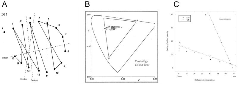
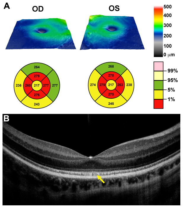
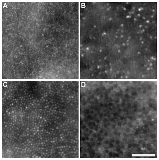
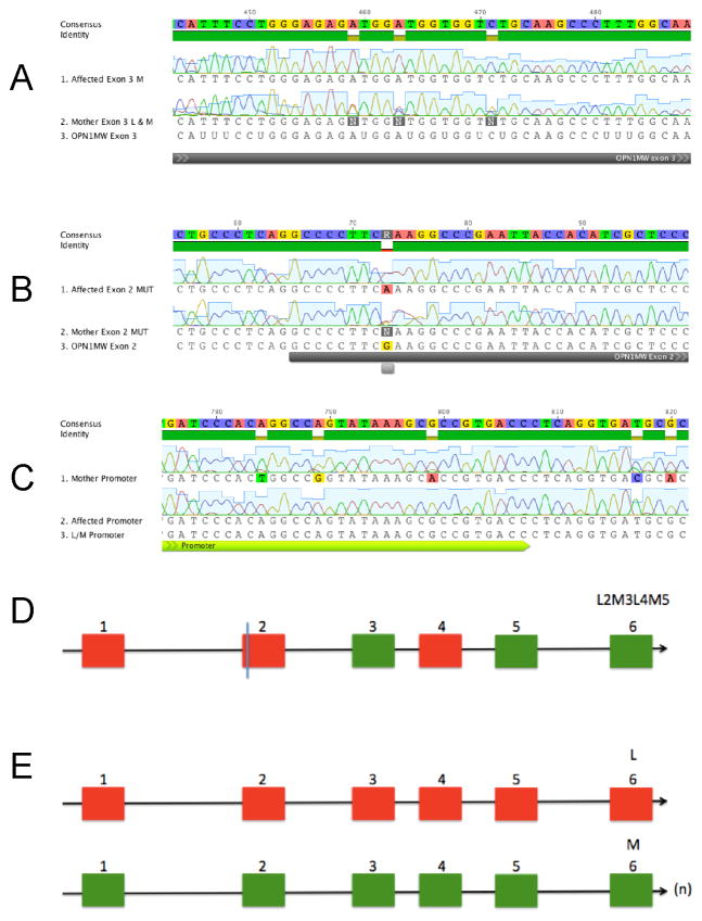
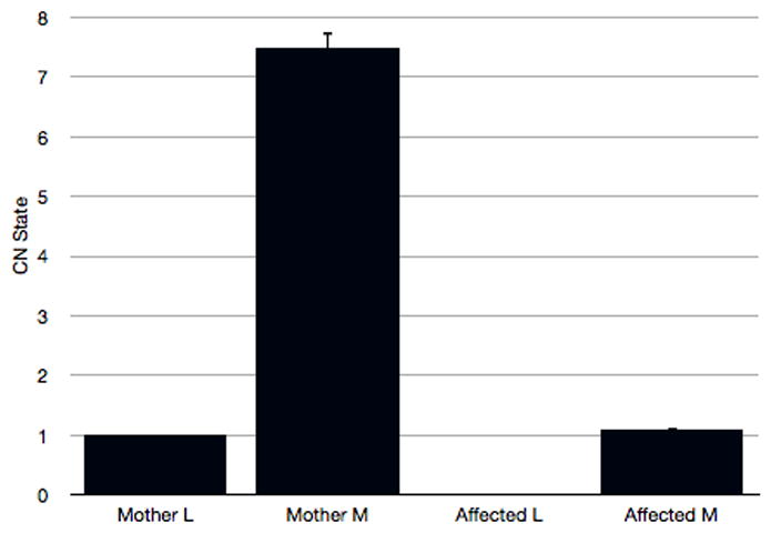
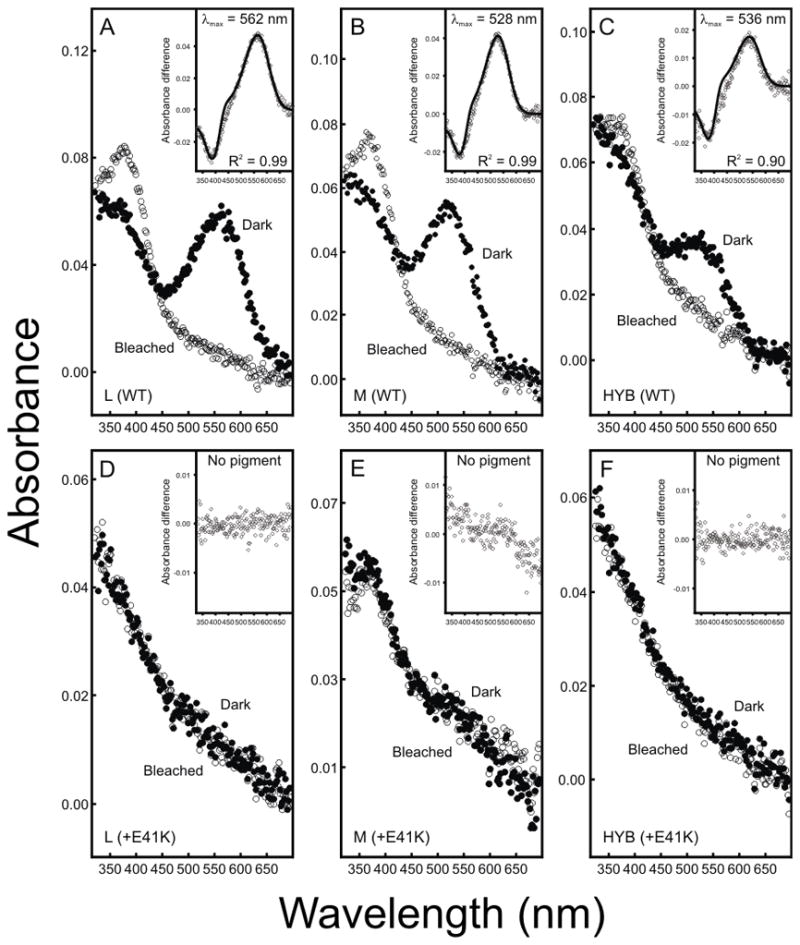
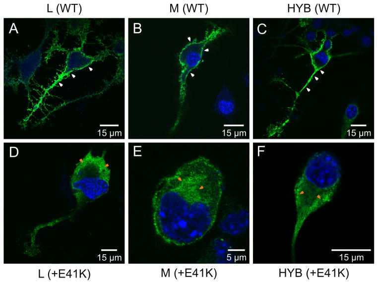
In this report, we describe a male subject who presents with a complex phenotype of myopia associated with cone dysfunction and a protan vision deficiency. Retinal imaging demonstrates extensive cone disruption, including the presence of non-waveguiding cones, an overall thinning of the retina, and an irregular mottled appearance of the hyper-reflective band associated with the inner segment ellipsoid portion of the photoreceptor. Mutation screening revealed a novel p.Glu41Lys missense mutation in a hybrid L/M opsin gene. Spectral analysis shows that the mutant opsin fails to form a pigment in vitro and fails to be trafficked to the cell membrane in transfected Neuro2a cells. Extensive sequence and quantitative PCR analysis identifies this mutant gene as the only gene present in the affected subject's L/M opsin gene array, yet the presence of protanopia indicates that the mutant opsin must retain some activity in vivo. To account for this apparent contradiction, we propose that a limited amount of functional pigment is formed within the normal cellular environment of the intact photoreceptor, and that this requires the presence of chaperone proteins that promote stability and normal folding of the mutant protein.


Figures

Figure 1
Figure 1
The L and M opsin genes are present in a tandem array on Xq28. The L opsin generally sits in the proximal position with multiple M genes downstream. The upstream promoter differs in sequence to the downstream promoters. Primer FG binds specifically to the upstream promoter whilst DG binds specifically to the downstream promoter. The reverse primer E6 binds to a common sequence in exon 6 of both L and M genes. The complete first gene in the array was amplified by use of primers FG and E6 and the amplicon generated used as template in subsequent exon-specific PCRs to determine the coding sequence. Any downstream genes present in the array were amplified together using primers DG and E6 and subsequent exon-specific PCRs identified whether the downstream genes were WT or L/M hybrids.
Figure 2
Figure 2
Psychophysical results for the propositus at age 9. (A) Arrangement of D15 tokens using right eye. The dotted lines indicate the direction of confusion expected for each type of dichromat. The propositus shows a predominantly protan pattern of confusions. (B) Discrimination ellipse on the Cambridge Colour Test. The data are plotted in the CIE u′v′ diagram and show how far the target has to differ in chromaticity from the grey background in order to be discriminated. The thresholds are extended in a classically protan direction, but discrimination in the orthogonal direction is good. The triangle marked RGB shows the gamut of chromaticities available on the monitor. (C) Brightness settings of the yellow field on the Nagel anomaloscope for right and left eyes. The propositus was able to make complete matches at all values of the red-green mixture. The dotted line marked P indicates typical settings of a protanope (from Pokorny, J. et al. 1979) and the dotted line marked A indicates typical settings for a rod achromat. The results show that the propositus has a photoreceptor with a spectral sensitivity similar to that of normal M cones.
Figure 3
Figure 3
Glu41Lys disrupts cone photoreceptor structure. Images from 0.5deg (A,B) and 2deg (C,D) reveal a disrupted mosaic in the patient with the p.Glu41Lys mutation (B,D) compared to that of a normal control (A,C). Near the fovea in the p.Glu41Lys retina (B), there was a sparse population of strongly waveguiding cones. In the parafoveal images of the same patient (D), there were a reduced number of cones and those present had severely diminished waveguiding. Normal-appearing rods are visible in-between the dark cone structures. Scale bar is 50 μm.
Figure 4
Figure 4
Altered retinal morphology in the affected subject assessed with OCT. (A) Topographical retinal thickness maps with early treatment diabetic retinopathy study (ETDRS) grids plotted below each thickness map showing moderate retinal thinning. (B) High resolution line scan through the fovea of the right eye, showing presence of all retinal layers, with a mottled appearance of the ISe layer (yellow arrow).
Figure 5
Figure 5
Mutation and opsin gene arrays in mother and son. (A) Sequence data revealing the exon 2 mutation c.121G>A (black line)) encoding a p.Glu41Lys substitution. (B) Exon 3 sequences. Note the variation in the maternal sequence at three sites that distinguish L and M exon 3. (C) Promoter sequences. Note the variation in the maternal sequence at five sites that distinguish L and M promoters. (D) Hybrid gene showing the position of the c.121G>A change in exon 2 found in affected male subject and in his mother. (E) Wild type L and M genes also present in the mother on her second X chromosome. (n) indicates multiple M cone opsin sequences in maternal L/M array.
Figure 6
Figure 6
Quantitative PCR analysis of L and M exon 5 sequences in the L/M opsin gene array of the affected male subject and his mother. Note the complete absence of L opsin exon 5 in the affected son.
Figure 7
Figure 7
Dark, bleached and difference spectra for pigments regenerated in vitro. Difference spectra shown in insets were obtained by subtracting bleached from dark spectra data. (A) L (WT), (B) M (WT), (C) L1-2M3L4M5-6 hybrid (HYB), (D) Mutant (E41K) L, (E) Mutant (E41K) M, (F) Mutant (E41K) L1-2M3L4M5-6 hybrid.
Figure 8
Figure 8
Confocal microscopy images of transfected Neuro2a cells expressing wild-type and mutant E41K opsins. In all cases, opsin was visualized by the green fluorescence of the Alexa Fluor® 488 dye conjugated to the secondary antibody. (A) L (WT), (B) M (WT), (C) L12M3L4M56 hybrid (HYB), (D) Mutant (E41K) L, (E) Mutant (E41K) M, (F) Mutant (E41K) L1-2M3L4M5-6 hybrid. Note the presence of membrane-associated fluorescence only in cells transfected with the wild-type and hybrid constructs (i.e. without the E41K substitution) (white arrows). Cytoplasmic-associated fluorescence is also indicated (orange arrows).

References

    1. Asenjo AB, Rim J, Oprian DD. Molecular determinants of human red/green color discrimination. Neuron. 1994;12(5):1131–1138. - PubMed
    1. Carroll J, Baraas RC, Wagner-Schuman M, Rha J, Siebe CA, Sloan C, Tait DM, Thompson S, Morgan JI, Neitz J, Williams DR, Foster DH, Neitz M. Cone photoreceptor mosaic disruption associated with Cys203Arg mutation in the M-cone opsin. Proc Natl Acad Sci U S A. 2009;106(49):20948–20953. - PMC - PubMed
    1. Carroll J, Dubra A, Gardner JC, Mizrahi-Meissonnier L, Cooper RF, Dubis AM, Nordgren R, Genead M, Connor TB, Jr, Stepien KE, Sharon D, Hunt DM, Banin E, Hardcastle AJ, Moore AT, Williams DR, Fishman G, Neitz J, Neitz M, Michaelides M. The effect of cone opsin mutations on retinal structure and the integrity of the photoreceptor mosaic. Invest Ophthalmol Vis Sci. 2012;53(13):8006–8015. - PMC - PubMed
    1. Carroll J, Neitz M, Hofer H, Neitz J, Williams DR. Functional photoreceptor loss revealed with adaptive optics: an alternate cause of color blindness. Proc Natl Acad Sci U S A. 2004;101(22):8461–8466. - PMC - PubMed
    1. Carroll J, Rossi EA, Porter J, Neitz J, Roorda A, Williams DR, Neitz M. Deletion of the X-linked opsin gene array locus control region (LCR) results in disruption of the cone mosaic. Vision Res. 2010;50(19):1989–1999. - PMC - PubMed

Publication types

Supplementary concepts