

Analysis of body composition in individuals with high bone mass reveals a marked increase in fat mass in women but not men
- PMID: 23337721
- PMCID: PMC3589712
- DOI: 10.1210/jc.2012-3342
Analysis of body composition in individuals with high bone mass reveals a marked increase in fat mass in women but not men
Abstract
Context: High bone mass (HBM), detected in 0.2% of dual-energy x-ray absorptiometry (DXA) scans, is characterized by raised body mass index, the basis for which is unclear.
Objective: To investigate why body mass index is elevated in individuals with HBM, we characterized body composition and examined whether differences could be explained by bone phenotypes, eg, bone mass and/or bone turnover.
Design, setting, and participants: We conducted a case-control study of 153 cases with unexplained HBM recruited from 4 UK centers by screening 219 088 DXA scans. A total of 138 first-degree relatives (of whom 51 had HBM) and 39 spouses were also recruited. Unaffected individuals served as controls.
Main outcome measures: We measured fat mass, by DXA, and bone turnover markers.
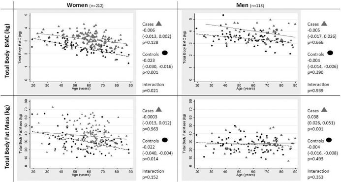
Results: Among women, fat mass was inversely related to age in controls (P = .01), but not in HBM cases (P = .96) in whom mean fat mass was 8.9 [95% CI 4.7, 13.0] kg higher compared with controls (fully adjusted mean difference, P < .001). Increased fat mass in male HBM cases was less marked (gender interaction P = .03). Compared with controls, lean mass was also increased in female HBM cases (by 3.3 [1.2, 5.4] kg; P < .002); however, lean mass increases were less marked than fat mass increases, resulting in 4.5% lower percentage lean mass in HBM cases (P < .001). Osteocalcin was also lower in female HBM cases compared with controls (by 2.8 [0.1, 5.5] μg/L; P = .04). Differences in fat mass were fully attenuated after hip bone mineral density (BMD) adjustment (P = .52) but unchanged after adjustment for bone turnover (P < .001), whereas the greater hip BMD in female HBM cases was minimally attenuated by fat mass adjustment (P < .001).
Conclusions: HBM is characterized by a marked increase in fat mass in females, statistically explained by their greater BMD, but not by markers of bone turnover.
Figures
References
-
- Reid IR, Plank LD, Evans MC. Fat mass is an important determinant of whole body bone density in premenopausal women but not in men. J Clin Endocrinol Metab. 1992;75:779–782 - PubMed
-
- Khosla S, Atkinson EJ, Riggs BL, Melton LJ. Relationship between body composition and bone mass in women. J Bone Miner Res. 1996;11:857–863 - PubMed
-
- Felson DT, Zhang Y, Hannan MT, Anderson JJ. Effects of weight and body mass index on bone mineral density in men and women: The Framingham study. J Bone Miner Res. 1993;8:567–573 - PubMed
-
- Reid IR, Legge M, Stapleton JP, Evans MC, Grey AB. Regular exercise dissociates fat mass and bone density in premenopausal women. J Clin Endocrinol Metab. 1995;80:1764–1768 - PubMed
-
- Reid I. Relationships between fat and bone. Osteoporos Int. 2008;19:595–606 - PubMed
Publication types
MeSH terms
Substances
Grants and funding
LinkOut - more resources
Full Text Sources
Other Literature Sources
Medical
