. 2013 Mar;182(3):828-40.
doi: 10.1016/j.ajpath.2012.11.029. Epub 2013 Jan 19.

Bullous pemphigoid IgG induces BP180 internalization via a macropinocytic pathway

Affiliations

Bullous pemphigoid IgG induces BP180 internalization via a macropinocytic pathway

Sho Hiroyasu et al. Am J Pathol. 2013 Mar.

Abstract

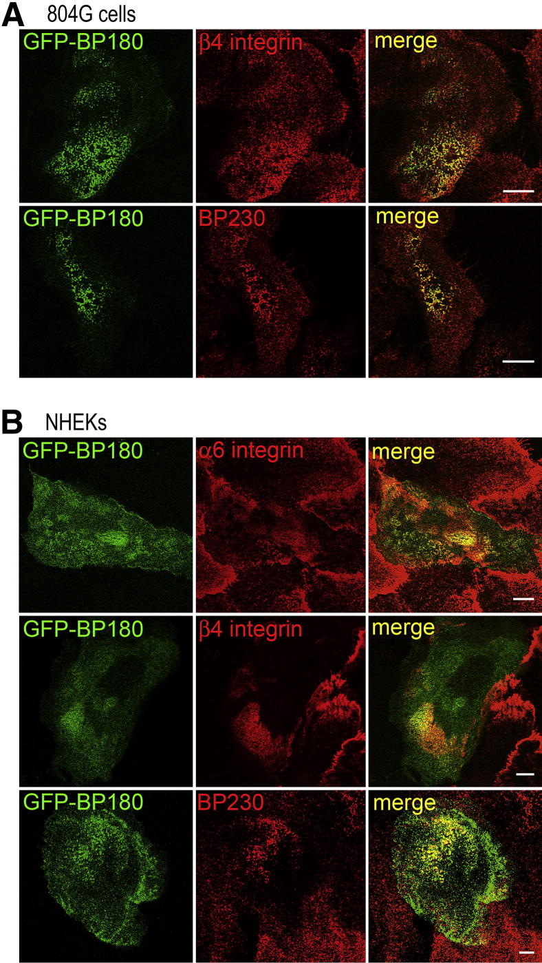
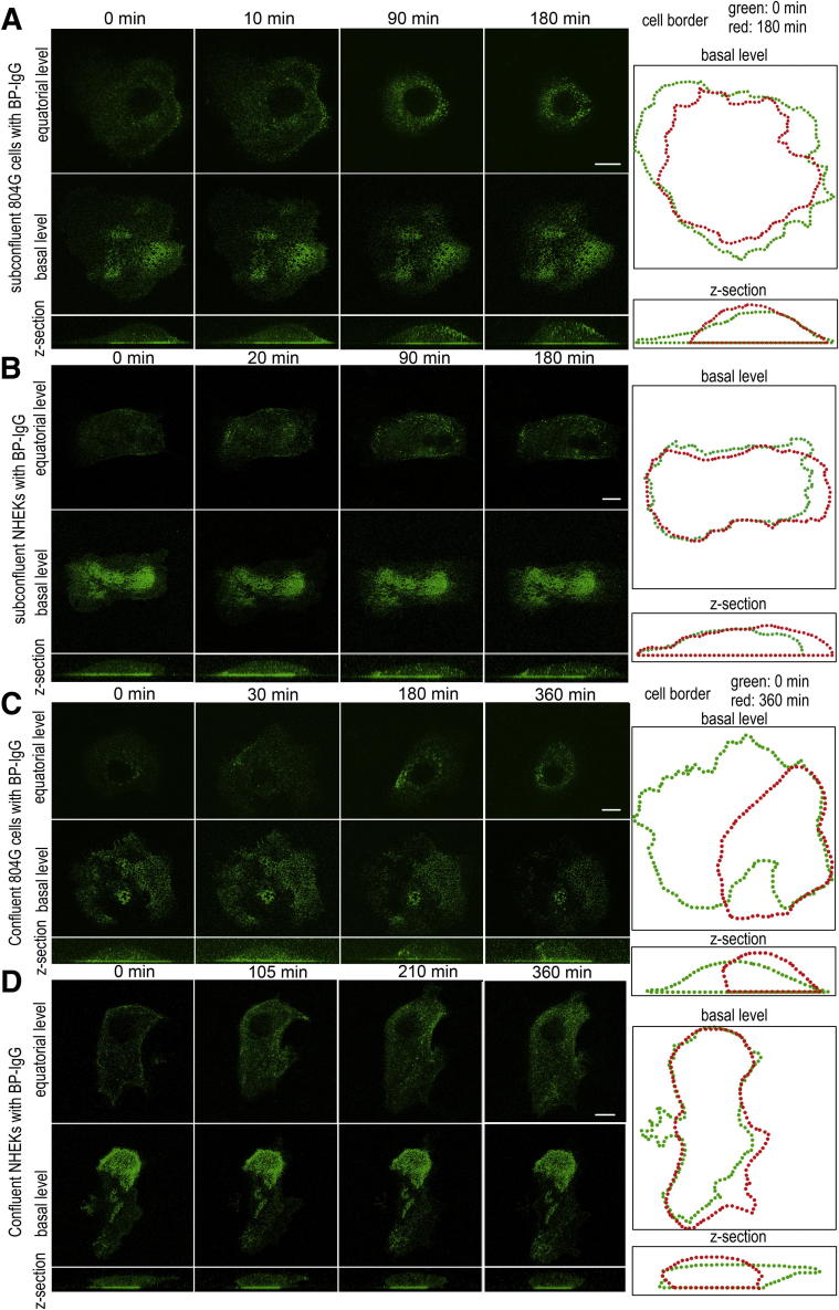
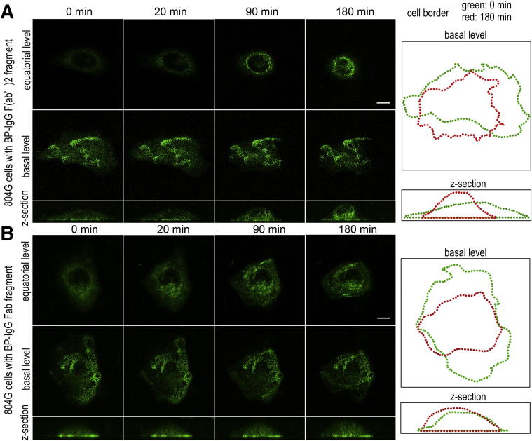
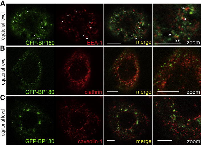
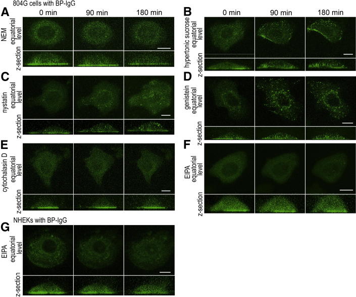
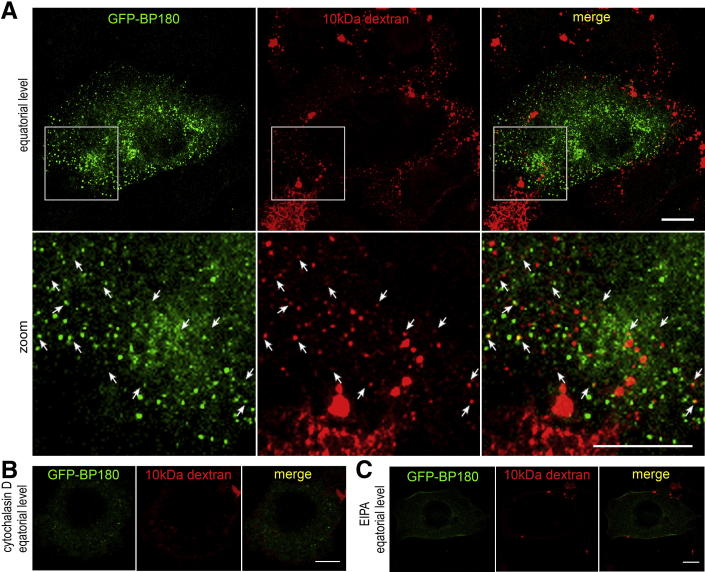
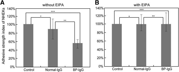
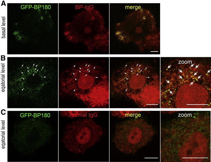
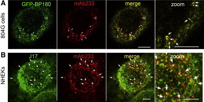
Bullous pemphigoid (BP) is an autoimmune blistering skin disease induced by pathogenic autoantibodies against a type II transmembrane protein (BP180, collagen type XVII, or BPAG2). In animal models, BP180 autoantibody-antigen interaction appears insufficient to develop blisters, but involvement of complement and neutrophils is required. However, cultured keratinocytes treated with BP-IgG exhibit a reduction in the adhesive strength and a loss of expression of BP180, suggesting that the autoantibodies directly affect epidermal cell-extracellular matrix integrity. In this study, we explored the consequences of two distinct epithelial cells treated with BP-IgG, particularly the fate of BP180. First, we followed the distribution of green fluorescent protein-tagged BP180 in an epithelial cell line, 804G, and normal human epidermal keratinocytes after autoantibody clustering. After BP-IgG treatment, the adhesive strength of the cells to their substrate was decreased, and BP180 was internalized in both cell types, together with the early endosomal antigen-1. By using various endocytosis inhibitors and a fluid-uptake assay, we demonstrated that BP-IgG-induced BP180 internalization is mediated via a macropinocytic pathway. Moreover, a macropinocytosis inhibitor rescued a BP-IgG-induced reduction in the adhesive strength of the cells from their substrate. The results of this study suggest that BP180 internalization induced by BP-IgG plays an important role in the initiation of disease pathogenesis.


Figures

Figure 1
Figure 1
GFP-BP180 incorporates into hemidesmosome-like structures in 804G cells and NHEKs. A: GFP-BP180–expressing 804G cells were stained using antibody against rat β4 integrin or 5E antibody against BP230. GFP-BP180 localized precisely with β4 integrin and BP230 in a cat paw–like pattern at the substratum-attached surface. B: Immunofluorescence analyses of GFP-BP180–expressing NHEKs using GoH3 mAb against human α6 integrin, antibody against human β4 integrin, or 5E antibody against BP230. GFP-BP180 colocalized with other hemidesmosomal components. Scale bars: 10 μm.
Figure 2
Figure 2
BP-IgG–induced GFP-BP180 internalization in live 804G cells and NHEKs. Live cell imaging studies of subconfluent (A) and confluent (C) cultures of 804G cells and subconfluent (B) and confluent (D) cultures of NHEKs. GFP-BP180–expressing cells were visualized by confocal microscopy, either along their substrate-attached surface (basal level) or at a higher focal plane (equatorial level), at various intervals after BP-IgG treatment. In both cell types and conditions, GFP-BP180 was internalized and redistributed centripetally toward the perinuclear region (AD). Z-sections of the cells are shown at the bottom of each column. At the right, images of the cell border at time 0 and the end of the observation are shown in green and red, respectively. In 804G cells, rounding up of the cells was observed (A and C). Scale bars: 10 μm.
Figure 3
Figure 3
BP-IgG F(ab′)2 and BP-IgG Fab fragments induce GFP-BP180 internalization in live 804G cells. Live cell imaging studies of subconfluent cultures of 804G cells treated with BP-IgG F(ab′)2 (A) and BP-IgG Fab (B) fragments. GFP-BP180 was internalized and redistributed centripetally toward the perinuclear region after treatment with both fragments. The fragments induced some cell rounding, as indicated by the outlines of the treated cells at times 0 and 180 minutes, shown to the right. Scale bars: 10 μm.
Figure 4
Figure 4
BP-IgG–induced GFP-BP180 internalization is mediated through endocytosis. A: After the incubation with BP-IgG for 1 hour, GFP-BP180–expressing 804G cells were stained for EEA-1, which showed some colocalization with GFP-BP180 in the cytoplasm (arrows). In contrast, after a 2-hour incubation with BP-IgG, GFP failed to colocalize with either clathrin (B) or caveolin-1 (C). Scale bars: 5 μm.
Figure 5
Figure 5
Pre-incubation with the inhibitors of general endocytosis and macropinocytosis prevents BP-IgG–induced GFP-BP180 internalization. Live cell imaging studies for GFP-BP180–expressing 804G cells (AF) and NHEKs (G) treated with BP-IgG and endocytosis inhibitors. A: Pre-incubation with NEM inhibited BP-IgG–induced GFP-BP180 internalization and cell morphological changes in 804G cells. Hypertonic sucrose (B), nystatin (C), and genistein (D) failed to prevent GFP-BP180 internalization in BP-IgG–treated 804G cells. Cytochalasin D (E) and EIPA (F) inhibited GFP-BP180 internalization in 804G cells. G: In NHEKs, EIPA prevented BP-IgG–induced GFP-BP180 internalization at 180 minutes. Scale bars:10 μm.
Figure 6
Figure 6
Internalized BP180 colocalizes with the fluid-phase marker. After incubation with BP-IgG and 10-kDa dextran–AF 594 for 10 minutes, GFP-BP180–expressing 804G cells were fixed and viewed. A: Boxed areas are shown at a higher power; 10-kDa dextran–AF 594 was internalized and colocalized with some GFP-BP180 as spot-like structures near the cell surface (arrows). Pre-incubation with cytochalasin D (B) and EIPA (C) inhibited the internalization of GFP-BP180 and dextran. Scale bars: 10 μm.
Figure 7
Figure 7
BP-IgG treatment increases the detachment of NHEKs from their substrate after vortex mixing, and the effect is inhibited by pre-incubation with EIPA. After pre-incubation with (B) or without (A) EIPA, NHEKs were treated with or without normal IgG and BP-IgG for 6 hours. The cells were vortex mixed, and the numbers of adherent cells were counted. The y axis depicts the percentage of adherent cells after agitation. The control index value without IgG addition was calculated as 100%. A: Without pre-incubation with EIPA, the adhesive strength of BP-IgG–treated cells was reduced to approximately 60% compared with normal IgG-treated cells. *P = 0.38 (not significant); **P = 0.0017; ***P = 0.0061. B: After pre-incubation with EIPA for 30 minutes, the adhesive strength of BP-IgG–treated cells was comparable to normal IgG-treated and non-treated cells. *P = 0.96; **P = 1.00; ***P = 0.96 (no significant differences).
Figure 8
Figure 8
HiLyte Fluor 647–conjugated BP-IgG is internalized along with BP180. HiLyte Fluor 647–conjugated BP-IgG (A and B) and HiLyte Fluor 647–conjugated normal IgG (C) were observed in GFP-BP180–expressing 804G cells after incubation for 30 minutes. A: HiLyte Fluor 647–conjugated BP-IgG colocalized with GFP-BP180 into hemidesmosome-like structures at the cell substratum–attached surface. B: At the equatorial plane, internalized BP-IgG was visible and colocalized with internalized GFP-BP180 (arrows). C: HiLyte Fluor 647–conjugated normal IgG failed to induce internalization of both IgG and GFP-BP180. Scale bars: 10 μm.
Figure 9
Figure 9
The intracellular domain of BP180 is internalized, along with the extracellular domain of BP180. A: GFP-BP180–expressing 804G cells were stained using anti-extracellular BP180 domain antibody mAb233, 2 hours after the incubation with BP-IgG. B: NHEKs were doubly stained using mAb233 and anti-cytoplasmic BP180 domain antibody J17. These studies showed that both intracellular and extracellular domains of internalized GFP-BP180 colocalized as cytoplasmic spots (arrows). Scale bars: 5 μm.

References

    1. Lever W.F. Pemphigus. Medicine (Baltimore) 1953;32:1–123. - PubMed
    1. Jordon R.E., Beutner E.H., Witebsky E., Blumental G., Hale W.L., Lever W.F. Basement zone antibodies in bullous pemphigoid. JAMA. 1967;200:751–756. - PubMed
    1. Labib R.S., Anhalt G.J., Patel H.P., Mutasim D.F., Diaz L.A. Molecular heterogeneity of the bullous pemphigoid antigens as detected by immunoblotting. J Immunol. 1986;136:1231–1235. - PubMed
    1. Diaz L.A., Ratrie H., 3rd, Saunders W.S., Futamura S., Squiquera H.L., Anhalt G.J., Giudice G.J. Isolation of a human epidermal cDNA corresponding to the 180-kD autoantigen recognized by bullous pemphigoid and herpes gestationis sera: immunolocalization of this protein to the hemidesmosome. J Clin Invest. 1990;86:1088–1094. - PMC - PubMed
    1. Stanley J.R., Hawley-Nelson P., Yuspa S.H., Shevach E.M., Katz S.I. Characterization of bullous pemphigoid antigen: a unique basement membrane protein of stratified squamous epithelia. Cell. 1981;24:897–903. - PubMed

Publication types

MeSH terms