. 2013 Apr;11(4):405-17.
doi: 10.1158/1541-7786.MCR-12-0551. Epub 2013 Jan 21.

Targeting constitutively activated β1 integrins inhibits prostate cancer metastasis

Affiliations

Targeting constitutively activated β1 integrins inhibits prostate cancer metastasis

Yu-Chen Lee et al. Mol Cancer Res. 2013 Apr.

Abstract

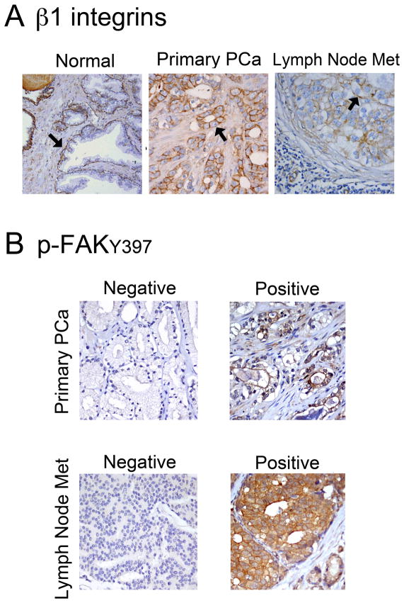
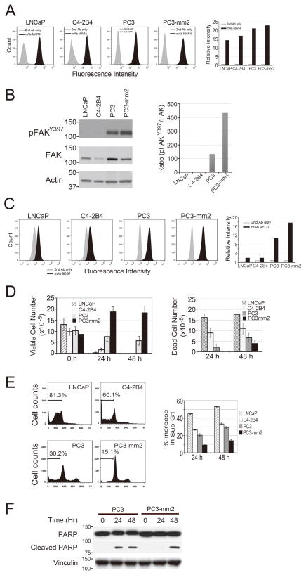
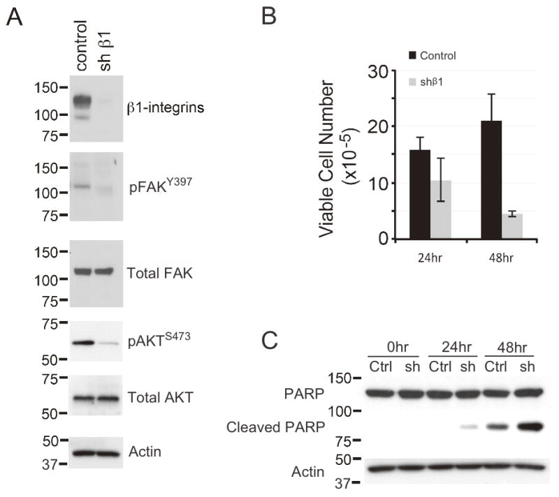
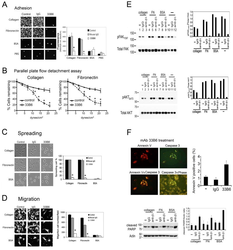
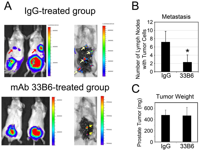
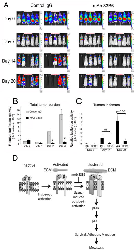
Disseminated prostate cancer cells must survive in circulation for metastasis to occur. Mechanisms by which these cells survive are not well understood. By immunohistochemistry of human tissues, we found that levels of β1 integrins and integrin-induced autophosphorylation of FAK (pFAK-Y397) are increased in prostate cancer cells in primary prostate cancer and lymph node metastases, suggesting that β1 integrin activation occurs in metastatic progression of prostate cancer. A conformation-sensitive antibody, 9EG7, was used to examine β1 integrin activation. We found that β1 integrins are constitutively activated in highly metastatic PC3 and PC3-mm2 cells, with less activation in low metastatic LNCaP and C4-2B4 cells. Increased β1 integrin activation as well as the anoikis resistance in prostate cancer cells correlated with metastatic potential in vivo. Knockdown of β1 integrin abrogated anoikis resistance in PC3-mm2 cells. In agreement with β1 integrin activation, PC3-mm2 cells strongly adhered to type I collagen and fibronectin, a process inhibited by the β1 integrin-neutralizing antibody mAb 33B6. mAb 33B6 also inhibited the phosphorylation of β1 integrin downstream effectors, focal adhesion kinase (FAK) and AKT, leading to a 3-fold increase in PC3-mm2 apoptosis. Systemic delivery of mAb 33B6 suppressed spontaneous metastasis of PC3-mm2 from the prostate to distant lymph nodes following intraprostatic injection and suppressed metastasis of PC3-mm2 to multiple organs following intracardiac injection. Thus, constitutively activated β1 integrins play a role in survival of PC3-mm2 cells in circulation and represent a potential target for metastasis prevention.


Conflict of interest statement

There are no conflicts of interests among all the authors.

Figures

Figure 1
Figure 1. Expression of β1 integrins and pFAK-Y397 in human PCa specimens
(A) Immunohistochemical staining of β1 integrins in PCa specimens. In normal prostate gland, β1 integrins are expressed on the basal cell layer (arrow). In PCa specimens, β1 integrins were localized on the membranes of tumor cells (arrow). In lymph node metastasis, β1 integrins are expressed on the membranes of metastatic cancer cells in lymph node (arrow). (B) Immunohistochemical staining of pFAK-Y397 in PCa specimens. Seven of 16 PCa specimens showed positive staining for pFAK-Y397 (right panel), while nine specimens were negative (left panel). Seven of 12 lymph node metastasis specimens showed positive staining for pFAK-Y397 (right panel), while the rest of five specimens were negative (left panel).
Figure 2
Figure 2. β1 integrin activation correlates with anoikis resistance in PCa cell lines
(A) FACS using anti-total β1-integrin antibody mAb MAR4. LNCaP, C4-2B4, PC3, and PC3-mm2 cells showed similar levels of binding with mAb MAR4. (B) Western blot of cell lysates for the expression of FAK and phosphorylated FAKY397. FAK is phosphorylated at Y397 in PC3 and PC3-mm2 cells, but not LNCaP or C4-2B4 cells. (C) FACS using conformation sensitive anti-β1-integrin antibody mAb 9EG7. High percentage of PC3 and PC3-mm2 showed specific binding with the conformation-sensitive mAb 9EG7, while LNCaP and C4-2B4 showed modest binding. (D) Cells were grown in anoikis condition as described in Materials and Methods. Viable and dead cell numbers were determined by Trypan Blue exclusion and propidium iodide staining, respectively. PC3 and PC3-mm2 are more resistant to anoikis-induced cell death than LNCaP and C4-2B4 cells. (E) FACS of propidium iodide labeled cells after the cells were grown in anoikis conditions for 24 or 48 hrs. LNCaP and C4-2B4 have higher sub-G1 fraction than PC3 or PC3-mm2 cells. (F) Western blot analysis of PARP and cleaved PARP of PC3 or PC3-mm2 grown under anoikis conditions for various lengths of times.
Figure 3
Figure 3. Knockdown of β1-integrins reduces FAK phosphorylation at Y397 and anoikis-resistance of PC3-mm2 cells
A stable PC3-mm2 cell line was derived in which β1-integrins are reduced by expression of shRNA as described in Materials and Methods. (A) Immunoblotting of β1-integrins, pFAK 397 and Total FAK, pAKT473 and total AKT. (B) Cells were grown in anoikis condition as described in Materials and Methods. Viable and dead cell numbers were determined by Trypan Blue exclusion. (C) Western blot analysis of PARP and cleaved PARP of PC3 or PC3-mm2 grown under anoikis conditions for various lengths of times.
Figure 4
Figure 4. Effect of mAb 33B6 on ECM-mediated cell adhesion, migration, apoptosis in vitro
(A) mAb 33B6 inhibits PC3-mm2 adhesion to ECM. PC3-mm2 cells were plated on type I collagen, fibronectin, or BSA coated plates in the presence or absence of mAb 33B6 and the fractions of cells that bound to the coated plates were determined by the fluorescence intensity. (B) Measurement of PC3-mm2 binding to fibronectin or type I collagen under flow. PC3-mm2 (2 × 10⁶) cells treated with or without mAb 33B6 were injected into the flow chambers coated with type I collagen or fibronectin as described in Materials and Methods. Shear stress calculations were determined every 50 seconds. (C) mAb 33B6 inhibits PC3-mm2 spreading on extracelluar matrix. Binding of PC3-mm2 to type I collagen or fibronectin, but not BSA, leads to a flattened cell shape, which was inhibited by mAb 33B6. (D) mAb 33B6 inhibits PC3-mm2 migration on type I collagen or fibronectin. *, p<0.05. (E) Lysates from PC3-mm2 cells treated with mAb 33B6 or control IgG for 24 hrs were immunoblotted with antibody against phospho-FAK, FAK, or phospho-AKT or AKT. The phosphorylated versus non-phosphorylated FAK or AKT ratios were determined by densitometry. The experiments were repeated twice. (F) PC3-mm2 cells were treated with mAb 33B6 or control IgG for 24 hrs and apoptosis was determined by staining with sulforhodamine 101-Annexin V and caspase 3 substrate DEVD-NucView 488. Cell lysates were immunoblotted with antibody against cleaved PARP. The cleaved PARP and actin ratios were determined by densitometry and the average of two experiments was shown.
Figure 5
Figure 5. Inhibition of PC3-mm2 metastasis to lymph node in vivo by mAb 33B6
Luciferase-labeled PC3-mm2 cells (1 × 10⁶) were injected into the prostate of SCID mice. Mice were treated with IgG or mAb 33B6 (1 mg/kg) before tumor cell inoculation and twice a week subsequently by intra-peritoneal injection. Bioluminescence imaging of mice was performed weekly afterwards to monitor tumor growth and metastasis in vivo. (A) Bioluminescence imaging of mice at 2-weeks post-injection. At termination, the prostates were removed before bioluminescence imaging. Arrows point to tumors in lymph nodes. (B) Average numbers of lymph node ± SD metastases detected at the termination of the study. (C) Average tumor sizes ± SD in the prostates at the termination of the study. *, p<0.05.
Figure 6
Figure 6. Inhibition of disseminated PC3-mm2 cell growth in vivo by mAb 33B6
Luciferase-labeled PC3-mm2 cells (1×10⁶) were injected into the left ventricle of SCID mice. Mice were treated with IgG or mAb 33B6 (1 mg/kg) before tumor cells inoculation and twice a week subsequently by intra-peritoneal injection. (A) Bioluminescence imaging of mice was performed within 30 min of injection and weekly afterwards to monitor tumor growth in vivo. (B) Overall tumor burden was recorded as photons per second. Average photon numbers ± SE of the control and mAb 33B6-treated mice are presented. (C) Tumor burden in the femurs of the control and mAb 33B6-treated mice. *p<0.05 (D) Model of activated β1 integrins enhancing PCa metastasis. In highly metastatic PCa cells, β1 integrins are present in an activated conformation through an inside-out activation mechanism, which increases its ligand binding affinity. Upon binding with ECM, β1 integrins are further activated by an outside-in mechanism that results in integrin clustering and activation of a downstream signaling cascade that enhances the metastatic potential of PCa cells.

References

    1. Siegel R, Ward E, Brawley O, Jemal A. Cancer statistics, 2011: the impact of eliminating socioeconomic and racial disparities on premature cancer deaths. CA Cancer J Clin. 2011;61:212–36. - PubMed
    1. Shah RB, Mehra R, Chinnaiyan AM, Shen R, Ghosh D, Zhou M, et al. Androgen-independent prostate cancer is a heterogeneous group of diseases: lessons from a rapid autopsy program. Cancer Res. 2004;64:9209–16. - PubMed
    1. Fidler IJ. Critical factors in the biology of human cancer metastasis: twenty-eighth G.H.A. Clowes memorial award lecture. Cancer Res. 1990;50:6130–8. - PubMed
    1. Zetter BR. The cellular basis of site-specific tumor metastasis. N Engl J Med. 1990;322:605–12. - PubMed
    1. Husemann Y, Geigl JB, Schubert F, Musiani P, Meyer M, Burghart E, et al. Systemic spread is an early step in breast cancer. Cancer Cell. 2008;13:58–68. - PubMed

Publication types