. 2013 Jan 22:11:4.
doi: 10.1186/1741-7007-11-4.

Enhanced 5-methylcytosine detection in single-molecule, real-time sequencing via Tet1 oxidation

Affiliations

Enhanced 5-methylcytosine detection in single-molecule, real-time sequencing via Tet1 oxidation

Tyson A Clark et al. BMC Biol. .

Abstract

Background: DNA methylation serves as an important epigenetic mark in both eukaryotic and prokaryotic organisms. In eukaryotes, the most common epigenetic mark is 5-methylcytosine, whereas prokaryotes can have 6-methyladenine, 4-methylcytosine, or 5-methylcytosine. Single-molecule, real-time sequencing is capable of directly detecting all three types of modified bases. However, the kinetic signature of 5-methylcytosine is subtle, which presents a challenge for detection. We investigated whether conversion of 5-methylcytosine to 5-carboxylcytosine using the enzyme Tet1 would enhance the kinetic signature, thereby improving detection.

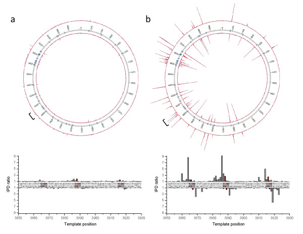
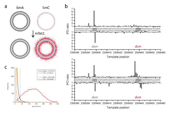
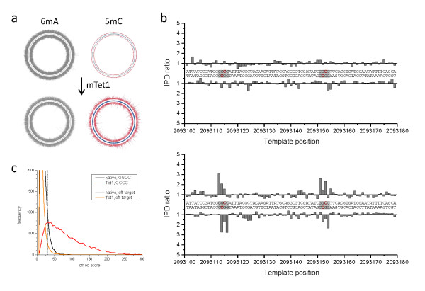
Results: We characterized the kinetic signatures of various cytosine modifications, demonstrating that 5-carboxylcytosine has a larger impact on the local polymerase rate than 5-methylcytosine. Using Tet1-mediated conversion, we show improved detection of 5-methylcytosine using in vitro methylated templates and apply the method to the characterization of 5-methylcytosine sites in the genomes of Escherichia coli MG1655 and Bacillus halodurans C-125.

Conclusions: We have developed a method for the enhancement of directly detecting 5-methylcytosine during single-molecule, real-time sequencing. Using Tet1 to convert 5-methylcytosine to 5-carboxylcytosine improves the detection rate of this important epigenetic marker, thereby complementing the set of readily detectable microbial base modifications, and enhancing the ability to interrogate eukaryotic epigenetic markers.


Figures

Figure 1
Figure 1
Kinetic signals from SMRT sequencing for the four epigenetic markers of cytosine 5mC, 5hmC, 5fC and 5caC. Synthetic oligonucleotides carrying two modified sites each (red bars) were subjected to SMRT sequencing and the polymerase kinetics compared by plotting the ratio of IPDs for each template position against a control template of identical sequence but lacking the modifications. The template is shown in the 5' to 3' direction from left to right, the polymerase movement is right to left across the template as indicated by the arrow. 5caC: 5-carboxylcytosine; 5fC: 5-formylcytosine; 5hmC: 5-hydroxymethylcytosine; 5mC: 5-methylcytosine; IPD: interpulse duration; SMRT: single-molecule: real-time.
Figure 2
Figure 2
Enhanced detection of 5mC through Tet1 oxidation using in vitro methylated plasmid samples. The Circos plots show the plasmid-wide view of IPD ratios for (a) untreated and (b) mouse Tet1-treated samples, with the outer and inner circle denoting the forward and reverse DNA strands, respectively, and the blue tick marks denoting all positions of the targeted M.HpaII in vitro methylated sequence motif of 5'-CCGG-3' (methylated base underlined). The bracket denotes a section of the plasmid which is shown at base resolution in the bottom graphs, containing three instances of the methylated motif (grey boxes). The methylated positions are highlighted in red. IPD: interpulse duration.
Figure 3
Figure 3
Genome-wide 5mC methyltransferase specificity detection in E. coli K12 MG1655. The Circos plots show the genome-wide view of IPD ratios for (a) untreated and (b) mTet1-treated samples, with the outer and inner circle denoting the forward and reverse DNA strands, respectively. The IPD ratios of the +2 position in 5'-CCWGG-3' sequence contexts are plotted in red, while IPD ratio data for all other contexts is plotted in grey. The graphs on the right show base-resolution IPD ratio views of a section of the genome containing one target site for adenine methylation by dam (5'-GATC-3') and one target site for cytosine methylation by dcm (5'-CCWGG-3'). (c) Kinetic score distributions before and after mTet1 conversion for all +2 positions of 5'-CCWGG-3' in the genome. An orthogonal off-target motif (5'-GGWCC-3') is also shown which was used to set a 1% false discovery rate threshold (dashed line, see Methods for details) for tabulation of detected methylated positions (Table 1). The blue tick marks in the Circos plots of (a) and (b) denote 5'-CCWGG-3' genomic positions detected as methylated using that threshold. 5mC: 5-methylcytosine; 6mA: 6-methyladenine; IPD: interpulse duration; mTet1: mouse Tet1.
Figure 4
Figure 4
Genome-wide 5mC methyltransferase specificity detection in B. halodurans. The Circos plots show the genome-wide view of IPD ratios for (a) untreated and (b) mTet1-treated samples, with the outer and inner circle denoting the forward and reverse DNA strands, respectively. The IPD ratios of the +2 position in 5'-GGCC-3' sequence contexts are plotted in red, while IPD ratio data for all other contexts is plotted in grey. The graphs on the right show base-resolution IPD ratio views of a section of the genome containing two target sites for cytosine methylation (5'-GGCC-3'). (c) Kinetic score distributions before and after Tet1 conversion for all +2 positions of 5'-GGCC-3' in the genome. An orthogonal off-target motif (5'-CCGG-3') is also shown which was used to set a 1% false discovery rate threshold (dashed line, see methods for details) for tabulation of detected methylated positions (Table 1). The blue tick marks in the Circos plots of (a) and (b) denote 5'-GGCC-3' genomic positions detected as methylated using that threshold. 5mC: 5-methylcytosine; 6mA: 6-methyladenine; IPD: interpulse duration; mTet1: mouse Tet1.

References

    1. Bird A. DNA methylation patterns and epigenetic memory. Genes Dev. 2002;16:6–21. doi: 10.1101/gad.947102. - DOI - PubMed
    1. Wu H, Zhang Y. Mechanisms and functions of Tet protein-mediated 5-methylcytosine oxidation. Genes Dev. 2011;25:2436–2452. doi: 10.1101/gad.179184.111. - DOI - PMC - PubMed
    1. Kriaucionis S, Heintz N. The nuclear DNA base 5-hydroxymethylcytosine is present in Purkinje neurons and the brain. Science. 2009;324:929–930. doi: 10.1126/science.1169786. - DOI - PMC - PubMed
    1. Tahiliani M, Koh KP, Shen Y, Pastor WA, Bandukwala H, Brudno Y, Agarwal S, Iyer LM, Liu DR, Aravind L, Rao A. Conversion of 5-methylcytosine to 5-hydroxymethylcytosine in mammalian DNA by MLL partner TET1. Science. 2009;324:930–935. doi: 10.1126/science.1170116. - DOI - PMC - PubMed
    1. Pfaffeneder T, Hackner B, Truss M, Munzel M, Muller M, Deiml CA, Hagemeier C, Carell T. The discovery of 5-formylcytosine in embryonic stem cell DNA. Angew Chem Int Ed Engl. 2011;50:7008–7012. doi: 10.1002/anie.201103899. - DOI - PubMed

MeSH terms