Review
. 2012;40(5):363-408.
doi: 10.1615/critrevbiomedeng.v40.i5.10.

Bone tissue engineering: recent advances and challenges

Affiliations
Review

Bone tissue engineering: recent advances and challenges

Ami R Amini et al. Crit Rev Biomed Eng. 2012.

Abstract

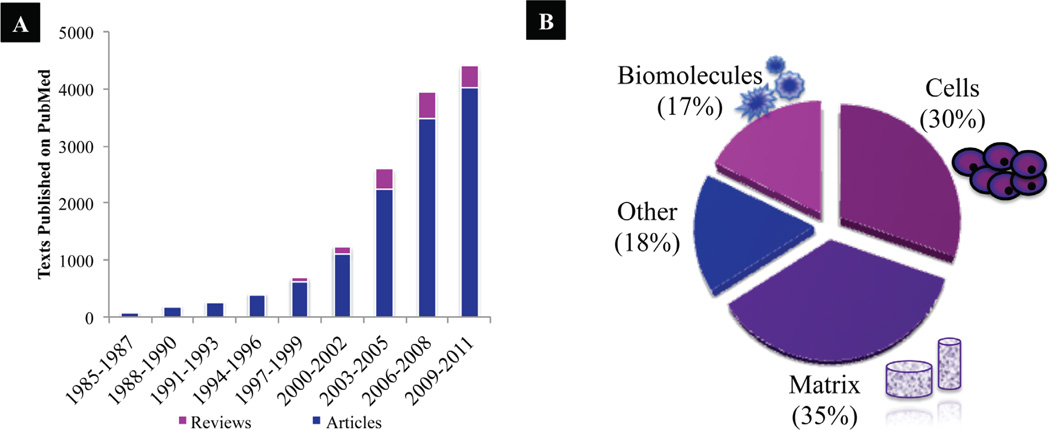
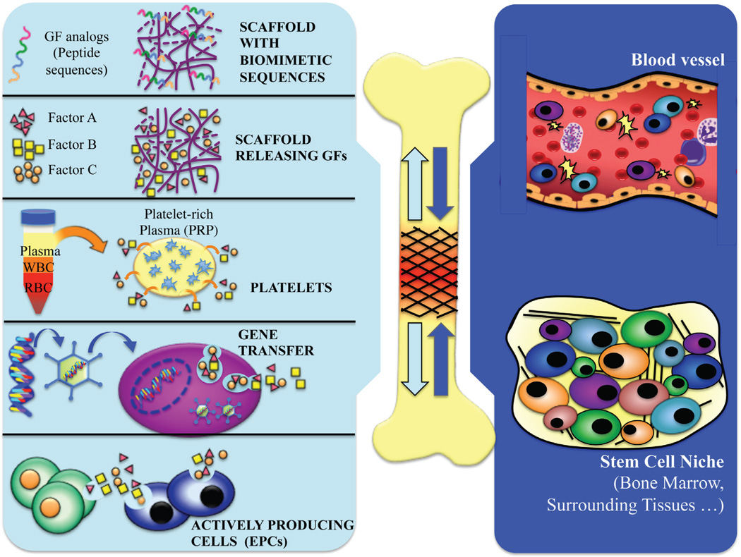
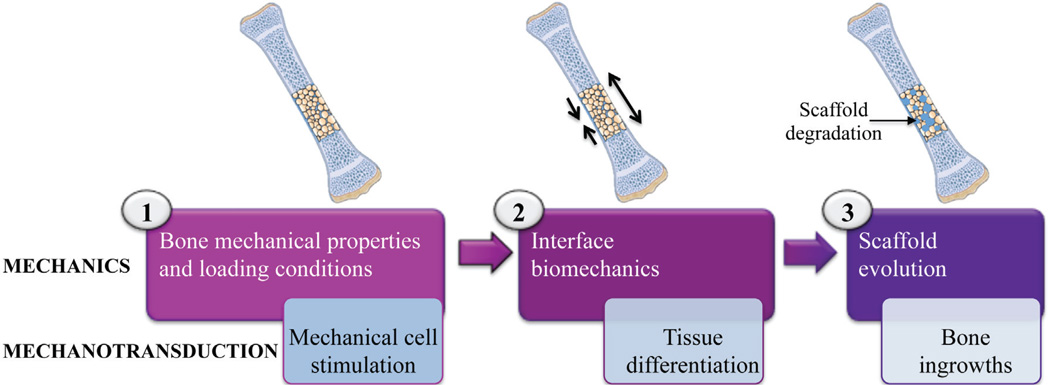
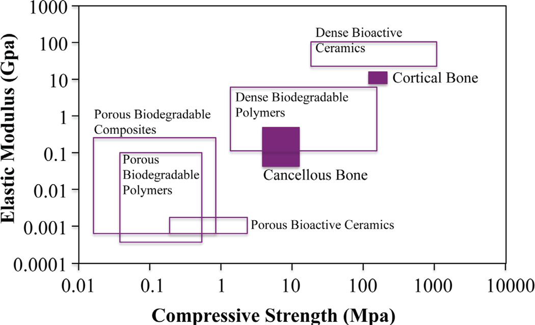
The worldwide incidence of bone disorders and conditions has trended steeply upward and is expected to double by 2020, especially in populations where aging is coupled with increased obesity and poor physical activity. Engineered bone tissue has been viewed as a potential alternative to the conventional use of bone grafts, due to their limitless supply and no disease transmission. However, bone tissue engineering practices have not proceeded to clinical practice due to several limitations or challenges. Bone tissue engineering aims to induce new functional bone regeneration via the synergistic combination of biomaterials, cells, and factor therapy. In this review, we discuss the fundamentals of bone tissue engineering, highlighting the current state of this field. Further, we review the recent advances of biomaterial and cell-based research, as well as approaches used to enhance bone regeneration. Specifically, we discuss widely investigated biomaterial scaffolds, micro- and nano-structural properties of these scaffolds, and the incorporation of biomimetic properties and/or growth factors. In addition, we examine various cellular approaches, including the use of mesenchymal stem cells (MSCs), embryonic stem cells (ESCs), adult stem cells, induced pluripotent stem cells (iPSCs), and platelet-rich plasma (PRP), and their clinical application strengths and limitations. We conclude by overviewing the challenges that face the bone tissue engineering field, such as the lack of sufficient vascularization at the defect site, and the research aimed at functional bone tissue engineering. These challenges will drive future research in the field.


Figures

FIGURE 1
FIGURE 1
(A) Published articles on BTE since mid-1980s on PubMed. (B) Break-down of the articles published in 2011 according to bone tissue engineering focus (i.e., biomolecules, cells, matrices, and other, including vascularization approaches and bioreactors).
FIGURE 2
FIGURE 2
Schematic illustration of bone tissue engineering paradigm. Factors from the implanted graft at the defect site that influence the host response may include growth factors (or their analogs, or from platelet-enriched plasma), and cells (genetically modified to release factors, or naturally produce factors). In response, cell homing and enhanced vascularization and bone regeneration will occur.
FIGURE 3
FIGURE 3
Illustration of a three-step biomechanical paradigm in BTE. In the first step, upon implantation, it is critical that the mechanical properties of the BTE scaffold should closely match that of the surrounding host bone tissue and loading conditions to reduce the stress-shielding effect. The second step involves interface biomechanics, and should allow for interface scaffold-bone mechanotransduction for enhanced osteointegration of the scaffold. Lastly, as the scaffold degrades, ingrowing bone tissue will begin to support the mechanical load of BTE scaffold. Adapted from Pioletti (97).
FIGURE 4
FIGURE 4
Elastic modulus versus compressive strength values of various BTE biomaterial classes compared to human bone. Adapted from Rezwan et al. (103).
FIGURE 5
FIGURE 5
SEM images of (A) PLGA (50/50) microsphere scaffolds, and (B) composite carbon nanotube/PLGA (50:50) microsphere scaffolds after 14 days in simulated body fluid. Crystalization is seen the joining areas of mi-crospheres in only composite CNT/PLGA scaffolds. Scale bar = 40 µm.
FIGURE 6
FIGURE 6
Optimally-porous, mechanically strong biodegradable scaffolds for enhanced bone regeneration. (A) Reconstructed MicroCT 3D porosity images demonstrating significantly increased interconnected pore sizes in optimally-porous scaffolds. (B) Schematic illustration of available oxygen levels throughout the scaffolds in vitro. (Scale from red to green demonstrating increasing oxygen levels.) (C) Pre-osteoblast cell viability in the center of the constructs after 14 days in vitro. Scaffolds with increasing porosity resulted significant cell survival in the interior of the macro-porous construct (right) compared to control scaffolds (left) (live cells = green; dead cells = red). Scale bar = 200 µm. (D) After 28 days in osteogenic medium, Alizarin Red staining was performed. Optimally-porous scaffolds displayed mineralization throughout the thickness of the scaffold, where as scaffolds with low pore sizes displayed mineralization to limited to the surface of the scaffolds. Scale bar = 1000 µm. Adapted and modified from Amini et al. (122).” (D) After 28 days in osteogenic medium, Alizarin Red staining was performed. Optimally-porous scaffolds displayed mineralization throughout the thickness of the scaffold, where as scaffolds with low pore sizes displayed mineralization to limited to the surface of the scaffolds. Scale bar = 1000 µm. Adapted and modified from Amini et al. (122).”
FIGURE 7
FIGURE 7
Tissue engineering of anatomically shaped bone grafts. (A–C) Scaffold preparation. (A, B) Clinical CT images were used to obtain high-resolution digital data for the reconstruction of exact geometry of human TMJ condyles. (C) These data were incorporated into MasterCAM software to machine TMJ-shaped scaffolds from fully decellularized trabecular bone. (D) A photograph illustrating the complex geometry of the final scaffolds that appear markedly different in each projection. Adapted from Grayson et al.
FIGURE 8
FIGURE 8
(A) Illustration of hybrid scaffolds composed of a mechanically strong component, and a hydrogel phase for enhanced bone regeneration abilities. (B) In vitro release kinetics of biotinylated BMP2. Amount of BMP2 released over time was measured by ELISA. Results show cumulative release of rhBMP2 for untethered groups (BMP2-biotin, BMP2) versus tethered group. (C) Survival of pre-osteoblastic MC3T3-E1 cells in hybrid scaffold. Images show live and dead cells cultured on hybrid scaffolds; green represents live cells. (D) Bone Sialoprotein (BSP) and RunX2 gene expression profile of pre-osteoblastic MC3T3-E1 grown in BMP2 untethered versus tethered SAP gel PLGA/nHA hybrid scaffolds (p < 0.05). Adapted and modified from Igwe et al.
FIGURE 9
FIGURE 9
(A) Image of extracted teeth that may be used to tooth-derived stem cell isolation. (B) Stem cells that may be isolated from primary teeth [i.e., stem cells from human exfoliated deciduous teeth (SHED)], and secondary teeth [i.e., dental pulp stem cells (DPSCs), periodontal ligament stem cells (PDLSC), and stem cells of apical papilla (SCAP)].
FIGURE 10
FIGURE 10
(A) Immunofluorescent staining of VEGF production by transfected adipose stem cells (ADSCs) cultured for 10 days on PLAG sintered microsphere scaffolds in vitro. ADSCs were stained with antibody directed against VEGF (green) and nuclei counterstained with DAPI (blue). Scale bar = 100 mm. Adapted from Jabbarzadeh et al. (B) Representative histological cross sections of transfected ADSCs with VEGF implanted with sintered microsphere scaffolds 21 days after subcutaneous implantation in SCID mice. Scale bar = 10 mm. Adapted from Jabbarzadeh et al. (C) Intravital microscopy images of control PLAG films (left) or S1P loaded films (right) in a dorsal skinfold window chamber at 7 days post-implantation. Significant lume-nal expansion of both arterioles (black) and venules (white) is induced by S1P over the course of 7 days (arrows). Scale bar = 500 µm. (241) (D) New bone volume formed within defect area following 6 weeks of healing. (*p<0.05) (238) (E, F) Micro-CT images of vascular (E) and bone (F) ingrowth several weeks after implantation of 70% L-lactide and 30% DL-lactide co-polymer (PLDL) scaffold loaded with recombinant human growth factors (combinations of BMP-2, TGF-β3, and VEGF). Adapted from Guldberg et al.
FIGURE 11
FIGURE 11
Schematic illustration of bioreactors utilized in BTE. Specifically, (A) spinner (red arrows show movement of the stir bar), (B) rotating wall (red arrows show movement of the vessel), and (C) perfusion bioreactors (red arrows show movement of the medium) are the most commonly used. (D) Comparison of bioreactor culture of bone constructs (right) versus static culture (middle). Bioreactors allow for increased nutrient perfusion throughout construct, and enhanced bone formation in vitro. Adapted from Martin et al. and Olivier et al.321,322
FIGURE 12
FIGURE 12
(A) Isolation of rabbit peripheral blood-derived endothelial progenitor cells (EPCs) via terminal exsan-guination. (B) Phase contrast image of PB-EPCs cultured in endothelial growth media. (C) Two-dimensional angio-genesis assay showing network formation by PB-EPCs (cells cultured on Matrigel for 8 hours in vitro). Scale bar = 500 mm. (B) Hematoxylin/eosin staining demonstrating capillary network and branch point formation of PB-EPCs cultured in Matrigel after 7 days in vitro. Scale bar = 250 mm. Adapted and modified from Amini et al.

References

    1. Laurencin CT, Ambrosio AM, Borden MD, Cooper JA. Tissue engineering: orthopedic applications. Annu Rev Biomed Eng. 1999;1:19–46. - PubMed
    1. Laurencin CT, Khan Y, El-Amin SF. Bone graft substitutes. Expert Rev Med Devices. 2006;3(1):49–57. - PubMed
    1. Nukavarapu SP, Wallace J, Elgendy H, Lieberman J, Laurencin CT. Bone and biomaterials. In: group TF, editor. An introduction to biomaterials and their applications. 2 ed. CRC Press; 2011. pp. 571–593.
    1. Baroli B. From natural bone grafts to tissue engineering therapeutics: brainstorming on pharmaceutical formulative requirements and challenges. J Pharm Sci. 2009;98(4):1317–1375. - PubMed
    1. Dimitriou R, Jones E, McGonagle D, Giannoudis PV. Bone regeneration: current concepts and future directions. BMC Med. 2011;9:66. - PMC - PubMed

Publication types

Substances