. 2013 Apr;33(7):1345-56.
doi: 10.1128/MCB.01411-12. Epub 2013 Jan 22.

The nucleus- and endoplasmic reticulum-targeted forms of protein tyrosine phosphatase 61F regulate Drosophila growth, life span, and fecundity

Affiliations

The nucleus- and endoplasmic reticulum-targeted forms of protein tyrosine phosphatase 61F regulate Drosophila growth, life span, and fecundity

Bree J Buszard et al. Mol Cell Biol. 2013 Apr.

Abstract

The protein tyrosine phosphatases (PTPs) T cell PTP (TCPTP) and PTP1B share a high level of catalytic domain sequence and structural similarity yet display distinct differences in substrate recognition and function. Their noncatalytic domains contribute to substrate selectivity and function by regulating TCPTP nucleocytoplasmic shuttling and targeting PTP1B to the endoplasmic reticulum (ER). The Drosophila TCPTP/PTP1B orthologue PTP61F has two variants with identical catalytic domains that are differentially targeted to the ER and nucleus. Here we demonstrate that the PTP61F variants differ in their ability to negatively regulate insulin signaling in vivo, with the nucleus-localized form (PTP61Fn) being more effective than the ER-localized form (PTP61Fm). We report that PTP61Fm is reliant on the adaptor protein Dock to attenuate insulin signaling in vivo. Also, we show that the PTP61F variants differ in their capacities to regulate growth, with PTP61Fn but not PTP61Fm attenuating cellular proliferation. Furthermore, we generate a mutant lacking both PTP61F variants, which displays a reduction in median life span and a decrease in female fecundity, and show that both variants are required to rescue these mutant phenotypes. Our findings define the role of PTP61F in life span and fecundity and reinforce the importance of subcellular localization in mediating PTP function in vivo.


Figures

Fig 1
Fig 1
Schematic representation of PTP61Fm and PTP61Fn. The two PTP61F variants are identical except for their extreme C termini. The PTP61Fm hydrophobic C-terminal tail that targets PTP61Fm to membranes, including the ER (35), is shown. Basic residues in the PTP61Fn C terminus shown in bold are thought to constitute a nuclear localization sequence (NLS) targeting PTP61Fn to the nucleus (35). The PTP catalytic domain and the PXXP motifs that are present in both variants and allow interaction with SH3 domain-containing proteins are also highlighted.
Fig 2
Fig 2
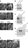
Differential regulation of IR signaling by PTP61Fm and PTP61Fn. (A) Scanning electron micrographs of a control Drosophila eye (GMR-Gal4/+) and those overexpressing IR alone using GMR-Gal4 (GMR>IR) and either N-terminal myc-tagged PTP61Fm (GMR>PTP61Fm) or PTP61Fn (GMR>PTP61Fn) alone using GMR-Gal4 or IR and either N-terminal myc-tagged PTP61Fm (GMR>IR+PTP61Fm) or PTP61Fn (GMR>IR+PTP61Fn) using GMR-Gal4. PTP61Fm attenuates and PTP61Fn almost completely suppresses the eye overgrowth induced by IR overexpression. Results shown are representative of two independent experiments (n = 4 or 5 per genotype). (B) The UAS-myc-PTP61Fm transgene is expressed at a higher level than UAS-myc-PTP61Fn. Protein extracts from the heads of GMR>PTP61Fm or GMR>PTP61Fn flies were resolved by SDS-PAGE and immunoblotted with anti-myc and antiactin. Results shown are representative of two independent experiments. (C) PTP61Fn suppresses IR phosphorylation level to that seen in controls. Protein extracts from the heads of GMR>IR or GMR>IR+PTP61Fn flies or from UAS-IR+PTP61Fn control flies were resolved by SDS-PAGE and immunoblotted with antibodies to the Y1553/Y1554 phosphorylated IR (p-IR) and actin. (D) PTP61Fm does not by itself suppress IR phosphorylation but does so in combination with Dock. Protein extracts from the heads of GMR>IR or GMR>IR+PTP61Fm flies or those overexpressing IR plus PTP61Fm and Dock (GMR>IR+PTP61Fm+Dock), or Dock alone (GMR>Dock) or from UAS-IR+PTP61Fn+Dock control flies were resolved by SDS-PAGE and immunoblotted with antibodies to the Y1553/Y1554 phosphorylated IR (p-IR) and actin. (E) Scanning electron micrographs show that compared to a control eye (GMR-Gal4/+), overexpression of the catalytic subunit of PI3K (dp110) using GMR-Gal4 (GMR>p110) results in significant eye overgrowth and this is not suppressed by coexpression of PTP61Fn (GMR>p110+PTP61Fn). Results shown are representative of two independent experiments (n = 4 or 5 per genotype).
Fig 3
Fig 3
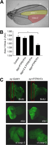
PTP61Fn is a negative regulator of growth and cellular proliferation. (A) The dpp-Gal4 driver was used to overexpress PTP61Fm versus PTP61Fn in the region of the Drosophila wing between longitudinal wing veins LIII and LIV bordered by the posterior cross vein and wing margin (area 1, yellow highlighting). The effect on growth was assessed by comparing the area of this region to the area of a region within the same wing where the dpp-Gal4 driver was not expressed, between wing veins LIV and LV (area 2, red highlighting). (B) Overexpression of PTP61Fn in the Drosophila wing significantly reduced growth (P = 0.02) compared to control (dpp-Gal4/+); however, overexpression of 1 or 2 copies of PTP61Fm had no significant effect. Results are means ± standard errors of the means (SEM) for n = 6 per genotype. (C) Immunostaining of Drosophila third-instar larval eye imaginal discs overexpressing PTP61Fn under ey-Gal4 and control flies (ey-Gal4/+), showing that PTP61Fn overexpression reduces cell proliferation as assessed by BrDu incorporation, without affecting cell differentiation (anti-elav) or apoptosis (anti-cleaved caspase-3 [cl.casp-3]).
Fig 4
Fig 4
PTP61FΔ mutant flies have a decreased life span. The life span of PTP61FΔ (arm-Gal4/+; PTP61FΔ) female flies is significantly reduced compared to that of w1118 female controls (P < 0.0001). Constitutive overexpression of PTP61Fm and PTP61Fn in the PTP61FΔ background using arm-Gal4 (arm>PTP61Fm+n; PTP61FΔ) is sufficient to rescue the life span defect of PTP61FΔ mutants; however, overexpression of PTP61Fn alone using arm-Gal4 (arm>PTP61Fn; PTP61FΔ) does not rescue the life span defect. n = 30 to 50. Statistical analysis was performed using the log rank test.
Fig 5
Fig 5
PTP61FΔ mutant flies have reduced female fecundity due to increased apoptosis in developing egg chambers. (A) Fecundity assays were performed using single-pair matings of females of the indicated genotype. Fecundity assays separated by dashed lines were performed independently of each other. PTP61FΔ females have significantly decreased fecundity compared to w1118 female controls (P = 9 × 10⁻⁵). When overexpressed together using arm-Gal4, PTP61Fm and PTP61Fn (arm>PTP61Fm+n) significantly rescued the fecundity defect of the PTP61FΔ mutant. PTP61Fm+n restored fecundity to a level not significantly different from that of control PTP61FΔ heterozygotes overexpressing PTP61Fm+n (P = 0.07). Overexpression of PTP61Fm or PTP61Fn individually in the PTP61FΔ mutant background resulted in only a partial rescue of fecundity, which was statistically different from what was observed in controls overexpressing the PTPs in the heterozygote background. Results are means ± standard errors of the means (SEM) for n = 8 to 10. **, P < 0.01; ***, P < 0.001. (B) Ovaries from w1118 and PTP61FΔ females were visualized under bright field and also immunostained with anti-vasa antibodies to identify germ line cells in the ovary. Ovaries from PTP61FΔ mutant females lack later-stage egg chambers (post-stage 10) compared to controls. Results shown are representative of 3 experiments. (C) TUNEL staining of egg chambers (magnification, ×100) from PTP61FΔ homozygous and heterozygous female ovaries demonstrated that in the PTP61FΔ mutants there was an approximate 8-fold increase in the number of apoptotic pre-stage 10 egg chambers compared to PTP61FΔ heterozygotes. Results are means ± SEM for n = 8. **, P < 0.01.
Fig 6
Fig 6
Heightened STAT92E signaling contributes to the female fecundity defect in PTP61FΔ mutant flies. (A) Protein extracts from ovarian tissue were immunoblotted with antibodies against the phosphorylated and activated forms of STAT (p-STAT92E) and the IR (p-IR). p-IR is increased in the ovaries of PTP61FΔ homozygous mutants. p-STAT92E is increased in both PTP61FΔ homozygotes and heterozygotes. Results shown are representative of at least 2 or 3 independent experiments. (B and C) Fecundity assays were performed using single-pair matings of females of the indicated genotype with w1118 males. Results are means ± SEM; n = 8 to 10. **, P < 0.01. (B) Reducing PI3K signaling in the PTP61FΔ mutant background by overexpressing dominant negative dp110 (dp110DN) using the arm-Gal4 driver did not restore fecundity (P = 0.36). Control, arm>dp110DN; PTP61FΔ/+. (C) Suppression of STAT92E in the PTP61FΔ homozygous background using in vivo RNAi (STATRNAi) resulted in a partial rescue of fecundity. Control, arm>STATRNAi; PTP61FΔ/+.

Similar articles

Cited by

References

    1. Tiganis T, Bennett AM. 2007. Protein tyrosine phosphatase function: the substrate perspective. Biochem. J. 402:1–15 - PMC - PubMed
    1. Tonks NK. 2006. Protein tyrosine phosphatases: from genes, to function, to disease. Nat. Rev. Mol. Cell Biol. 7:833–846 - PubMed
    1. Andersen JN, Mortensen OH, Peters GH, Drake PG, Iversen LF, Olsen OH, Jansen PG, Andersen HS, Tonks NK, Moller NP. 2001. Structural and evolutionary relationships among protein tyrosine phosphatase domains. Mol. Cell. Biol. 21:7117–7136 - PMC - PubMed
    1. Iversen LF, Moller KB, Pedersen AK, Peters GH, Petersen AS, Andersen HS, Branner S, Mortensen SB, Moller NP. 2002. Structure determination of T cell protein tyrosine phosphatase. J. Biol. Chem. 277:19982–19990 - PubMed
    1. Salmeen A, Andersen JN, Myers MP, Tonks NK, Barford D. 2000. Molecular basis for recognition and dephosphorylation of the activation segment of the insulin receptor by protein tyrosine phosphatase 1B. Mol. Cell 6:1401–1412 - PubMed

Publication types

MeSH terms

Substances

LinkOut - more resources