. 2013 Jan-Feb;4(1):39-51.
doi: 10.4161/trns.22874.

Mediator MED23 regulates basal transcription in vivo via an interaction with P-TEFb

Affiliations

Mediator MED23 regulates basal transcription in vivo via an interaction with P-TEFb

Wei Wang et al. Transcription. 2013 Jan-Feb.

Abstract

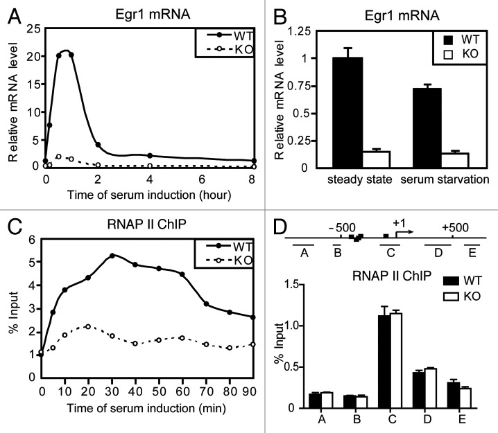
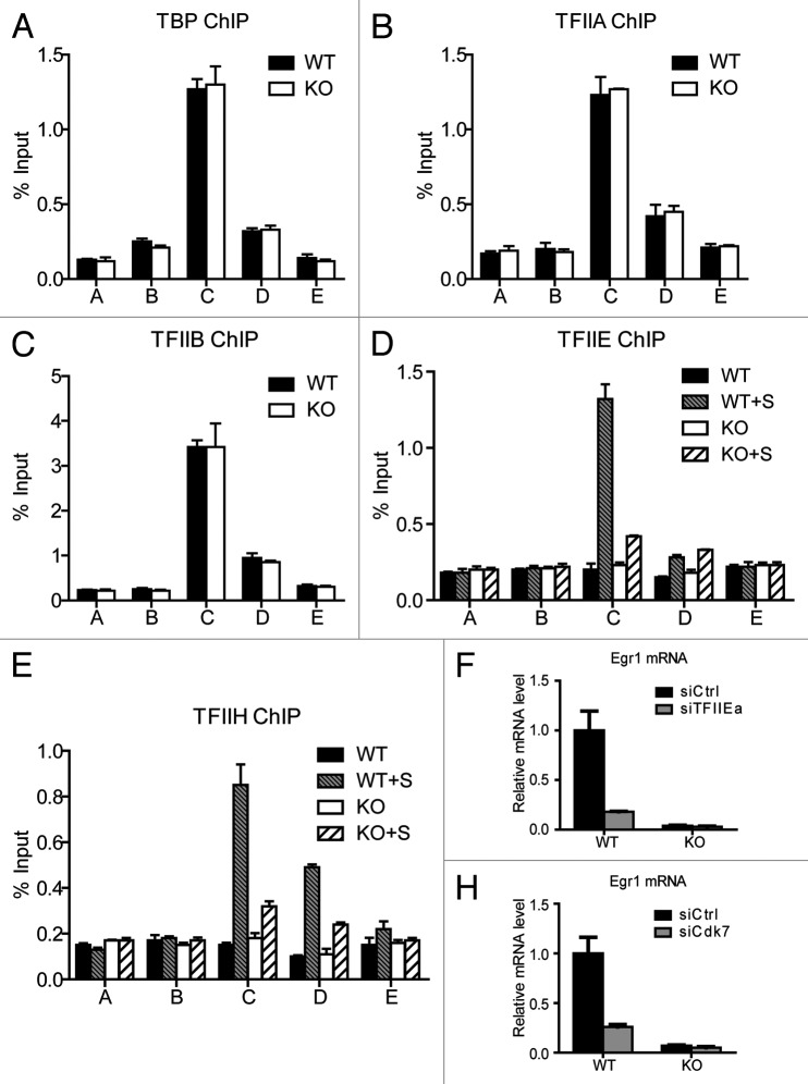
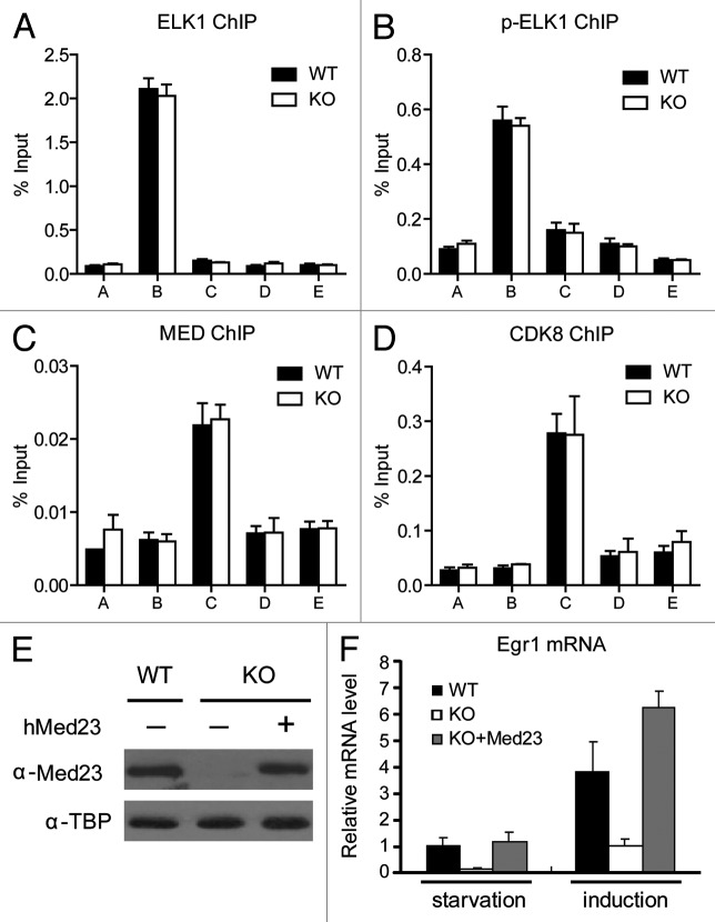
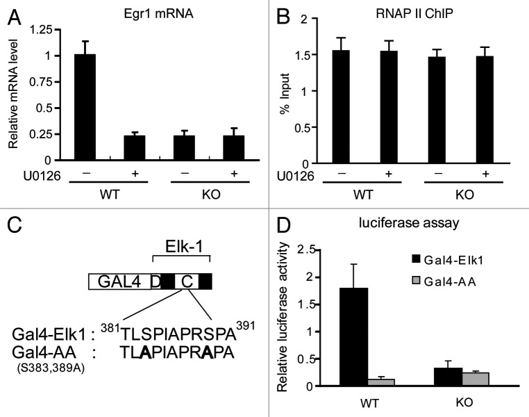
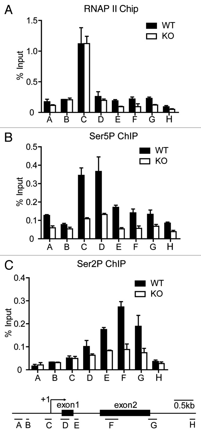
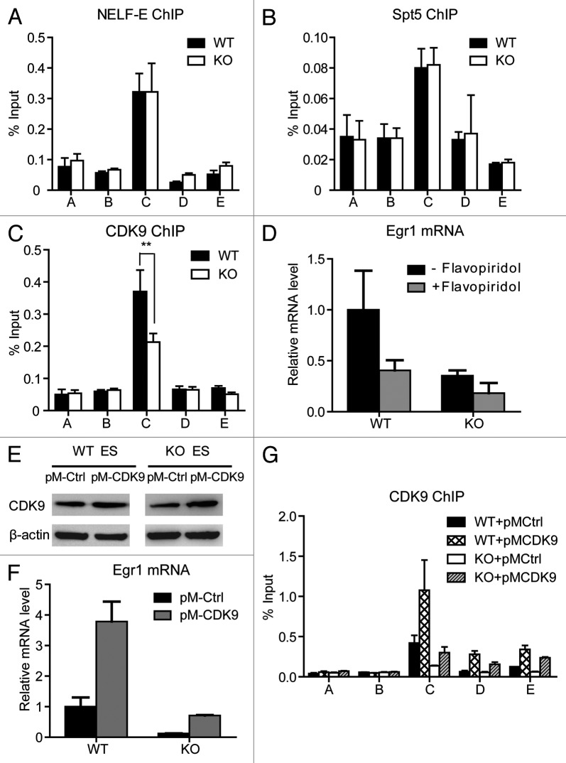
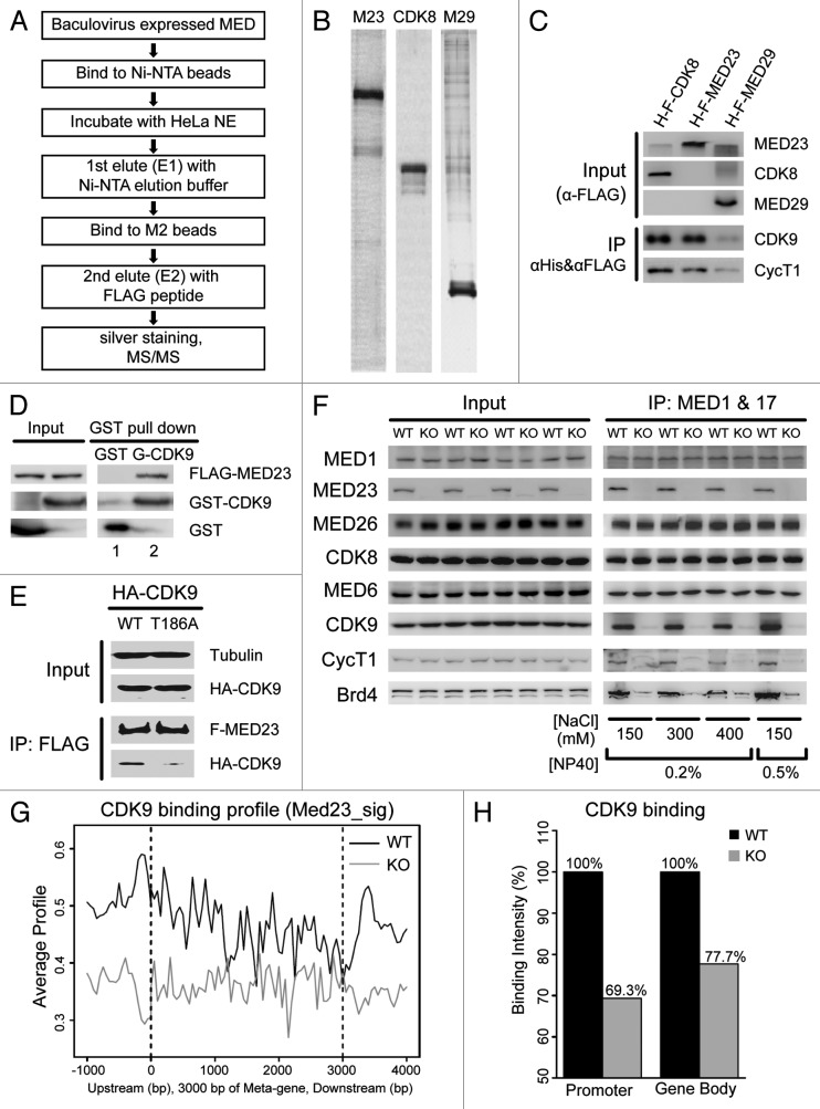
The Mediator is a multi-subunit complex that transduces regulatory information from transcription regulators to the RNA polymerase II apparatus. Growing evidence suggests that Mediator plays roles in multiple stages of eukaryotic transcription, including elongation. However, the detailed mechanism by which Mediator regulates elongation remains elusive. In this study, we demonstrate that Mediator MED23 subunit controls a basal level of transcription by recruiting elongation factor P-TEFb, via an interaction with its CDK9 subunit. The mRNA level of Egr1, a MED23-controlled model gene, is reduced 4-5 fold in Med23 (-/-) ES cells under an unstimulated condition, but Med23-deficiency does not alter the occupancies of RNAP II, GTFs, Mediator complex, or activator ELK1 at the Egr1 promoter. Instead, Med23 depletion results in a significant decrease in P-TEFb and RNAP II (Ser2P) binding at the coding region, but no changes for several other elongation regulators, such as DSIF and NELF. ChIP-seq revealed that Med23-deficiency partially reduced the P-TEFb occupancy at a set of MED23-regulated gene promoters. Further, we demonstrate that MED23 interacts with CDK9 in vivo and in vitro. Collectively, these results provide the mechanistic insight into how Mediator promotes RNAP II into transcription elongation.

Keywords: Egr1; Mediator MED23; P-TEFb; basal transcription; elongation.


Figures

None
Figure 1.Med23 modulates Egr1 basal transcription in vivo without changing RNAP II recruitment. (A) Time-course analysis of Egr1 expression after serum addition to serum-starved WT and KO ES cells. The expression was normalized to EF2 mRNAs, and the normalized value in serum-starved WT cells at time 0 was defined as 1. (B) Real-time PCR analysis of the Egr1 expression at steady-state and under serum starvation. The expression was normalized to EF2 mRNA expression, and the normalized value in WT cells at steady-state was defined as 1. In this and subsequent figures, results are expressed as the mean ± s.d. of n = 3 independent experiments. (C) Time-course ChIP analysis of RNAP II binding to the Egr1 promoter after serum addition. Values from real-time PCR were normalized to the percentage of input chromatin. (D) ChIP analysis of RNAP II binding to the Egr1 gene using primers for the indicated fragments. The scale is in base pairs.
None
Figure 2.Med23 deficiency does not alter the occupancies of GTFs at the Egr1 promoter under an unstimulated condition. ChIP experiments were performed using antibodies against TBP (A), TFIIA (B), TFIIB (C), TFIIE (D), and TFIIH (E). The precipitated DNA was analyzed by real-time PCR with primers described in Figure 1D. WT+S/KO+S: 30 min after serum addition. (E) and (F), Egr1 mRNA levels in siCtrl and siTFIIEa/siCdk7 cells. The expression was normalized to EF2 mRNA expression, and the normalized value in siCtrl WT cells was defined as 1.
None
Figure 3. ELK1 and Mediator occupancies at the Egr1 promoter are not affected by the presence or absence of Med23. ChIP experiments were performed using antibodies against ELK1 (A), p-ELK1 (B), the Mediator Complex (Med1 and Med17) (C), and CDK8 (D). (E) KO MEF cells were infected with retroviruses encoding hMED23 and subsequently selected for hygromycin resistance. Western blotting was used to detect the protein levels of MED23 in WT, KO, and KO+Med23 cells. The anti-TBP blot was included as an internal control. (F) Real-time PCR analysis of the Egr1 expression in states of serum starvation and serum induction (30 min). The expression was normalized to EF2 mRNA expression, and the normalized value in serum-starved WT cells was defined as 1.
None
Figure 4. The interaction between ELK1 and Med23 is necessary for maintaining Egr1 basal transcription. (A) The MEK1/2 inhibitor U0126 was added to the culture medium for 30 min, and total RNA samples were harvested and analyzed by real-time PCR. (B) WT and KO ES cells were treated or not treated by U0126 for 30 min. ChIP assays were performed using RNAP II antibody. (C) The structures of Gal4-Elk1 and Gal4-AA (S383,389A) are schematically depicted. The sequences are shown below. D: D-box domain; C: C-box domain. (D) 293T cells were transfected with a 5 × Gal-E1B-TATA-luciferase reporter construct and a plasmid encoding the Gal4-Elk1 activation domain or Gal4-Elk1 mutant, with a MEKK expression plasmid. Firefly luciferase activity was normalized to Renilla luciferase activity.
None
Figure 5. MED23 is required for RNAP II CTD phosphorylation and RNAP II elongation but not RNAP II recruitment. ChIP experiments were performed using antibodies against total RNAP II (A), phosphor-Ser5 CTD (Ser5P) (B), and phospho-Ser2 (Ser2P) (C) at the Egr1 locus using primers as indicated below.
None
Figure 6. MED23 is partially required for the recruitment of CDK9 to the Egr1 promoter. ChIP experiments were performed using antibodies against NELF-E (A), Spt5 (B), and CDK9 (C) at the Egr1 locus, as described in Figure 1. (D) Egr1 mRNA levels were measured by real-time PCR in WT and KO ES cells with or without 1 h of flavopiridol (250 nM) pre-treatment. The expression was normalized to the EF2 mRNA expression. (E) WT and KO ES cells were infected with retroviruses encoding CDK9. Stable cell lines were selected for hygromycin resistance, and the CDK9 protein level was analyzed by western blotting. β-actin was used as an internal control. (F) Egr1 mRNA levels were measured by real-time PCR in control- and CDK9-overexpressed WT and KO ES cells. (G) ChIP analysis using an antibody against CDK9 at the Egr1 locus in control- and CDK9-overexpressed WT and KO ES cells. The mean of at least three separate experiments is shown, and the standard deviation is indicated. Student’s t test, **p < 0.001.
None
Figure 7. MED23 interacts with CDK9 in vitro and in vivo. (A) Scheme for tandem affinity purification and MS/MS to identify Mediator subunit binding proteins. (B) Baculovirus-expressed His-Flag-MED23 (M23), His-Flag-CDK8 (CDK8) and His-Flag-MED29 (M29) were purified by Ni-NTA beads and then FLAG M2 beads sequentially, as described in (A). The proteins eluted from FLAG M2 beads were analyzed by SDS- PAGE followed by silver staining. (C) The proteins eluted from FLAG M2 beads were analyzed by SDS- PAGE followed by immunoblotting with antibodies against CDK9 and Cyclin T1. (D) Soluble His-Flag-MED23 was incubated at 4 degree overnight with immobilized GST or GST-CDK9. After washing, the bound proteins were eluted by boiling and immunoblotted with the indicated antibodies. (E) Flag-Med23 plasmid was co-transfected with HA-cdk9 or its mutant HA-cdk9 (T186A) into 293T cells. Whole-cell extracts were used for immunoprecipitation with the anti-FLAG M2 beads, followed by immunoblotting using antibodies against Tubulin, FLAG or HA. (F) Nuclear extracts prepared from wild type (WT) or Med23−/− (KO) mouse embryonic stem cells were subjected to co-IP with MED1 and MED17 antibodies under various stringent wash conditions as indicated. The immunoprecipitated proteins were detected with indicated antibodies by immunoblotting. (G) ChIP-seq analysis of CDK9 enrichment on the set of genes whose expression level are regulated by MED23, in WT (black) and KO (gray) ES cells. All these genes were normalized to 3 kb for Mata-gene, with 1 kb extended upstream from TSS, and 1 kb downstream from TTS for analyzing average profile in 50 bp bins. (H) Quantitation of CDK9 binding intensity on promoter region (1 kb upstream from TSS) and gene body region (from TSS to TTS). Both are normalized to the values derived from the WT ES cells.

Similar articles

Cited by

References

    1. Ptashne M, Gann A. Transcriptional activation by recruitment. Nature. 1997;386:569–77. doi: 10.1038/386569a0. - DOI - PubMed
    1. Roeder RG. Transcriptional regulation and the role of diverse coactivators in animal cells. FEBS Lett. 2005;579:909–15. doi: 10.1016/j.febslet.2004.12.007. - DOI - PubMed
    1. Core LJ, Waterfall JJ, Lis JT. Nascent RNA sequencing reveals widespread pausing and divergent initiation at human promoters. Science. 2008;322:1845–8. doi: 10.1126/science.1162228. - DOI - PMC - PubMed
    1. Margaritis T, Holstege FC. Poised RNA polymerase II gives pause for thought. Cell. 2008;133:581–4. doi: 10.1016/j.cell.2008.04.027. - DOI - PubMed
    1. Gilmour DS, Lis JT. RNA polymerase II interacts with the promoter region of the noninduced hsp70 gene in Drosophila melanogaster cells. Mol Cell Biol. 1986;6:3984–9. - PMC - PubMed

Publication types

MeSH terms

LinkOut - more resources