

Occlusal effects on longitudinal bone alterations of the temporomandibular joint
- PMID: 23340211
- PMCID: PMC6728563
- DOI: 10.1177/0022034512473482
Occlusal effects on longitudinal bone alterations of the temporomandibular joint
Abstract
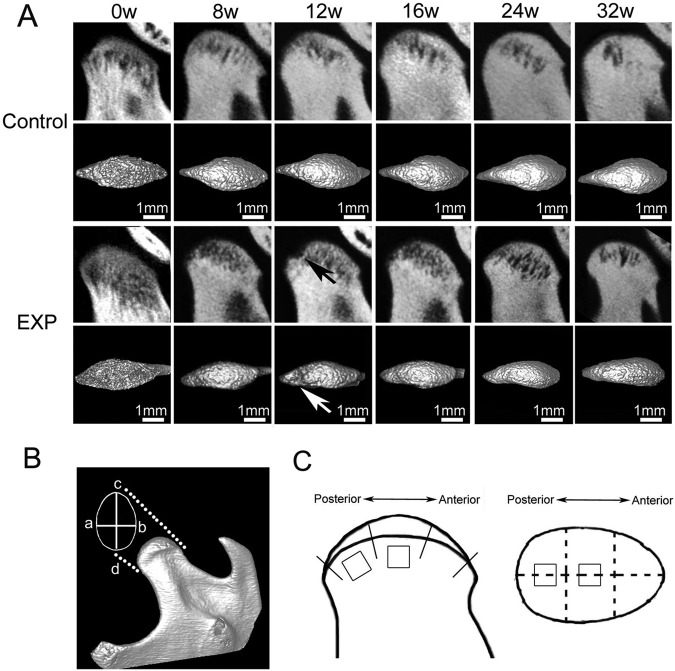
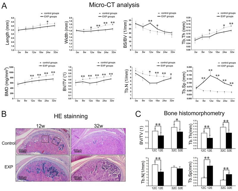
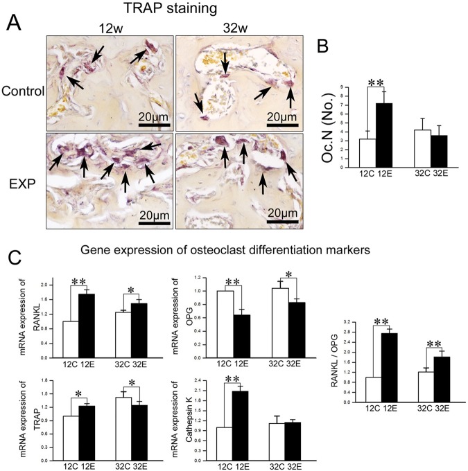
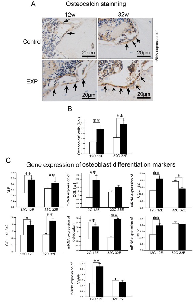
The pathological changes of subchondral bone during osteoarthritis (OA) development in the temporomandibular joint (TMJ) are poorly understood. In the present study, we investigated the longitudinal alterations of subchondral bone using a rat TMJ-OA model developed in our laboratory. Changes in bone mass were examined by micro-CT, and changes in osteoblast and osteoclast activities were analyzed by real-time PCR, immunohistochemistry, and TRAP staining. Subchondral bone loss was detected from 8 weeks after dental occlusion alteration and reached the maximum at 12 weeks, followed by a repair phase until 32 weeks. Although bone mass increased at late stages, poor mechanical structure and lower bone mineral density (BMD) were found in these rats. The numbers of TRAP-positive cells were increased at 12 weeks, while the numbers of osteocalcin-expressing cells were increased at both 12 and 32 weeks. Levels of mRNA expression of TRAP and cathepsin K were increased at 12 weeks, while levels of ALP and osteocalcin were increased at both 12 and 32 weeks. These findings demonstrated that there is an active bone remodeling in subchondral bone in TMJs in response to alteration in occlusion, although new bone was formed with lower BMD and poor mechanical properties.
Conflict of interest statement
The authors declare no potential conflicts of interest with respect to the authorship and/or publication of this article.
Figures
References
-
- Bailey AJ, Buckland-Wright C, Metz D. (2001). The role of bone in osteoarthritis. Age Ageing 30:374-378. - PubMed
-
- Bailey AJ, Sims TJ, Knott L. (2002). Phenotypic expression of osteoblast collagen in osteoarthritic bone: production of type I homotrimer. Int J Biochem Cell Biol 34:176-182. - PubMed
-
- Bouchgua M, Alexander K, Carmel EN, d’Anjou MA, Beauchamp G, Richard H, et al. (2009). Use of routine clinical multimodality imaging in a rabbit model of osteoarthritis—Part II: bone mineral density assessment. Osteoarthritis Cartilage 17:197-204. - PubMed
-
- Chiba K, Ito M, Osaki M, Uetani M, Shindo H. (2011). In vivo structural analysis of subchondral trabecular bone in osteoarthritis of the hip using multi-detector row CT. Osteoarthritis Cartilage 19:180-185. - PubMed
-
- Cowin SC. (2004). Tissue growth and remodeling. Annu Rev Biomed Eng 6:77-107. - PubMed
Publication types
MeSH terms
Substances
Grants and funding
LinkOut - more resources
Full Text Sources
Other Literature Sources
Medical
