Glucocorticoids reduce intracellular calcium concentration and protects neurons against glutamate toxicity
- PMID: 23340218
- PMCID: PMC4208294
- DOI: 10.1016/j.ceca.2012.12.006
Glucocorticoids reduce intracellular calcium concentration and protects neurons against glutamate toxicity
Abstract
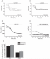
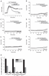
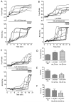
Glucocorticoids are steroid hormones which act through the glucocorticoid receptor. They regulate a wide variety of biological processes. Two glucocorticoids, the naturally occurring corticosterone and chemically produced dexamethasone, have been used to investigate the effect of glucocorticoids on Ca(2+)-signalling in cortical co-cultures of neurons and astrocytes. Dexamethasone and to a lesser degree corticosterone both induced a decrease in cytosolic Ca(2+) concentration in neurons and astrocytes. The effect of both compounds can be blocked by inhibition of the plasmamembrane ATPase, calmodulin and by application of a glucocorticoid receptor antagonist, while inhibition of NMDA receptors or the endoplasmic reticulum calcium pump had no effect. Glucocorticoid treatment further protects against detrimental calcium signalling and cell death by modulating the delayed calcium deregulation in response to glutamate toxicity. At the concentrations used dexamethasone and corticosterone did not show cell toxicity of their own. Thus, these results indicate that dexamethasone and corticosterone might be used for protection of the cells from calcium overload.
Copyright © 2013 Elsevier Ltd. All rights reserved.
Figures


Similar articles
-
Inorganic Polyphosphate Regulates AMPA and NMDA Receptors and Protects Against Glutamate Excitotoxicity via Activation of P2Y Receptors.J Neurosci. 2019 Jul 31;39(31):6038-6048. doi: 10.1523/JNEUROSCI.0314-19.2019. Epub 2019 May 30. J Neurosci. 2019. PMID: 31147524 Free PMC article.
-
Imaging of mitochondrial Ca2+ dynamics in astrocytes using cell-specific mitochondria-targeted GCaMP5G/6s: mitochondrial Ca2+ uptake and cytosolic Ca2+ availability via the endoplasmic reticulum store.Cell Calcium. 2014 Dec;56(6):457-66. doi: 10.1016/j.ceca.2014.09.008. Epub 2014 Sep 30. Cell Calcium. 2014. PMID: 25443655 Free PMC article.
-
Glucocorticoids exacerbate peroxynitrite mediated potentiation of glucose deprivation-induced death of rat primary astrocytes.Brain Res. 2001 Dec 27;923(1-2):163-71. doi: 10.1016/s0006-8993(01)03212-7. Brain Res. 2001. PMID: 11743984
-
The active role of astrocytes in synaptic transmission.Cell Mol Life Sci. 1999 Dec;56(11-12):991-1000. doi: 10.1007/s000180050488. Cell Mol Life Sci. 1999. PMID: 11212330 Free PMC article. Review.
-
In vitro studies of glucocorticoid effects on neurons and astrocytes.Ann N Y Acad Sci. 1994 Nov 30;746:243-58; discussion 258-9, 289-93. doi: 10.1111/j.1749-6632.1994.tb39241.x. Ann N Y Acad Sci. 1994. PMID: 7825881 Review.
Cited by
-
Role of Glia in Stress-Induced Enhancement and Impairment of Memory.Front Integr Neurosci. 2016 Jan 11;9:63. doi: 10.3389/fnint.2015.00063. eCollection 2015. Front Integr Neurosci. 2016. PMID: 26793072 Free PMC article. Review.
-
In vitro modeling of the neurobiological effects of glucocorticoids: A review.Neurobiol Stress. 2023 Feb 23;23:100530. doi: 10.1016/j.ynstr.2023.100530. eCollection 2023 Mar. Neurobiol Stress. 2023. PMID: 36891528 Free PMC article. Review.
-
Improvement of dexamethasone sensitivity by chelation of intracellular Ca2+ in pediatric acute lymphoblastic leukemia cells through the prosurvival kinase ERK1/2 deactivation.Oncotarget. 2017 Apr 18;8(16):27339-27352. doi: 10.18632/oncotarget.16039. Oncotarget. 2017. PMID: 28423696 Free PMC article.
-
Amylin Protein Expression in the Rat Brain and Neuro-2a Cells.Int J Mol Sci. 2022 Apr 14;23(8):4348. doi: 10.3390/ijms23084348. Int J Mol Sci. 2022. PMID: 35457166 Free PMC article.
-
Corticosteroids and perinatal hypoxic-ischemic brain injury.Drug Discov Today. 2018 Oct;23(10):1718-1732. doi: 10.1016/j.drudis.2018.05.019. Epub 2018 May 17. Drug Discov Today. 2018. PMID: 29778695 Free PMC article. Review.
References
-
- Kim M, Lee G, Jung E, Choi K, Oh G, Jeung E. Dexamethasone differentially regulates renal and duodenal calcium-processing genes in calbindin-D9k and D28k knockout mice. Experimental Physiology. 2009;94:138–151. - PubMed
-
- Chen S, Wang X, Zhang X, Wang W, Liu D, Long Z, Dai W, Chen Q, Xu M, Zhou J. High-dose glucocorticoids induce decreases calcium in hypothalamus neurons via plasma membrane Ca2+ pumps. NeuroReport. 2011;22:660–663. - PubMed
-
- Reul JMHM, Bosch J.R.V.d., de ERK. Relative occupation of type 1 and type 2 corticosteroid receptors in rat brain following stress and dexamethasone treatment: functional implications. Journal of Endocrinology. 1987;115:459–467. - PubMed
-
- Beato M, Klug J. Steroid hormone receptors: an update. Human Reproduction Update. 2000;6:225–236. - PubMed
Publication types
MeSH terms
Substances
Grants and funding
LinkOut - more resources
Full Text Sources
Other Literature Sources
Medical
Miscellaneous

