Review
. 2013 Sep 26:249:53-62.
doi: 10.1016/j.neuroscience.2013.01.023. Epub 2013 Jan 20.

Translational developmental studies of stress on brain and behavior: implications for adolescent mental health and illness?

Affiliations
Review

Translational developmental studies of stress on brain and behavior: implications for adolescent mental health and illness?

M Malter Cohen et al. Neuroscience. .

Abstract

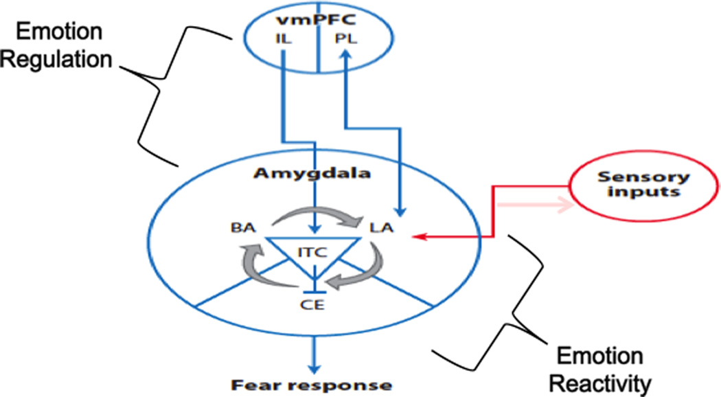
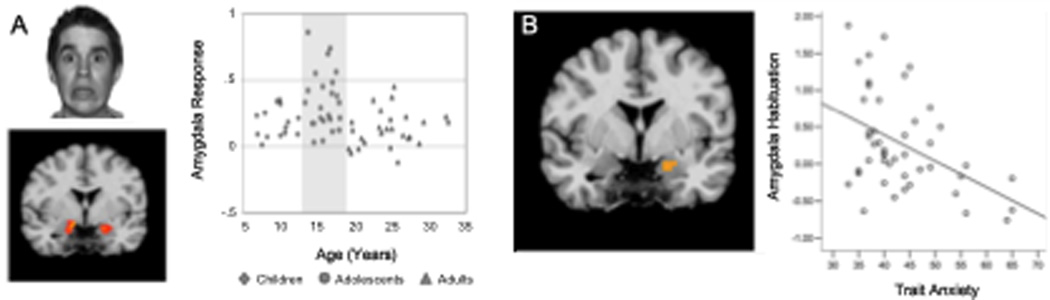
Adolescence is the transition from childhood to adulthood, with onset marked by puberty and the offset by relative independence from parents. Across species, it is a time of incredible change that carries increased risks and rewards. The ability of the individual to respond adequately to the mental, physical and emotional stresses of life during this time is a function of both their early environment and their present state. In this article, we focus on the effects that acute threat and chronic stress have on the brain and behavior in humans and rodents. First, we highlight developmental changes in frontolimbic function as healthy individuals transition into and out of adolescence. Second, we examine genetic factors that may enhance susceptibility to stress in one individual over another using translation from genetic mouse models to human neuroimaging. Third, we examine how the timing and nature of stress varies in its impact on brain and behavior. These findings are discussed in the context of implications for adolescent mental health and illness.

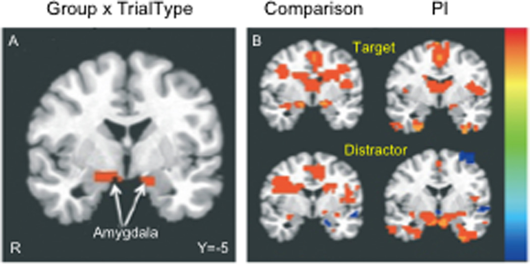
Keywords: BDNF; BDNFmet; MRI; Met; P; PI; SCR; Val; Val66met; adolescence; anxiety; brain-derived neurotrophic factor; emotion regulation; fMRI; fear; functional magnetic resonance imaging; magnetic resonance imaging; methionine; methionine in codon 66 of the BDNF protein; postnatal day; previously institutionalized; skin conductance response; stress; valine; valine-to-methionine substitution at codon 66; ventromedial prefrontal cortex; vmPFC.


Figures

Fig. 1
Fig. 1. Frontoamygdala circuit of Fear
A simplified cartoon of the brain circuitry involved in emotion reactivity and regulation related to threat processing. The amygdala receives multimodal sensory signals that may initiate a fear response, while top-down input from the infralimbic prefrontal cortex to the amygdala can dampen or extinguish fear responses generated there. Abbreviations: BA, basal amygdala; CE, central amygdala; IL, infralimbic prefrontal cortex; ITC, intercalated; LA, lateral amygdala; PL, prefrontal cortex; vmPFC, ventromedial prefrontal cortex. Adapted from Casey et al 2013.
Fig. 2
Fig. 2. Amygdala response to empty threat as a function of age and symptoms of anxiety
(A) Depiction of threat stimulus and location of activation in the amygdala. Middle: Amygdala activity to empty threat (fearful faces) plotted as a function of age. (B) Scatter plot of the correlation between Spielberger trait anxiety scores and habituation (decrease from early to late trials) of amygdala activity for teens and adults (note: anxiety scale was not appropriate for under 13 years) r = −.447, p < 0.001. Adapted from Hare et al., 2008.
Fig. 3
Fig. 3. Developmental variation in fear extinction learning
(B) Extinction learning is attenuated during adolescence in the human as measured by less change in galvanic skin response with repeated presentation of the conditioned stimulus alone during extinction trials. (C) This finding is paralleled in the mouse as measured by less change in freezing behavior. Reproduced with permission from (Pattwell et al 2012).
Fig. 4
Fig. 4. Genetic variation in fear extinction learning and limbic activity
(A) Extinction learning is attenuated in mice with the BNDF Met (M) allele relative to non-Met allele (V) carriers as measured by less change in freezing behavior with repeated presentation of the conditioned stimulus alone during extinction trials. (B) This finding is paralleled in the human as measured by less change in galvanic skin response. (C) Brain activity as indexed by percent change in magnetic resonance (MR) signal during extinction in the ventromedial prefrontal cortex (vmPFC) by genotype (x, y, z = −4, 24, 3), with Met allele carriers having significantly less activity than Val/Val homozygotes (VM < VV is blue), image threshold P < 0.05, corrected. (D) Genotypic differences in left amygdala activity during extinction (x, y, z = −25, 2, −20) in 70 humans, with Met allele carriers having significantly greater activity than Val/Val homozygotes (VM > VV is orange), image threshold P < 0.05, corrected. *P < 0.05. **MM were included in the analysis with VM, but plotted separately to see the dose response. All results are presented as mean +/− SEM. VV, Val/Val; VM, Val/Met; MM, Met/Met. Adapted from Soliman et al 2010.
Fig. 5
Fig. 5. Structural changes in limbic structures with early adversity
(A) Anatomical segmentation of the amygdala. (B) Children institutionalized for more than 15 months had larger amygdala volumes than those institutionalized for less than 15 months, or control children. No differences in hippocampal volume were observed between groups. Adapted from Tottenham et al., 2010.
Fig. 6
Fig. 6. Frontoamygdala activity to distracting emotional stimuli with early adversity
(A) Previously institutionalized (PI) children exhibited greater amygdala activity in response to emotional distractors than their typically reared counterparts, suggesting an inability to suppress emotionally laden irrelevant information. (B) Post-hoc t-tests of activity vs. baseline. Adapted from Tottenham et al., 2011.

References

    1. Akirav I, Maroun M. Ventromedial prefrontal cortex is obligatory for consolidation and reconsolidation of object recognition memory. Cereb Cortex. 2006;16:1759–1765. - PubMed
    1. APA. 2010
    1. Arnsten AF. Development of the cerebral cortex: XIV. Stress impairs prefrontal cortical function. J Am Acad Child Adolesc Psychiatry. 1999;38:220–222. - PubMed
    1. Bland ST, Schmid MJ, Watkins LR, Maier SF. Prefrontal cortex serotonin, stress, and morphine-induced nucleus accumbens dopamine. Neuroreport. 2004;15:2637–2641. - PubMed
    1. Bremner JD, Randall P, Scott TM, Bronen RA, Seibyl JP, Southwick SM, Delaney RC, McCarthy G, Charney DS, Innis RB. MRI-based measurement of hippocampal volume in patients with combat-related posttraumatic stress disorder. Am J Psychiatry. 1995;152:973–981. - PMC - PubMed

Publication types