. 2013 Mar;27(3):511-23.
doi: 10.1210/me.2012-1307. Epub 2013 Jan 22.

IGF-I signaling is essential for FSH stimulation of AKT and steroidogenic genes in granulosa cells

Affiliations

IGF-I signaling is essential for FSH stimulation of AKT and steroidogenic genes in granulosa cells

Ping Zhou et al. Mol Endocrinol. 2013 Mar.

Abstract

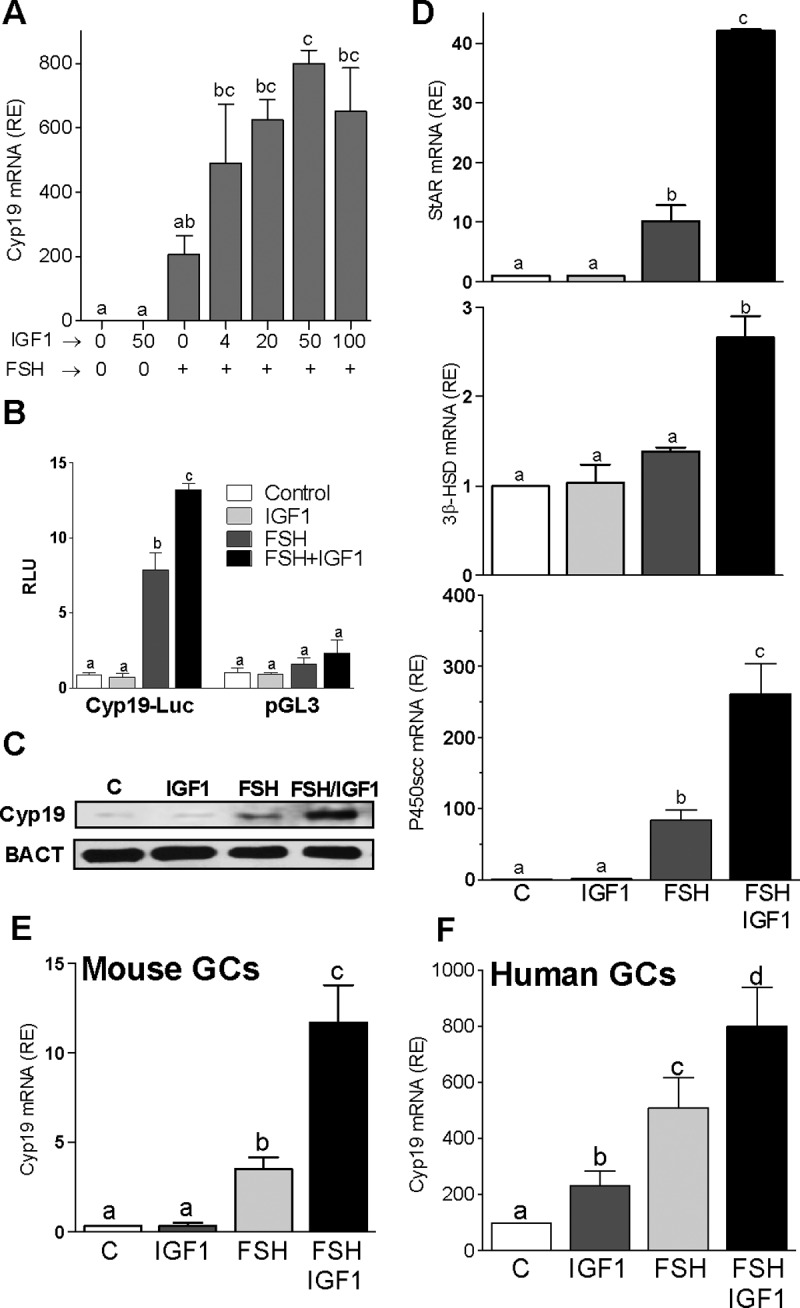
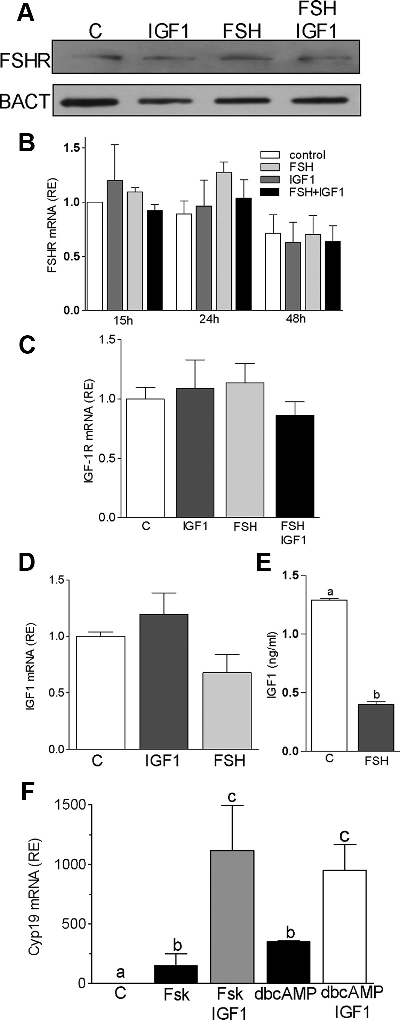
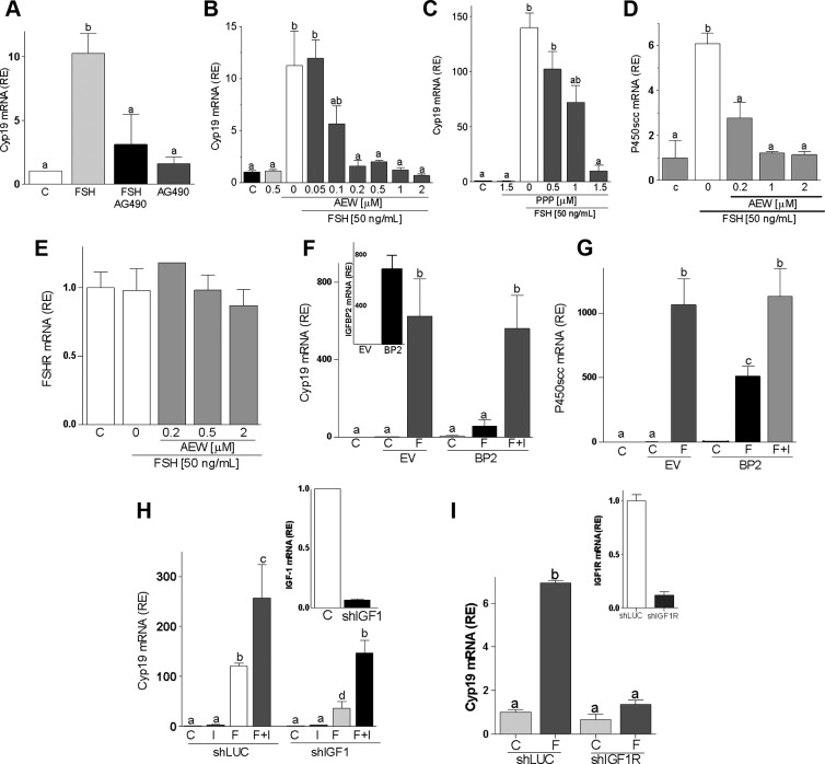
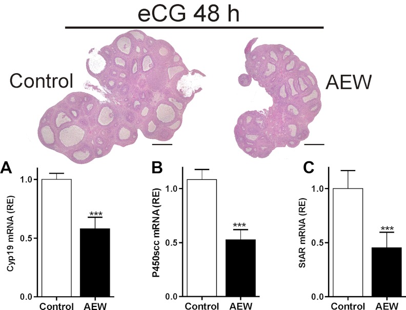
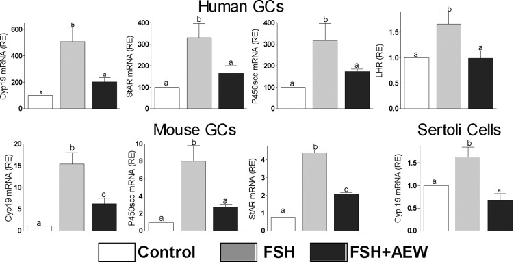
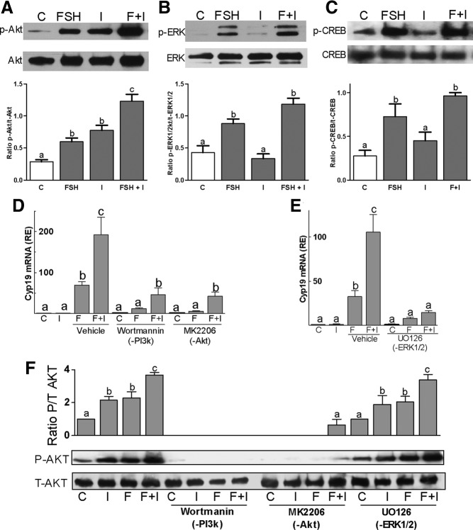
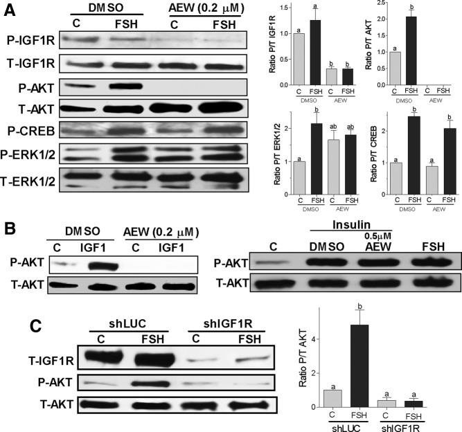
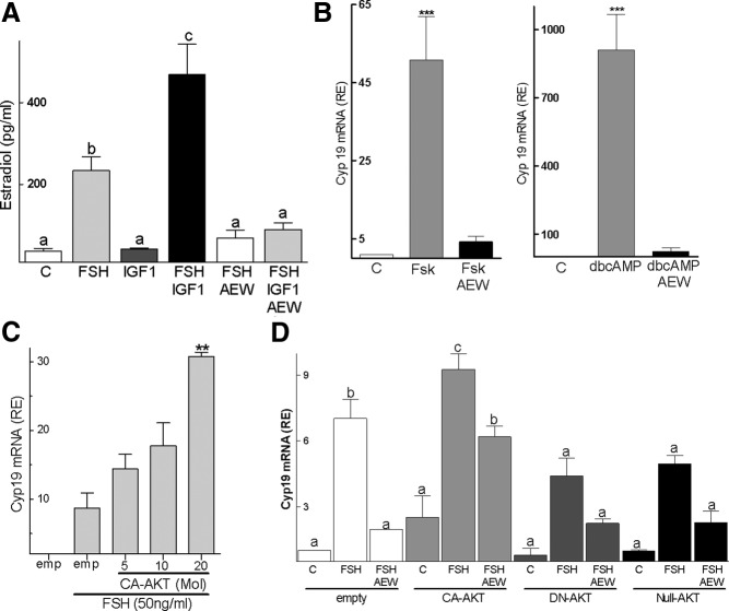
FSH and IGF-I synergistically stimulate gonadal steroid production; conversely, silencing the FSH or the IGF-I genes leads to infertility and hypogonadism. To determine the molecular link between these hormones, we examined the signaling cross talk downstream of their receptors. In human and rodent granulosa cells (GCs), IGF-I potentiated the stimulatory effects of FSH and cAMP on the expression of steroidogenic genes. In contrast, inhibition of IGF-I receptor (IGF-IR) activity or expression using pharmacological, genetic, or biochemical approaches prevented the FSH- and cAMP-induced expression of steroidogenic genes and estradiol production. In vivo experiments demonstrated that IGF-IR inactivation reduces the stimulation of steroidogenic genes and follicle growth by gonadotropins. FSH or IGF-I alone stimulated protein kinase B (PKB), which is also known as AKT and in combination synergistically increased AKT phosphorylation. Remarkably, blocking IGF-IR expression or activity decreased AKT basal activity and abolished AKT activation by FSH. In GCs lacking IGF-IR activity, FSH stimulation of Cyp19 expression was rescued by overexpression of constitutively active AKT. Our findings demonstrate, for the first time, that in human, mouse, and rat GCs, the well-known stimulatory effect of FSH on Cyp19 and AKT depends on IGF-I and on the expression and activation of the IGF-IR.


Figures

Figure 1.
Figure 1.
IGF-I synergizes with FSH to stimulate steroidogenic genes. A–F, Rat (A and D), mouse (E), or human (F) primary GCs were cultured in the presence of 50 ng/ml FSH with increasing concentrations (A) or 50 ng/ml (D–F) of IGF-I. Relative mRNA levels for Cyp19 (A, E, and F) and StAR, 3β-HSD, and P450scc (D) expression are shown. B, Rat GCs were transfected with a Cyp19 promoter reporter construct or an empty vector (pGL3) and treated with 50 ng/ml FSH and/or 50 ng/ml IGF-I. C, GCs were treated with 50 ng/ml FSH and/or 50 ng/ml IGF-I for 48 hours, and Cyp19 protein and β-actin (BACT) levels were determined by Western blot. Each experiment was repeated at least three times. Columns represent the mean ± SEM; n = 3. Columns with different letters differ significantly (P < .05). RE, relative expression.
Figure 2.
Figure 2.
IGF-I does not affect FSHR expression in GCs in culture. Rat GCs were cultured in the presence of FSH (50 ng/ml) and/or IGF-I (50 ng/ml) for 48 hours. Cells were harvested for protein or RNA isolation, and the culture medium was collected for IGF-I determination. A, FSHR and β-actin (BACT) protein levels were quantified by using Western blot. B, Rat GCs were cultured for 15, 24, or 48 hours before FSHR mRNA quantification. C and D, IGF-IR (C) and IGF-I (D) mRNA expression levels after treatment for 48 hours as indicated in the graph. E, IGF-I levels were quantified by ELISA after 48 hours treatment with FSH. F, Rat GCs were cultured in the presence of forskolin (Fsk, 5μM) or dbcAMP (2mM) with or without IGF-I (50ng/ml) for 48 hours before Cyp19 mRNA quantification. Columns with different letters differ significantly (P < .05). Error bars are ±SEM; n = 3.
Figure 3.
Figure 3.
Inhibition of IGF-IR activation blocks FSH-induced differentiation. Rat GCs were cultured with FSH plus AG490 (25μM, panel A) or different concentrations of AEW (panels B, D, and E) or PPP (panel C). mRNA levels for Cyp19 (panels A–C), P450scc (panel D), or FSHR (panel E) were determined after 48 hours of treatment. Panels F and G, GCs transfected with a plasmid expressing IGFBP2 (BP2) or the empty vector (EV) were treated with FSH (50 ng/ml) or vehicle for 48 hours before Cyp19 and P450scc mRNA level quantification. The inset in panel F shows IGFBP2 expression levels in cells transfected with EV or BP2. Panels H and I, GCs were infected with a lentivirus carrying a control shRNA (shLUC or C), anti–IGF-I shRNA (shIGF-I), or anti–IGF-IR shRNA (shIGF-IR). At 24 hours after infection, cells were treated with FSH (50 ng/ml) and/or IGF-I (50 ng/ml), and Cyp19 mRNA levels were quantified 48 hours later. The inset shows IGF-I (panel H) or IGF-IR (panel I) mRNA levels in cells infected with shLUC, shIGF-I, or shIGF-IR. Each experiment was performed three times, and mean and SEM are shown. Columns with different letters differ significantly (a-b and b-c P < .05; a-c P < .01).
Figure 4.
Figure 4.
In vivo IGF-IR inhibition blocks FSH-induced follicle growth. AEW (50 μg per ovary) or vehicle (control) was injected into the ovarian bursa of immature rats 2 hours before the administration of 10 IU eCG, and 48 hours later, one ovary was processed for hematoxylin and eosin staining and the other was used to isolate total RNA for mRNA quantification. A, Hhistology of a representative ovary (bar, 0.5 mm). B, Mean and SEM of relative mRNA levels. *** P < .01 vs control. Three animals were included in each group.
Figure 5.
Figure 5.
FSH reliance on IGF-IR activation is species and cell-type independent. Human or mouse GCs and Sertoli cells were cultured in presence of FSH (50 ng/ml) with or without AEW (0.5μM) for 48 hours before the quantification of Cyp19, P450scc, StAR, and LHR mRNA levels (n = 4). Controls (C) received vehicle. Columns with different letters differ significantly (P < .05).
Figure 6.
Figure 6.
FSH and IGF-I signaling cross talk. Rat primary GCs were treated with FSH (50 ng/ml) and/or IGF-I (50 ng/ml) for 1 hour. A–C, Phosphorylated and total forms of AKT (panel A), ERK1/2 (panel B), and CREB (panel C) levels were determined by Western blot. The bar graphs under each blot show the mean ± SEM of the ratio of phosphorylated to total protein of three or more experiments. Panels D and E, Rat GCs were pretreated with wortmannin (100nM), an inhibitor of PI3K; MK2206 (1μM), an inhibitor of AKT; or UO126 (5μM), which inhibits ERK1/2, for 1 hour followed by treatment with FSH and/or IGF-I for 48 hours. Cyp19 levels were quantified by real-time PCR. Panel F, GCs were pretreated for 1 hour with wortmannin, MK2206, or UO126 followed by 1 hour treatment with vehicle or FSH and/or IGF-I before protein isolation. Phosphorylated and total AKT protein levels were evaluated by Western blot. Each experiment was repeated 6 times. Columns with different letters differ significantly (a-b and b-c P < .05; a-c P < .01). Abbreviations: C, control; F, FSH; I, IGF-I; p, phospho.
Figure 7.
Figure 7.
IGF-IR expression and activity are obligatory for FSH activation of AKT. Panel A, Rat GCs were pretreated with vehicle (DMSO) or with AEW (0.2μM) for 1 hour followed by treatment with FSH (50 ng/ml) or buffer (control [C]) for 1 hour. Protein extracts were subjected to Western blot for phosphorylated (P-) IGF-IR, AKT, ERK1/2, and CREB. Total (T-) IGF-IR, AKT, and ERK were used as a loading control. The graphs show the ratio of phosphorylated to total (P/T) for each band as the mean ± SEM of the densitometry quantification of three separated experiments. Panel B, GCs were pretreated with vehicle (DMSO) or AEW (0.2μM or 0.5μM) for 30 minutes before the addition of IGF-I (20 ng/mL) or insulin (20 ng/mL). Controls (C) were treated with buffer. Total and phospho-AKT were determined 1 hour later. This experiment was performed three times with identical results. Panel C, GCs were infected with a lentivirus carrying a control shRNA (shLUC) or anti–IGF-IR shRNAs (shIGF-IR). At 24 hours after infection, cells were treated with FSH (50 ng/ml) for 1 hour and total IGF-IR and phosphorylated and total AKT levels were measured by Western blot. On the right, the ratio of phosphorylated to total AKT is shown as the mean ± SEM of three different experiments. Columns with different letters differ significantly (ab P < .05; a P < .01).
Figure 8.
Figure 8.
CA-AKT overcomes the inhibitory effect of AEW. Panel A, Cells were left untreated or treated with FSH (50 ng/ml) and/or IGF-I (50 ng/ml) for 48 hours. Androstenedione was added to the medium at a final concentration of 50nM for the last 4 hours of culture. The concentration of estradiol in the medium was measured by ELISA. Panel B, Rat GCs were pretreated with AEW (0.5μM) for 1 hour before the addition of forskolin (Fsk, 5μM) or dbcAMP (2mM), and 48 hours later, Cyp19 mRNA levels were determined. *** P < .001 vs control (C) and AEW (n = 4). Panel C, Rat GCs were infected with an empty virus (emp) or with a virus that expresses CA-AKT at an increasing multiplicity of infection, and 24 hours later, vehicle or FSH was added to the medium. Cyp19 expression was quantified 48 hours after the initiation of FSH treatment. ** P < .01 vs empty (n = 3). Panel D, Rat GCs were infected with lentiviruses expressing CA-AKT, DN-AKT, or null-AKT. Vehicle (control [C]), FSH, and/or AEW were added to the medium 24 hours later. Cyp19 expression was quantified 48 hours after the initiation of FSH treatment. Columns represent the mean ± SEM (n = 4). Columns with different letters differ significantly (a-b and b-c, P < .05). RE, relative expression.

Similar articles

Cited by

References

    1. Ohlsson C, Mohan S, Sjogren K, et al. The role of liver-derived insulin-like growth factor-I. Endocr Rev. 2009;30:494–535 - PMC - PubMed
    1. Baker J, Hardy MP, Zhou J, et al. Effects of an Igf1 gene null mutation on mouse reproduction. Mol Endocrinol. 1996;10:903–918 - PubMed
    1. Daftary SS, Gore AC. IGF-1 in the brain as a regulator of reproductive neuroendocrine function. Exp Biol Med. 2005;230:292–306 - PubMed
    1. Hikake T, Hayashi S, Iguchi T, Sato T. The role of IGF1 on the differentiation of prolactin secreting cells in the mouse anterior pituitary. J Endocrinol. 2009;203:231–240 - PubMed
    1. Srivastava VK, Hiney JK, Dees WL. Hypothalamic actions and interactions of alcohol and IGF-1 on the expression of glial receptor protein tyrosine phosphatase-β during female pubertal development. Alcohol Clin Exp Res. 2011;35:1812–1821 - PMC - PubMed

Publication types

MeSH terms

Substances