Submillisievert median radiation dose for coronary angiography with a second-generation 320-detector row CT scanner in 107 consecutive patients
- PMID: 23340461
- PMCID: PMC3606544
- DOI: 10.1148/radiol.13122621
Submillisievert median radiation dose for coronary angiography with a second-generation 320-detector row CT scanner in 107 consecutive patients
Abstract
Purpose: To (a) use a new second-generation wide-volume 320-detector row computed tomographic (CT) scanner to explore optimization of radiation exposure in coronary CT angiography in an unselected and consecutive cohort of patients referred for clinical purposes and (b) compare estimated radiation exposure and image quality with that from a cohort of similar patients who underwent imaging with a previous first-generation CT system.
Materials and methods: The study was approved by the institutional review board, and all subjects provided written consent. Coronary CT angiography was performed in 107 consecutive patients with a new second-generation 320-detector row unit. Estimated radiation exposure and image quality were compared with those from 100 consecutive patients who underwent imaging with a previous first-generation scanner. Effective radiation dose was estimated by multiplying the dose-length product by an effective dose conversion factor of 0.014 mSv/mGy ⋅ cm and reported with size-specific dose estimates (SSDEs). Image quality was evaluated by two independent readers.
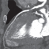
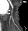
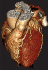
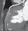
Results: The mean age of the 107 patients was 55.4 years ± 12.0 (standard deviation); 57 patients (53.3%) were men. The median body mass index was 27.3 kg/m(2) (range, 18.1-47.2 kg/m(2)); however, 71 patients (66.4%) were overweight, obese, or morbidly obese. A tube potential of 100 kV was used in 97 patients (90.6%), single-volume acquisition was used in 104 (97.2%), and prospective electrocardiographic gating was used in 106 (99.1%). The mean heart rate was 57.1 beats per minute ± 11.2 (range, 34-96 beats per minute), which enabled single-heartbeat scans in 100 patients (93.4%). The median radiation dose was 0.93 mSv (interquartile range [IQR], 0.58-1.74 mSv) with the second-generation unit and 2.67 mSv (IQR, 1.68-4.00 mSv) with the first-generation unit (P < .0001). The median SSDE was 6.0 mGy (IQR, 4.1-10.0 mGy) with the second-generation unit and 13.2 mGy (IQR, 10.2-18.6 mGy) with the first-generation unit (P < .0001). Overall, the radiation dose was less than 0.5 mSv for 23 of the 107 CT angiography examinations (21.5%), less than 1 mSv for 58 (54.2%), and less than 4 mSv for 103 (96.3%). All studies were of diagnostic quality, with most having excellent image quality. Three of four image quality indexes were significantly better with the second-generation unit compared with the first-generation unit.
Conclusion: The combination of a gantry rotation time of 275 msec, wide volume coverage, iterative reconstruction, automated exposure control, and larger x-ray power generator of the second-generation CT scanner provides excellent image quality over a wide range of body sizes and heart rates at low radiation doses.
Supplemental material: http://radiology.rsna.org/lookup/suppl/doi:10.1148/radiol.13122621/-/DC1.
RSNA, 2013
Figures


Comment in
-
[Coronary angiography and radiation dose - generation 2 devices provide high image quality at reduced radiation dose].Rofo. 2013 Jul;185(7):609-10. doi: 10.1055/s-0032-1319584. Epub 2013 Jun 26. Rofo. 2013. PMID: 23804130 German. No abstract available.
-
Radiation dose of second-generation 320-detector row CT.Radiology. 2013 Sep;268(3):927-8. doi: 10.1148/radiol.13130791. Radiology. 2013. PMID: 23970515 Free PMC article. No abstract available.
-
Response.Radiology. 2013 Sep;268(3):928. Radiology. 2013. PMID: 24137710 No abstract available.
References
-
- Bluemke DA, Achenbach S, Budoff M, et al. Noninvasive coronary artery imaging: magnetic resonance angiography and multidetector computed tomography angiography—a scientific statement from the American Heart Association Committee on Cardiovascular Imaging and Intervention of the Council on Cardiovascular Radiology and Intervention, and the Councils on Clinical Cardiology and Cardiovascular Disease in the Young. Circulation 2008;118(5):586–606 - PubMed
-
- American College of Cardiology Foundation Task Force on Expert Consensus Documents. Mark DB, Berman DS, et al. ACCF/ACR/AHA/NASCI/SAIP/SCAI/SCCT 2010 expert consensus document on coronary computed tomographic angiography: a report of the American College of Cardiology Foundation Task Force on Expert Consensus Documents. Circulation 2010;121(22):2509–2543 - PubMed
-
- Budoff MJ, Dowe D, Jollis JG, et al. Diagnostic performance of 64-multidetector row coronary computed tomographic angiography for evaluation of coronary artery stenosis in individuals without known coronary artery disease: results from the prospective multicenter ACCURACY (Assessment by Coronary Computed Tomographic Angiography of Individuals Undergoing Invasive Coronary Angiography) trial. J Am Coll Cardiol 2008;52(21):1724–1732 - PubMed
-
- Miller JM, Rochitte CE, Dewey M, et al. Diagnostic performance of coronary angiography by 64-row CT. N Engl J Med 2008;359(22):2324–2336 - PubMed
-
- Goldstein JA, Chinnaiyan KM, Abidov A, et al. The CT-STAT (Coronary Computed Tomographic Angiography for Systematic Triage of Acute Chest Pain Patients to Treatment) trial. J Am Coll Cardiol 2011;58(14):1414–1422 - PubMed
Publication types
MeSH terms
Grants and funding
LinkOut - more resources
Full Text Sources
Other Literature Sources
Medical

