

Scapula fractures
- PMID: 23341034
- PMCID: PMC3702760
- DOI: 10.1007/s12178-012-9151-x
Scapula fractures
Abstract
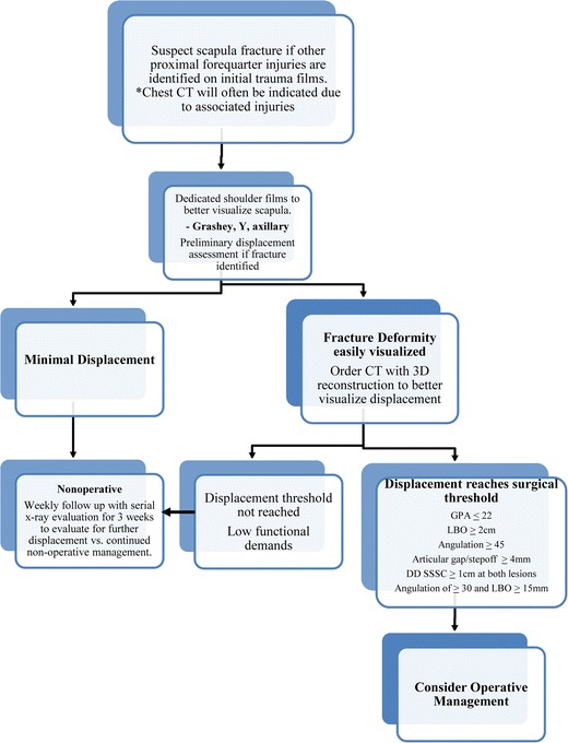
Over the past decade, there has been an increased interest in understanding the operative indications and techniques in treating scapular fractures and tracking their outcomes. Multiple studies have documented poor functional outcomes following nonoperative management of displaced scapular fractures. There is a groundswell of recognition that severe deformity from scapular malunion is associated with functional consequences for patients. This has led to a growing recognition that scapular fractures should be held to the same standards as other bodily fractures with regard to fracture fixation principles, including anatomic articular reduction, proper alignment, and stable internal fixation. Through research, there has been an improved understanding of scapular fracture patterns and the relevant surgical approaches and exposures used for fracture fixation. As with many bones, however, there still remains the absence of a compelling study that defines thresholds for surgical indication based on degrees of deformity and amounts of displacement.
Figures

References
-
- Court-Brown CM, Aitken SA, Forward DR, et al. The epidemiology of fractures. In: Bucholz RW, et al., editors. Fractures in adults. Wilkins: Lippincott Williams; 2009.
-
- ROWE CR. Fractures of the scapula. Surg Clin North Am. 1963;43:1565–71. - PubMed
-
- Butters KP. The scapula. In: Rockwood CA Jr, Matsen FA II, editors. The shoulder. Philadelphia: WB Saunders; 1990.
-
- McClure PW, Michener LA, Karduna AR. Shoulder function and 3-dimensional scapular kinematics in people with and without shoulder impingement syndrome. Phys Ther. 2006;86:1075–90. - PubMed
-
- Warner JJ, Micheli LJ, Arslanian LE, et al. Scapulothoracic motion in normal shoulders and shoulders with glenohumeral instability and impingement syndrome. A study using moire topographic analysis. Clin Orthop Relat Res. 1992;285:191–9. - PubMed
LinkOut - more resources
Full Text Sources
Other Literature Sources
Research Materials
Miscellaneous
