. 2013 May;31(5):928-40.
doi: 10.1002/stem.1332.

The four and a half LIM-domain 2 controls early cardiac cell commitment and expansion via regulating β-catenin-dependent transcription

Affiliations
Free PMC article

The four and a half LIM-domain 2 controls early cardiac cell commitment and expansion via regulating β-catenin-dependent transcription

Anke Renger et al. Stem Cells. 2013 May.
Free PMC article

Abstract

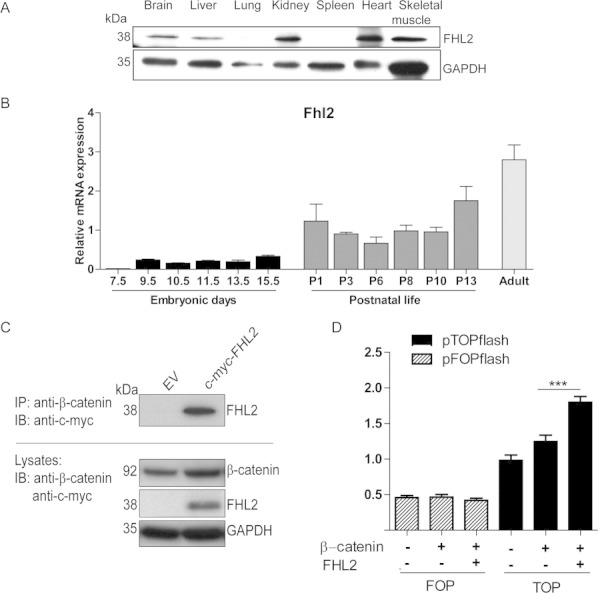
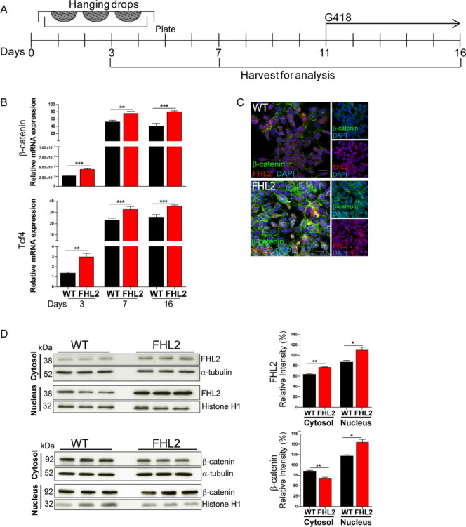
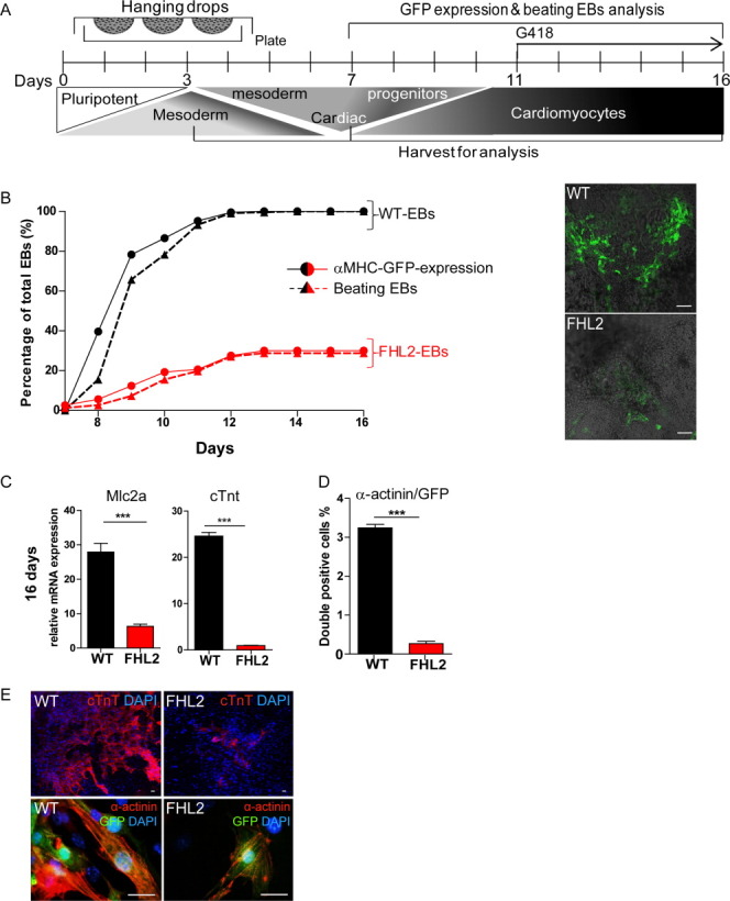
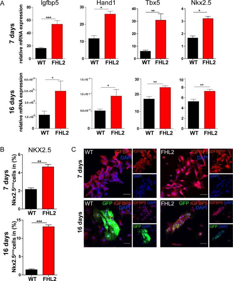
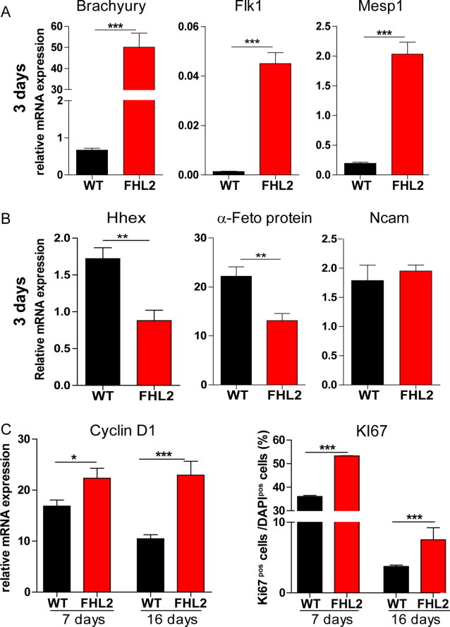
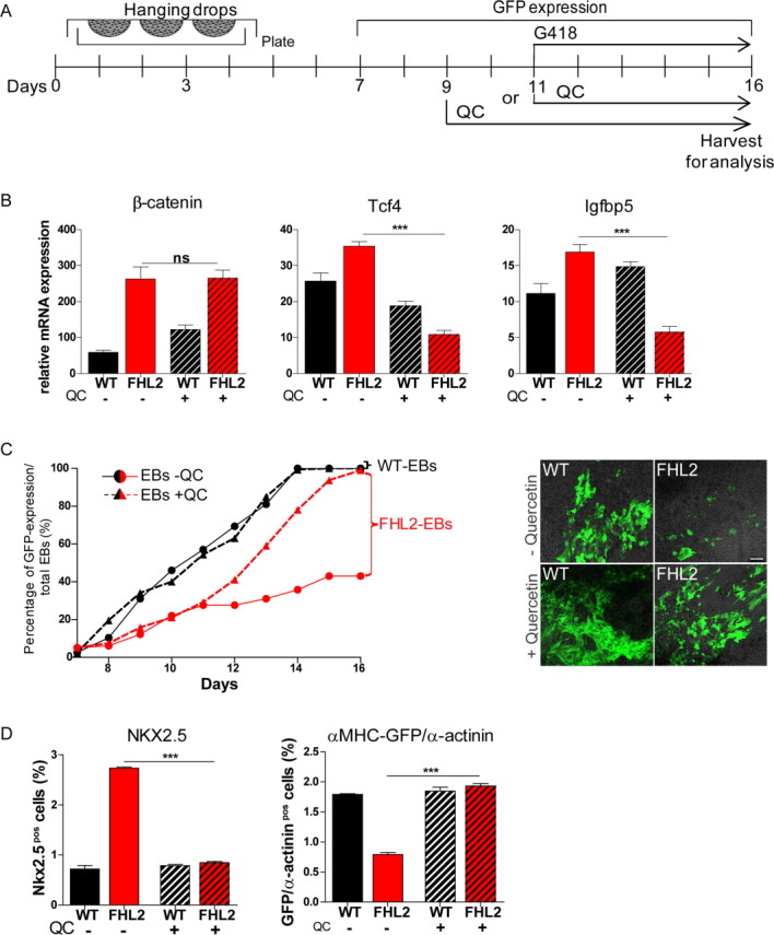
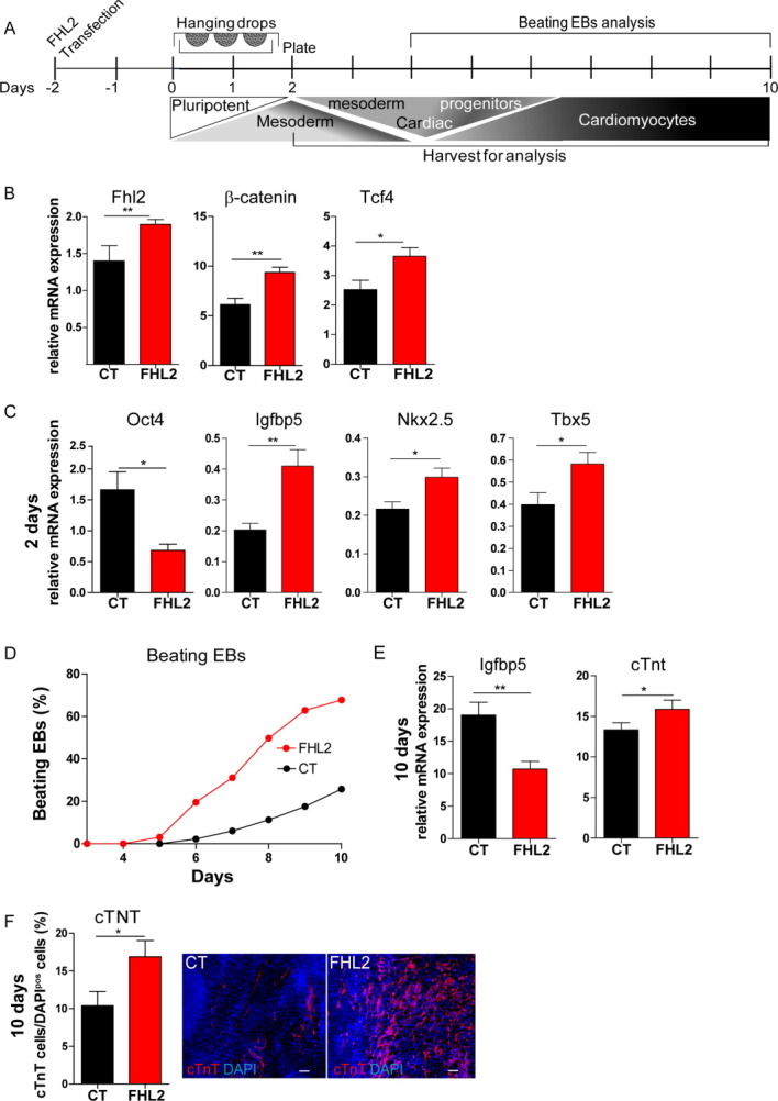
The multiphasic regulation of the Wnt/β-catenin canonical pathway is essential for cardiogenesis in vivo and in vitro. To achieve tight regulation of the Wnt/β-catenin signaling, tissue- and cell-specific coactivators and repressors need to be recruited. The identification of such factors may help to elucidate mechanisms leading to enhanced cardiac differentiation efficiency in vitro as well as promote regeneration in vivo. Using a yeast-two-hybrid screen, we identified four-and-a-half-LIM-domain 2 (FHL2) as a cardiac-specific β-catenin interaction partner and activator of Wnt/β-catenin-dependent transcription. We analyzed the role of this interaction for early cardiogenesis in an in vitro model by making use of embryoid body cultures from mouse embryonic stem cells (ESCs). In this model, stable FHL2 gain-of-function promoted mesodermal cell formation and cell proliferation while arresting cardiac differentiation in an early cardiogenic mesodermal progenitor state. Mechanistically, FHL2 overexpression enhanced nuclear accumulation of β-catenin and activated Wnt/β-catenin-dependent transcription leading to sustained upregulation of the early cardiogenic gene Igfbp5. In an alternative P19 cell model, transient FHL2 overexpression led to early activation of Wnt/β-catenin-dependent transcription, but not sustained high-level of Igfbp5 expression. This resulted in enhanced cardiogenesis. We propose that early Wnt/β-catenin-dependent transcriptional activation mediated by FHL2 is important for the transition to and expansion of early cardiogenic mesodermal cells. Collectively, our findings offer mechanistic insight into the early cardiogenic code and may be further exploited to enhance cardiac progenitor cell activity in vitro and in vivo.


Figures

Figure 1
Figure 1
FHL2 is expressed in cardiac tissue, interacts with β-catenin, and enhances its transcriptional activity in heart cells. (A): Protein expression analysis of FHL2 via Western blot in different adult organs in mice normalized to GAPDH. (B): qPCR analysis of Fhl2 in embryonic cardiac developing tissue, early postnatal, and adult mouse heart. Relative mRNA levels were normalized to tumor protein, translationally controlled 1 (Tpt1), a gene expressed with insignificant variation along development and adulthood. (C): FHL2 and β-catenin interact in neonatal rat cardiomyocyte (NRCM) cultures. NRCM cultures were transfected with a c-myc-FHL2-expressing plasmid and an EV (as control), subsequently endogenous β-catenin was co-IP and FHL2 was IB with a c-myc-antibody. Both proteins were also detected in protein lysates from NRCM cultures. (D): FHL2 activates β-catenin-dependent transcription in NRCM cultures. Luciferase activity measurement demonstrated significant activation of the pTOPflash luciferase LEF/TCF reporter activity upon FHL2 and β-catenin coexpression in NRCM. The FOPflash-plasmid containing mutated TCF binding sites served as control and Renilla luciferase was used for normalization. Data represent mean ± SEM, n = 3, ***, p < .001 (ANOVA; Bonferroni's multiple comparison test). Abbreviations: EV, empty vector; FHL2, four-and-a-half-LIM-domain 2; IB, immunodetected; IP, immunoprecipitated.
Figure 2
Figure 2
Nuclear accumulation of β-catenin and enhanced transcriptional activity mediated by FHL2 in mouse embryonic stem cell-embryoid body cultures (ESC-EBs). (A): Schematic representation of the 16 days differentiation protocol for in vitro cardiogenesis using ESCs. Differentiating ESCs were harvested for analysis following 3, 7, and 16 days of differentiation. At day 11 of differentiation, αMHC cells were selected with the neomycin derivate G418. (B): β-Catenin/TCF transcriptional activation under FHL2 gain-of-function. Significantly increased expression of β-catenin and Tcf4 following 3, 7, and 16 days as revealed by qPCR in ESC-EBs. mRNA levels are displayed as relative expression to Gapdh. (C): Confocal immunofluorescence analysis showing upregulated β-catenin signal (green) and FHL2 (red) in undifferentiated ESC-overexpressing FHL2. DAPI (blue) was used for nuclear staining. (D): Nuclear β-catenin accumulation upon FHL2 gain-of-function. Immunoblot showing increased FHL2 expression as well as β-catenin nuclear accumulation along with cytosolic depletion in FHL2-ESCs. Semiquantitative assessment via densitometry in three independent experiments. Normalization was performed with α-tubulin for the cytosolic and Histone H1 for the nuclear fraction. Data represent mean ± SEM; (B): n = 6; (D): n = 3, *, p < .05; **, p < .01; ***, p < .001 (two-tailed Student's t test). Scale bar = 20 μm. Abbreviations: DAPI, 4′,6-diamidino-2-phenylindole; FHL2, four-and-a-half-LIM-domain 2.
Figure 3
Figure 3
FHL2 gain-of-function blocks cardiac differentiation in embryonic stem cell (ESC)-EBs. (A): Schematic representation of the 16-day differentiation protocol for in vitro cardiogenesis. Differentiating ESC-EBs were harvested for analysis on culture days 3, 7, and 16. (B): ESC-EBs overexpressing FHL2 showed decreased cardiac differentiation. Beating EBs (triangles and discontinues lines) and αMHC-GFP expression (dots and continuous lines) were measured as percentage of total EBs. Both parameters were significantly reduced in FHL2- (red lines) in comparison to wild-type ESC-EBs (black lines) during cardiac differentiation in vitro. Representative immunofluorescence pictures showing αMHC-GFP expression in both cell lines are shown (right). (C): Reduced gene expression of cardiomyocytes genes in ESC-EBs overexpressing FHL2. FHL2-ESC-EBs showed significant reduction in myosin light chain 2a and cardiac Troponin T expression as measured by qPCR; mRNA levels are normalized to Gapdh. (D): Reduction in α-sarcomeric actinin (α-actinin)/GFP cell number as measured by flow cytometry following 16 days of differentiation. (E): Representative immunofluorescence pictures with lower cTNT and α-actinin/αMHC-GFP expression in FHL2- in comparison to wild-type-ESC-EBs. DAPI (blue) was used for nuclear staining. Data represent mean ± SEM; (B): n = 12; (C): n = 6, (D): n = 3, ***, p < .001 (two-tailed Student's t test). Scale bar = (B): 100 μm and (E): 20 μm. Abbreviations: DAPI, 4′,6-diamidino-2-phenylindole; EB, embryoid body; FHL2, four-and-a-half-LIM-domain 2; GFP, green fluorescent protein.
Figure 4
Figure 4
FHL2 mediates enhanced activation of early cardiogenic gene expression in embryonic stem cell-embryoid bodies (ESC-EBs). (A): Expression of the early cardiogenic lineage genes Igfbp5, Hand1, Nkx2.5, and Tbx5 following 7 and 16 days of differentiation upon FHL2 gain-of-function in ESC-EBs as measured by qPCR; mRNA levels are normalized to Gapdh. (B): FHL2 gain-of-function in ESC-EBs resulted in an augmented early cardiogenic cell population as revealed by increased NKX2.5 cell number analyzed by flow cytometry on culture days 7 and 16. (C): Sustained upregulated IGFBP5 expression in FHL2-ESC-EBs accompanied by decreased αMHC-GFP-expressing cells. Increased nuclear localized IGFBP5 expression at 7 days of differentiation in FHL2-ESC-EBs is observed via confocal immunofluorescence analysis. Increased IGFBP5 along with decreased αMHC-GFP expression detected at 16 days of differentiation in FHL2-ESC-EBs. DAPI (blue) was used for nuclear staining. Data represent mean ± SEM; (A): n = 12, (B): n = 3, *, p < .05; **, p < .01; ***, p < .001 (two-tailed Student's t test). Scale bar = 50 μm. Abbreviations: DAPI, 4′,6-diamidino-2-phenylindole; FHL2, four-and-a-half-LIM-domain 2; GFP, green fluorescent protein.
Figure 5
Figure 5
FHL2 gain-of-function promotes mesodermal cell specification and proliferation in embryonic stem cell-embryoid bodies (ESC-EBs). (A): At day 3 of differentiation, FHL2 gain-of-function in ESC-EBs increases expression of the early panmesodermal, mesodermal, and cardiac mesodermal markers Brachyury, Flk1, and Mesp1, respectively, as shown by qPCR analysis. (B): FHL2-ESC-EBs showed decreased expression of the endodermal markers Hhex and α-Feto protein; no change in expression of the neuroectodermal marker Ncam was observed in comparison to wild-type-ESC-EBs at day 3 of differentiation. (C): FHL2 overexpression promoted significantly augmented expression of the cell cycling marker Cyclin D1 as shown by qPCR as well as increased Ki67 expression as demonstrated by immunofluorescence microscopy (percentage of total DAPI-positive cells) following 7 and 16 days of differentiation. mRNA level was normalized to Gapdh. DAPI (blue) was used for nuclear staining. Data represent mean ± SEM; n = 6, *, p < .05; **, p < .01; ***, p < .001 (two-tailed Student's t test). Abbreviation: FHL2, four-and-a-half-LIM-domain 2.
Figure 6
Figure 6
Inhibition of β-catenin-dependent transcription rescues cardiac differentiation in FHL2-ESC-EBs. (A): Schematic representation of in vitro cardiogenesis in embryonic stem cell (ESC)-EBs upon 5 μmol/l QC application from day 9 or 11 of differentiation. Cells were harvested at day 16 of differentiation for analysis. (B): Transcriptional effect of quercetin treatment on differentiating FHL2-ESCs. qPCR analysis showed unchanged β-catenin expression but reduced levels of upregulated Tcf4 and Igfbp5 expression in FHL2-ESC-EBs upon quercetin treatment in comparison to untreated FHL2-ESC-EBs following 16 days of differentiation. mRNA levels were normalized to Gapdh. (C): Blocking β-catenin transcriptional upregulation via quercetin increased the expression of cardiomyocyte genes in FHL2-ESC-EBs. Quercetin treatment at day 11 reversed the decreased αMHC-GFP expression during cardiac differentiation in FHL2-ESC-EBs (triangles and discontinues red lines) in comparison to untreated FHL2-ESC-EBs (dots and continues red lines) expressed as percentages of total EBs. Quercetin did not affect expression of αMHC-GFP in wild-type-ESC-EBs (black lines). Representative confocal immunofluorescence pictures of the αMHC-GFP expression are shown (right). (D): Flow cytometry shows a rescue in upregulation of NKX2.5 as well as in downregulation of α-MHC-GFP/α-sarcomeric-actinin-expressing cells in FHL2-ESC-EBs compared to untreated FHL2-ESC-EBs at 16 days of differentiation. Data represent mean ± SEM; (B): n = 6, (C): n = 10, (D): n = 3, ***, p < .001 (ANOVA; Bonferroni's multiple comparison test). Scale bar = 100 μm. Abbreviations: EB, embryoid body; FHL2, four-and-a-half-LIM-domain 2; GFP, green fluorescent protein; QC, quercetin.
Figure 7
Figure 7
Transient overexpression of FHL2 enhances cardiogenesis in P19 cells in vitro. (A): Schematic representation of differentiation protocol for in vitro cardiogenesis. P19 cells were differentiated in the presence of Dimethyl sulfoxide (DMSO) and harvested for analysis following 1, 2, and 10 days of differentiation. (B): Transient upregulation of FHL2, β-catenin, and its target gene Tcf4 in P19 cells transfected with a FHL2-expressing plasmid (FHL2) as demonstrated by qPCR analysis 24 hours post-transfection. (C): Increased early cardiogenic commitment in FHL2-P19-EBs transiently expressing cells. Significant loss of the pluripotent marker Oct4 and increase of the early cardiogenic markers Igfbp5, Nkx2.5, and Tbx5 following 2 days of differentiation in FHL2-EBs compared to control-EBs (CT) as shown by qPCR analysis. mRNA levels were normalized to Gapdh. (D): Increased cardiogenesis in P19-EBs transiently expressing FHL2. Augmentation of beating EB numbers was observed along cardiac differentiation of FHL2-P19 cells in comparison to CT. (E): Significant downregulation of Igfbp5 expression and augmentation of cTnt expression normalized to Gapdh was observed in FHL2-P19-EBs following 10 days of differentiation. (F): cTNT (red) expression analyzed by immunofluorescence microscopy following 10 days of differentiation showing increased cTNT cell number (as percentage of total DAPI-positive cells) in FHL2-P19 cells. Representative pictures are depicted. DAPI (blue) was used for nuclear staining. Data represent mean ± SEM; B, C, E, and F: n = 6, D: n = 9, *, p < .05; **, p < .01 (two-tailed Student's t test). Scale bar = 50 μm. Abbreviations: cTNT, cardiac troponin T; DAPI, 4′,6-diamidino-2-phenylindole; EB, embryoid body; FHL2, four-and-a-half-LIM-domain 2.

Similar articles

Cited by

References

    1. Brembeck FH, Rosario M, Birchmeier W. Balancing cell adhesion and Wnt signaling, the key role of beta-catenin. Curr Opin Genet Dev. 2006;16:51–59. - PubMed
    1. Cadigan KM. Wnt-beta-catenin signaling. Curr Biol. 2008;18:R943–R947. - PubMed
    1. Gordon MD, Nusse R. Wnt signaling: Multiple pathways, multiple receptors, and multiple transcription factors. J Biol Chem. 2006;281:22429–22433. - PubMed
    1. Tam PP, Loebel DA. Gene function in mouse embryogenesis: Get set for gastrulation. Nat Rev Genet. 2007;8:368–381. - PubMed
    1. Grapin-Botton A, Constam D. Evolution of the mechanisms and molecular control of endoderm formation. Mech Dev. 2007;124:253–278. - PubMed

Publication types

MeSH terms