. 2013 Mar 8;288(10):7169-81.
doi: 10.1074/jbc.M112.406108. Epub 2013 Jan 22.

β-Arrestin recruitment and G protein signaling by the atypical human chemokine decoy receptor CCX-CKR

Affiliations

β-Arrestin recruitment and G protein signaling by the atypical human chemokine decoy receptor CCX-CKR

Anne O Watts et al. J Biol Chem. .

Abstract

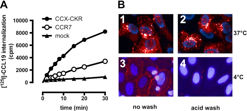
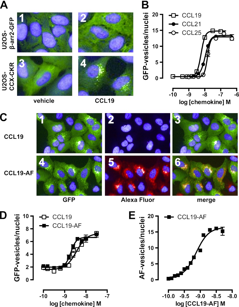
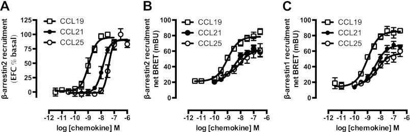
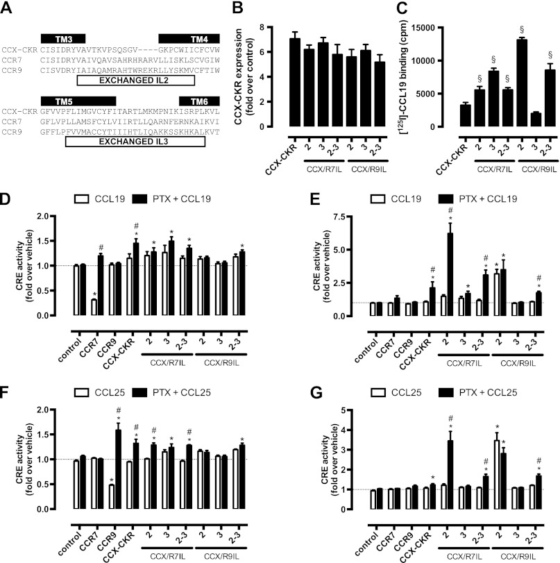
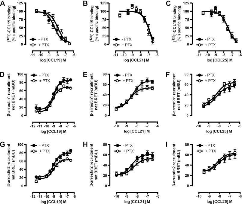
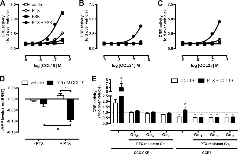
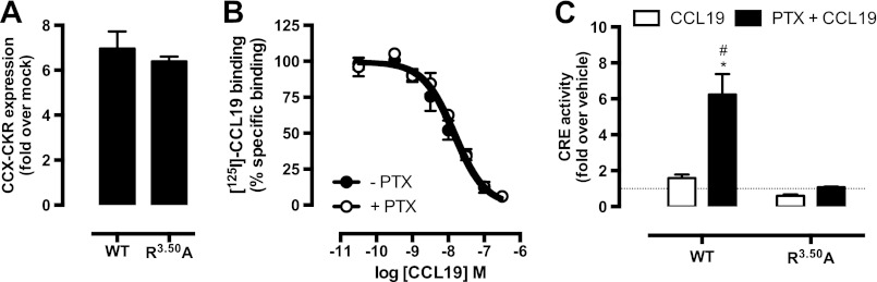
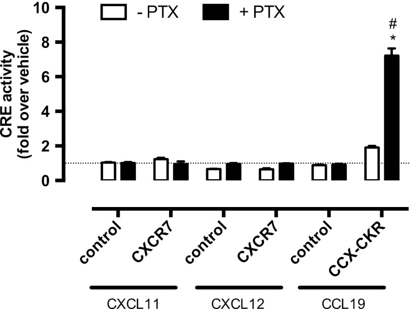
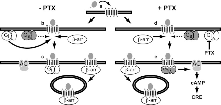
Chemokine receptors form a large subfamily of G protein-coupled receptors that predominantly activate heterotrimeric Gi proteins and are involved in immune cell migration. CCX-CKR is an atypical chemokine receptor with high affinity for CCL19, CCL21, and CCL25 chemokines, but is not known to activate intracellular signaling pathways. However, CCX-CKR acts as decoy receptor and efficiently internalizes these chemokines, thereby preventing their interaction with other chemokine receptors, like CCR7 and CCR9. Internalization of fluorescently labeled CCL19 correlated with β-arrestin2-GFP translocation. Moreover, recruitment of β-arrestins to CCX-CKR in response to CCL19, CCL21, and CCL25 was demonstrated using enzyme-fragment complementation and bioluminescence resonance energy transfer methods. To unravel why CCX-CKR is unable to activate Gi signaling, CCX-CKR chimeras were constructed by substituting its intracellular loops with the corresponding CCR7 or CCR9 domains. The signaling properties of chimeric CCX-CKR receptors were characterized using a cAMP-responsive element (CRE)-driven reporter gene assay. Unexpectedly, wild type CCX-CKR and a subset of the chimeras induced an increase in CRE activity in response to CCL19, CCL21, and CCL25 in the presence of the Gi inhibitor pertussis toxin. CCX-CKR signaling to CRE required an intact DRY motif. These data suggest that inactive Gi proteins impair CCX-CKR signaling most likely by hindering the interaction of this receptor with pertussis toxin-insensitive G proteins that transduce signaling to CRE. On the other hand, recruitment of the putative signaling scaffold β-arrestin to CCX-CKR in response to chemokines might allow activation of yet to be identified signal transduction pathways.


Figures

FIGURE 1.
FIGURE 1.
CCX-CKR internalizes CCL19. A, HEK293T cells transiently transfected with CCX-CKR, CCR7, or empty plasmid (mock) were incubated with 125I-CCL19 for the indicated time. Surface bound 125I-CCL19 was removed by an acid wash, and internalized 125I-CCL19 was quantified. B, U2OS osteosarcoma cells expressing β-arrestin2 fused to green fluorescent protein (U2OS-β-arr2-GFP) were stably transfected with vector coding for CCX-CKR (U2OS-CCX-CKR cells). U2OS-CCX-CKR cells were treated with Alexa Fluor 647-coupled CCL19 (CCL19-AF) at 37 (upper panels) or 4 °C (lower panels) for 45 min. Then, cells were washed with an acidic buffer (0.2 m glycine (pH 3.0), 0.5 m NaCl) or PBS for 5 min before fixation and staining. Alexa Fluor 647 and Hoechst signals are depicted in red and blue, respectively.
FIGURE 2.
FIGURE 2.
Internalized CCL19 and β-arrestin co-localize in endocytic vesicles in cells expressing CCX-CKR. A, U2OS-β-arr2-GFP and U2OS-CCX-CKR cells were treated with 100 nm CCL19 for 45 min. Cells were then fixed, stained with Hoechst, and imaged using fluorescence microscopy for GFP (green) and nuclei (blue). B, U2OS-CCX-CKR cells were stimulated with increasing concentrations of CCL19, CCL21, and CCL25 for 45 min. The recruitment of β-arrestin2 to CCX-CKR was quantified as the amount of GFP-containing vesicles per nucleus. C, U2OS-CCX-CKR cells were treated with 100 nm CCL19 or CCL19-AF for 45 min, followed by fluorescence microscopy. GFP is shown in green, Alexa Fluor 647 is in red, and nuclei are stained blue. D, U2OS-CCX-CKR cells were treated with increasing doses of CCL19 and CCL19-AF for 45 min followed by determination of GFP-containing vesicle formation. E, quantification of AF-containing vesicle formation of U2OS-CCX-CKR cells treated with increasing concentrations of CCL19-AF.
FIGURE 3.
FIGURE 3.
CCX-CKR recruits β-arrestin1 and β-arrestin2 in a chemokine-dependent manner. A, CHO cells stably expressing CCX-CKR fused to a peptide fragment of β-galactosidase and β-arrestin2 coupled to a complementary β-galactosidase mutant (CHO-CCX-CKR cells) were treated with ascending concentrations of CCL19, CCL21, and CCL25 for 90 min before measurement of β-galactosidase activity. B and C, potencies of β-arrestin2 (B) and β-arrestin1 (C) recruitment induced by CCL19 (white squares), CCL21 (black circles), and CCL25 (white circles) were determined using a BRET-based assay in HEK293T cells transiently co-transfected with CCX-CKR-Rluc and β-arrestin-EYFP. Data are presented as averages ± S.E. of 2–4 independent experiments performed in triplicate.
FIGURE 4.
FIGURE 4.
CCX-CKR does not activate G protein-dependent or β-arrestin-dependent intracellular signaling pathways. A and B, CHO-CCX-CKR cells were stimulated with 100 nm CCL19 or vehicle for several time periods. Control cells were treated with 1 μm PMA (A) or 100 ng/ml of insulin (B). Cell lysates were then analyzed for levels of (A) phosphorylated Thr202/Tyr204-ERK1/2 (pERK) and total ERK1/2 (tERK) or (B) phosphorylated Ser473-Akt (pAkt) and total Akt (tAkt). C, CHO-CCX-CKR cells were treated with 100 nm CCL19, CCL21, and CCL25 in the presence or absence of 0.5 μm FSK for 60 min. Subsequently, cells were lysed and intracellular cAMP concentrations were measured. D, CHO-CCX-CKR cells were stimulated with 100 nm CCL19, CCL21, and CCL25 for 90 min before measurement of intracellular IPone concentrations. As a positive control, cells were treated with 100 μm ATP.
FIGURE 5.
FIGURE 5.
IL2 and IL3 of functional chemokine receptors do not convey Gαi-activating properties to CCX-CKR. A, CCX-CKR, CCR7, and CCR9 amino acid sequences of TM3-IL2-TM4 and TM5-IL3-TM6 are aligned on the basis of highly conserved residues. The IL2 and IL3 sequences that have been exchanged in the CCX-CKR chimeras are indicated with white boxes. HEK293T cells were co-transfected with a CRE-driven reporter gene and CCX-CKR, CCX/R7IL2, CCX/R7IL3, CCX/R7IL2IL3, CCX/R9IL2, CCX/R9IL3, or CCX/R9IL2IL3 as indicated. B, cell surface expression was measured by ELISA. C, 125I-CCL19 binding was performed on intact cells. Cells were pre-treated with PTX (100 ng/ml) overnight (filled bars). Cells were treated with vehicle, 100 nm CCL19 (D and E), or 100 nm CCL25 (F and G) for 5 h in the presence (D and F) or absemce (E and G) of 1 μm FSK. Statistical comparisons were performed using one-way analysis of variance followed by the Bonferroni test. Significant difference in cell surface expression and 125I-CCL19 binding as compared with wild type CCX-CKR is indicated by §. B and C, significant difference (p < 0.05) in CRE activity between vehicle and corresponding chemokine-treated cells is indicated by *, whereas # indicates significant difference in chemokine-induced CRE activity between cells pretreated with or without PTX (D–G). Data are shown as averages ± S.E. of fold over vehicle data from 4 independent experiments performed in duplicate (B–E) or 2 independent experiments performed in triplicate (F and G).
FIGURE 6.
FIGURE 6.
CCX-CKR chemokine binding affinities are not dependent on Gαi proteins. 125I-CCL19 displacement curves were performed for CCL19 (A), CCL21 (B), and CCL25 (C) in the absence (empty symbols) and presence (filled symbols) of 100 ng/ml of PTX in HEK293T cells transiently transfected with CCX-CKR. β-Arrestin1 (D–F) and β-arrestin2 (G–I) recruitment induced by CCL19 (D and G), CCL21 (E and H), and CCL25 (F and I) were determined using a BRET-based assay in HEK293T cells transiently co-transfected with CCX-CKR-Rluc and β-arrestin-EYFP without (empty symbols) and with (filled symbols) PTX (100 ng/ml) pretreatment. Data are shown as averages ± S.E. of normalized specific binding of two independent experiments performed in duplicate.
FIGURE 7.
FIGURE 7.
PTX potentiates ligand-induced CRE activation mediated by CCX-CKR. A–C, HEK293T cells were co-transfected with a CRE-driven reporter gene and CCX-CKR. Concentration-response curves of CCL19 (A), CCL21 (B), and CCL25 (C) were obtained in the absence of PTX and FSK (□), in the presence of 1 μm FSK (■), in the presence of 100 ng/ml of PTX (○), or the presence of both PTX and FSK (●). D, HEK293T cells were transiently transfected with the CCX-CKR-coding plasmid along with a plasmid encoding the CAMYEL cAMP biosensor. Cells were pre-treated or not with 100 ng/ml of PTX overnight. Cells were stimulated with 100 nm CCL19 in the presence of 1 μm FSK for 10 min prior to measurement of the intramolecular BRET signal. Elevation of cAMP levels is detected as decreased BRET ratio. Data are shown as averages ± S.E. of normalized and baseline corrected BRET ratios from three independent experiments performed in duplicate. E, HEK293T cells were co-transfected with a CRE-driven reporter gene, CCX-CKR, or CCR7 and the PTX-insensitive mutants of Gαi subunit Gαi1/C351I, Gαi2/C352I, and Gαi3/C351I as indicated. Cells were pre-treated (black bars) or not (empty bars) with 100 ng/ml of PTX overnight. Cells were incubated with vehicle or 100 nm CCL19 in the presence of 1 μm FSK for 6 to 8 h. Statistical comparisons to identify significant differences between vehicle and chemokine-stimulated cells (*) as well as between control and PTX-pretreated cells (#) were performed using one-way analysis of variance followed by the Bonferroni test. p value < 0.05 indicate significant difference. Graphs (A-C and E) show averages ± S.E. of fold over vehicle data from three to six independent experiments performed in duplicate.
FIGURE 8.
FIGURE 8.
Potentiation of CCX-CKR-mediated CRE activation is G protein-dependent. A, cell surface expression of wild type CCX-CKR and the DRY mutant CCX/R3.50A in the reporter gene assay was determined using ELISA with anti-CCX-CKR antibody. Graph shows averages ± S.E. of fold over mock data from two independent experiments performed in duplicate. B, 125I-CCL19 displacement curves with CCL19 were obtained in the absence (empty symbols) or presence (filled symbols) of 100 ng/ml of PTX in HEK293T cells transfected with CCX/R3.50A. Data are shown as averages ± S.E. of normalized specific binding of one independent experiment performed in duplicate. C, HEK293T cells were co-transfected with a CRE-driven reporter gene and wild type CCX-CKR or CCX/R3.50A as indicated. Cells were pre-treated with 100 ng/ml of PTX (empty and black bars) overnight. Cells were incubated with vehicle (empty bars) or 100 nm CCL19 (filled bars) in the presence of 1 μm FSK for 5 h. Statistical comparisons to identify significant differences between vehicle and chemokine-stimulated cells (*) as well as between control and PTX-pretreated cells (#) were performed using one-way analysis of variance followed by the Bonferroni test. p value < 0.05 indicate significant difference. Graph show averages ± S.E. of fold over vehicle data from two independent experiments performed in duplicate.
FIGURE 9.
FIGURE 9.
CXCR7 does not mediate CRE activation. HEK293T cells were co-transfected with a CRE-driven reporter gene and empty vector (−), CXCR7, or CCX-CKR as indicated. Cells were pre-treated (black bars) or not (empty bars) with 100 ng/ml of PTX overnight. Cells were incubated with vehicle, CXCL11, CXCL12, or CCL19 (100 nm chemokine) as indicated in the presence of 1 μm FSK. Data are shown as averages ± S.E. of fold over vehicle data from two independent experiments performed in duplicate. Statistical comparisons to identify significant differences between vehicle and chemokine-stimulated cells (*) as well as between control and PTX-pretreated cells (#) were performed using one-way analysis of variance followed by the Bonferroni test. p value < 0.05 indicate significant difference.
FIGURE 10.
FIGURE 10.
Proposed model of CCX-CKR interaction with β-arrestins and G proteins. Chemokine binding to CCX-CKR (a) recruits Gi proteins and β-arrestin (β-arr) with high affinity (b), consequently hindering the low affinity interaction between CCX-CKR and Gs proteins. Inactive Gi protein may stay bound to CCX-CKR, whereas the chemokine-bound CCX-CKR internalizes with β-arrestin (c). Inhibition of Gi coupling to CCX-CKR by PTX pre-treatment did not affect β-arrestin recruitment but allows Gs to interact with CCX-CKR (d), which results in stimulation of adenylyl cyclase (AC) and CRE activity (e).

Similar articles

Cited by

References

    1. Scholten D. J., Canals M., Maussang D., Roumen L., Smit M. J., Wijtmans M., de Graaf C., Vischer H. F., Leurs R. (2012) Pharmacological modulation of chemokine receptor function. Br. J. Pharmacol. 165, 1617–1643 - PMC - PubMed
    1. O'Hayre M., Salanga C. L., Handel T. M., Allen S. J. (2008) Chemokines and cancer. Migration, intracellular signalling and intercellular communication in the microenvironment. Biochem. J. 409, 635–649 - PubMed
    1. Ulvmar M. H., Hub E., Rot A. (2011) Atypical chemokine receptors. Exp. Cell Res. 317, 556–568 - PMC - PubMed
    1. Gosling J., Dairaghi D. J., Wang Y., Hanley M., Talbot D., Miao Z., Schall T. J. (2000) Cutting edge. Identification of a novel chemokine receptor that binds dendritic cell- and T cell-active chemokines including ELC, SLC, and TECK. J. Immunol. 164, 2851–2856 - PubMed
    1. Förster R., Davalos-Misslitz A. C., Rot A. (2008) CCR7 and its ligands. Balancing immunity and tolerance. Nat. Rev. Immunol. 8, 362–371 - PubMed

Publication types

MeSH terms