. 2013 Mar 8;288(10):7282-93.
doi: 10.1074/jbc.M112.438374. Epub 2013 Jan 22.

The polysialyltransferases interact with sequences in two domains of the neural cell adhesion molecule to allow its polysialylation

Affiliations

The polysialyltransferases interact with sequences in two domains of the neural cell adhesion molecule to allow its polysialylation

Matthew G Thompson et al. J Biol Chem. .

Abstract

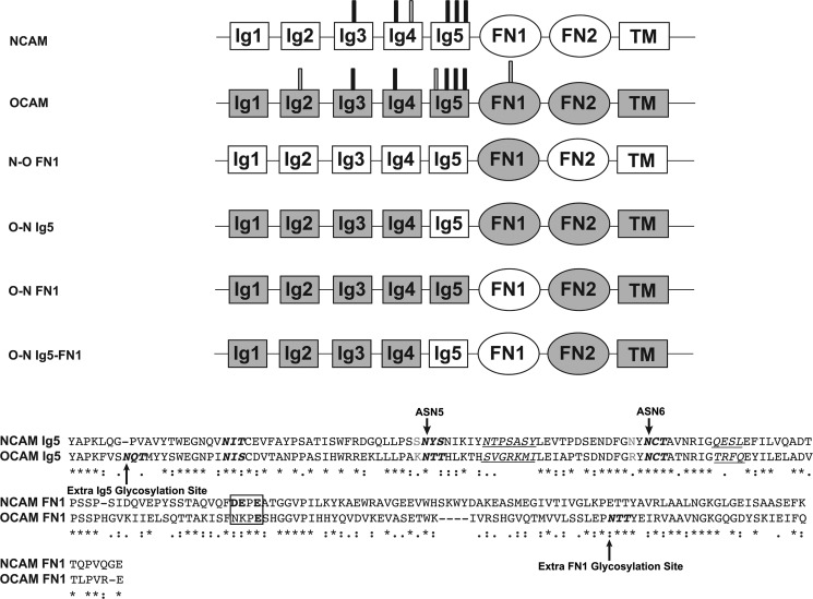
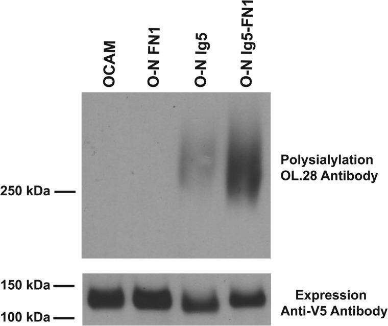
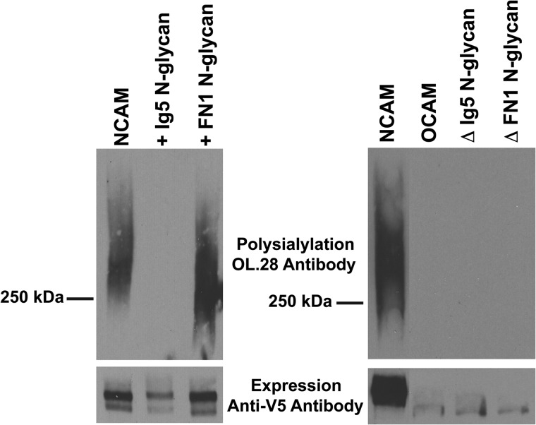
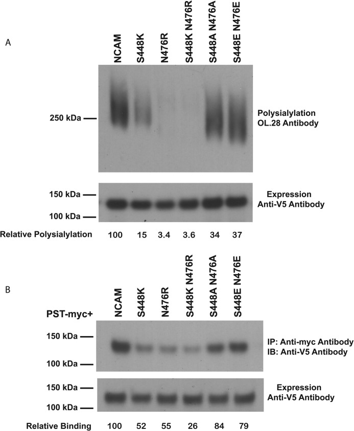
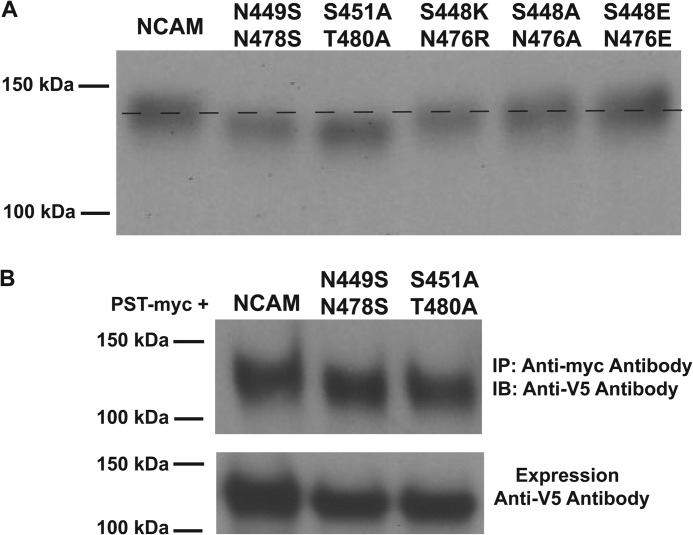
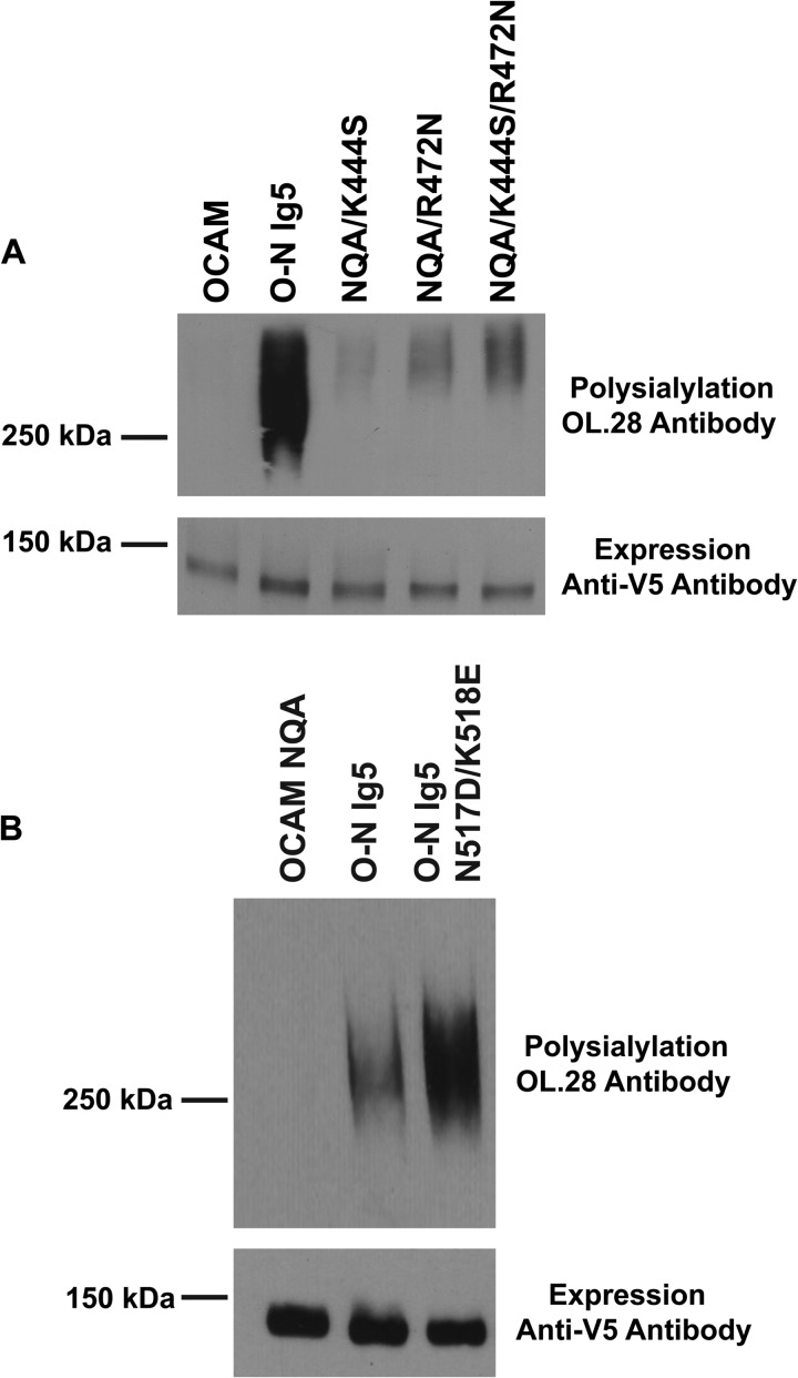
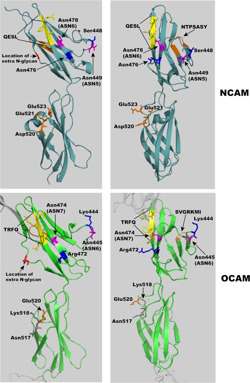
The neural cell adhesion molecule (NCAM) is the major substrate for the polysialyltransferases (polySTs), ST8SiaII/STX and ST8SiaIV/PST. The polysialylation of NCAM N-glycans decreases cell adhesion and alters signaling. Previous work demonstrated that the first fibronectin type III repeat (FN1) of NCAM is required for polyST recognition and the polysialylation of the N-glycans on the adjacent Ig5 domain. In this work, we highlight the importance of an FN1 acidic patch in polyST recognition and also reveal that the polySTs are required to interact with sequences in the Ig5 domain for polysialylation to occur. We find that features of the Ig5 domain of the olfactory cell adhesion molecule (OCAM) are responsible for its lack of polysialylation. Specifically, two basic OCAM Ig5 residues (Lys and Arg) found near asparagines equivalent to those carrying the polysialylated N-glycans in NCAM substantially decrease or eliminate polysialylation when used to replace the smaller and more neutral residues (Ser and Asn) in analogous positions in NCAM Ig5. This decrease in polysialylation does not reflect altered glycosylation but instead is correlated with a decrease in polyST-NCAM binding. In addition, inserting non-conserved OCAM sequences into NCAM Ig5, including an "extra" N-glycosylation site, decreases or completely blocks NCAM polysialylation. Taken together, these results indicate that the polySTs not only recognize an acidic patch in the FN1 domain of NCAM but also must contact sequences in the Ig5 domain for polysialylation of Ig5 N-glycans to occur.


Figures

FIGURE 1.
FIGURE 1.
Sequence comparison and schematic of NCAM, OCAM, and chimeric proteins used in this study. Top, schematics of the domain arrangement of NCAM (white), OCAM (gray), and the chimeric proteins made and analyzed in this study. The N-linked glycosylation sites that are conserved between NCAM and OCAM are indicated with black bars, and those that are not conserved are indicated by gray bars. The FN1 domain of NCAM was replaced with OCAM FN1 to generate N-O FN1. The Ig5, FN1, or Ig5-FN1 domains of OCAM were replaced with the NCAM Ig5, FN1, or Ig5-FN1 domains to generate O-N Ig5, O-N FN1, or O-N Ig5-FN1, respectively. TM, transmembrane domain. Bottom, comparison of NCAM and OCAM Ig5-FN1 sequences. Asterisks below the sequences indicate identity, double dots indicate strong homology, and single dots indicate weak homology. Consensus N-linked glycosylation sites within the Ig5 and FN1 sequences are in boldface italic type, with NCAM ASN5 and ASN6 and the extra OCAM N-glycosylation sites labeled. Non-conserved residues adjacent to ASN5 and ASN6 in NCAM Ig5 and the analogous residues in OCAM Ig5 are shown in gray. Stretches of nonconserved Ig5 amino acids analyzed in this study are italicized and underlined. The three core acidic path residues of NCAM FN1 (DEPE; Asp520, Glu521, and Glu523) and the analogous residues in OCAM FN1 (NKPE; Asn517, Lys518, Pro519, and Glu520) are boxed, and the acidic patch residues are in boldface type. Note that mouse OCAM was used in this study with Glu at position 520, whereas the structure shown in Fig. 7 is of human OCAM with an Asp at position 520.
FIGURE 2.
FIGURE 2.
The Ig5 domain of OCAM does not support polysialylation. Top, OCAM or chimeric proteins containing NCAM domains (O-N FN1, O-N Ig5, and O-N Ig5-FN1) were co-expressed with ST8SiaIV/PST-Myc in COS-1 cells. OCAM proteins were immunoprecipitated from cell lysates, and polysialylation was determined by immunoblotting with the anti-polySia antibody, OL.28, as described under “Experimental Procedures.” Bottom, relative protein expression levels were determined by immunoblotting cell lysate aliquots with anti-V5 epitope tag antibody.
FIGURE 3.
FIGURE 3.
The additional N-linked glycosylation site in OCAM Ig5 prevents polysialylation when inserted into NCAM Ig5, but removing this site from OCAM Ig5 is not sufficient to allow OCAM polysialylation. Top panels, COS-1 cells were co-transfected with V5-tagged wild type or mutated NCAM or OCAM and ST8SiaIV/PST-Myc. NCAM, OCAM, or mutant proteins were immunoprecipitated from cell lysates using anti-V5 epitope tag antibody, and polysialylation was measured using the anti-polySia antibody, OL.28. The polysialylation of NCAM mutants with additional OCAM N-glycosylation sites inserted is shown on the left, and the polysialylation of OCAM mutants with these N-glycosylation sites removed is shown on the right. Bottom panels, relative protein expression levels were determined by immunoblotting cell lysate aliquots with anti-V5 epitope tag antibody.
FIGURE 4.
FIGURE 4.
Inserting the basic residues found adjacent to Ig5 glycosylation sites in OCAM into analogous positions in NCAM Ig5 reduces or eliminates NCAM polysialylation and polyST-NCAM interactions. A, top, wild type NCAM or mutated NCAM proteins with Ser448 and Asn476 replaced with Lys or Arg (as found in OCAM Ig5), alanine, or glutamic acid residues were co-expressed with ST8SiaIV/PST-Myc in COS-1 cells. NCAM proteins were immunoprecipitated from cell lysates, and polysialylation was determined by immunoblotting with the OL.28 anti-polySia antibody. A, bottom, relative protein expression levels were determined by immunoblotting cell lysate aliquots with an anti-V5 epitope tag antibody. B, top, Lec 2 CHO cells were co-transfected with V5-tagged NCAM or its S448/N476 mutants and ST8SiaIV/PST-Myc. The polyST was immunoprecipitated (IP) from cell lysates using an anti-Myc tag antibody, and co-precipitating NCAM proteins were detected by immunoblotting (IB) with an anti-V5 epitope tag antibody. B, bottom, the relative expression levels of wild type and mutant NCAM proteins were determined by immunoblotting cell lysate aliquots with an anti-V5 epitope tag antibody. Relative polysialylation (top) and relative binding (bottom) was determined by densitometry.
FIGURE 5.
FIGURE 5.
NCAM Ser448 and Asn476 mutants do not compromise the glycosylation of ASN5 and ASN6, and these N-glycans are not required for polyST-NCAM interactions. A, the molecular masses of S448K/N476R, S448A/N476A, and S448E/N476E mutants were compared with those of NCAM glycosylation mutants in which ASN5 (Asn449) and ASN6 (Asn478) were replaced with serine residues (N449S/N478S), or the third position of each of these glycosylation sites was replaced with alanine (S451A/T480A). Following expression in COS-1 cells, cell lysates (100 μl, or 10% of total cell lysate) were separated on a 4–15% gradient gel, and proteins were detected by immunoblotting with an anti-V5 epitope tag antibody. B, top, the ability of NCAM and NCAM glycosylation mutants, N449S/N478S and S451A/T480A, that lack N-glycans on ASN5 and ASN6 to bind to the polyST ST8SiaIV/PST-Myc was compared by co-immunoprecipitation (IP). NCAM glycosylation mutants (V5-tagged) were co-expressed with ST8SiaIV/PST-Myc in COS-1 cells, the polyST was immunoprecipitated using an anti-Myc antibody, and co-precipitating NCAM proteins were detected by immunoblotting (IB) with an anti-V5 epitope tag antibody. B, bottom, relative protein expression levels were determined by immunoblotting cell lysate aliquots with anti-V5 epitope tag antibody.
FIGURE 6.
FIGURE 6.
Replacing OCAM Ig5 Lys444 and Arg472 with NCAM residues in the absence of the extra OCAM Ig5 N-glycan allows only low level OCAM polysialylation, whereas creating the core FN1 acidic patch in the O-N Ig5 chimera substantially enhances this protein's polysialylation. A, top, to determine whether the extra N-glycan in OCAM Ig5 plus the two basic residues (Lys444 and Arg472) adjacent to OCAM N-glycosylation sites in this domain serve to block OCAM polysialylation, the polysialylation of OCAM with K444S, R472N, or both mutations in the absence of the extra OCAM Ig5 N-glycosylation site (third position mutant, NQA) was determined by OL.28 immunoblotting following expression in COS-1 cells with ST8SiaIV/PST-Myc and immunoprecipitation of OCAM proteins using the anti-V5 epitope tag antibody. The polysialylations of wild type OCAM and the O-N Ig5 chimera are shown for comparison. A, bottom, the relative expression levels of wild type and mutant OCAM proteins were determined by immunoblotting cell lysate aliquots with an anti-V5 epitope tag antibody. B, top, the polysialylations of OCAM, the O-N Ig5 chimera, and this chimera plus the N517D/K518E mutant that recreates the core FN1 acidic patch were compared by expressing these proteins with ST8SiaIV/PST in COS-1 cells and evaluating polysialylation by immunoblotting immunoprecipitated OCAM or O-N chimeric proteins with the OL.28 anti-polySia antibody. B, bottom, relative protein expression levels were determined by immunoblotting cell lysate aliquots with an anti-V5 epitope tag antibody.
FIGURE 7.
FIGURE 7.
Comparison of key residues in NCAM and OCAM Ig5-FN1 tandems. Shown are two views of the structures of the human NCAM (Protein Data Bank entry 3MTR) and human OCAM (Protein Data Bank entry 2JLL) Ig5-FN1 tandems. Indicated are common glycosylation sites in pink: NCAM Asn449 (ASN5) and Asn478 (ASN6), which carry the polysialylated N-glycans, and the analogous Asn445 (ASN6) and Asn474 (ASN7) in OCAM. Residues near these glycosylation sites in OCAM (Lys444 and Arg472) prohibit NCAM polysialylation when used to replace analogous residues in NCAM (Ser448 and Asn476) (residues in blue). Orange residues in the two FN1 domains represent the core acidic patch residues for NCAM (Asp520, Glu521, and Glu523) and an analogous acidic residue, Glu520, in mouse OCAM. Note that mouse OCAM was used in this study with Glu at position 520, whereas the structure shown is of human OCAM with an Asp at position 520. The two OCAM FN1 non-acidic residues, Asn517 and Lys518, replaced in Fig. 6 are shown in gray. We have labeled the OCAM structure according to the mouse amino acid sequence. Shown in the right panels only, replacing a sequence on the strand adjacent to that carrying ASN6 in NCAM (QESL; yellow) with sequences from OCAM (TRFQ; yellow) leads to a decrease in polysialylation. Notably, a large phenylalanine residue from OCAM (Phe485 in TRFQ) replaces a smaller serine residue (Ser489 in QESL). Arg472, Lys444, and Phe485 protrude from the surface of OCAM Ig5 and may serve to block polyST access and decrease or prevent OCAM polysialylation. Replacing a second non-conserved sequence (NTPSASY) in NCAM Ig5 with analogous residues from OCAM (SVGRKMI) leads to a smaller decrease in NCAM polysialylation (both sequences shown in orange). Shown in the left panels only, the location of the extra glycosylation site in OCAM Ig5 and its location when inserted into NCAM Ig5 are indicated (red).

Similar articles

Cited by

References

    1. Varki A. (1993) Biological roles of oligosaccharides. All of the theories are correct. Glycobiology 3, 97–130 - PMC - PubMed
    1. Haltiwanger R. S., Lowe J. B. (2004) Role of glycosylation in development. Annu. Rev. Biochem. 73, 491–537 - PubMed
    1. Johnson C. P., Fujimoto I., Rutishauser U., Leckband D. E. (2005) Direct evidence that neural cell adhesion molecule (NCAM) polysialylation increases intermembrane repulsion and abrogates adhesion. J. Biol. Chem. 280, 137–145 - PubMed
    1. Hildebrandt H., Mühlenhoff M., Weinhold B., Gerardy-Schahn R. (2007) Dissecting polysialic acid and NCAM functions in brain development. J. Neurochem. 103, 56–64 - PubMed
    1. Rutishauser U. (2008) Polysialic acid in the plasticity of the developing and adult vertebrate nervous system. Nat. Rev. Neurosci. 9, 26–35 - PubMed

Publication types

MeSH terms