

Small angle X-ray scattering analysis of Clostridium thermocellum cellulosome N-terminal complexes reveals a highly dynamic structure
- PMID: 23341454
- PMCID: PMC3597834
- DOI: 10.1074/jbc.M112.408757
Small angle X-ray scattering analysis of Clostridium thermocellum cellulosome N-terminal complexes reveals a highly dynamic structure
Abstract
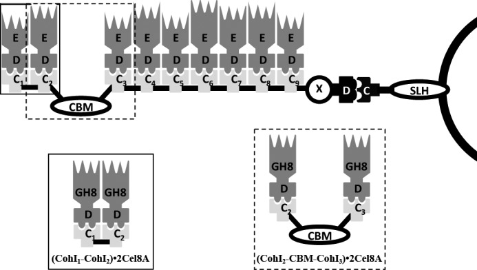
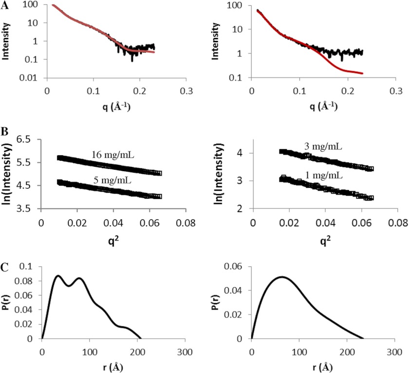
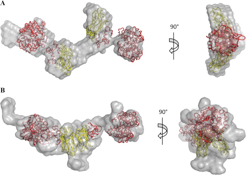
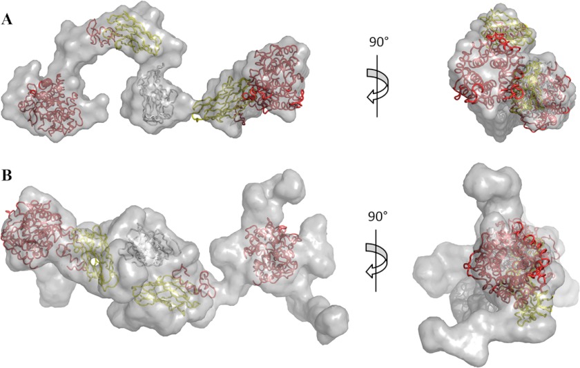
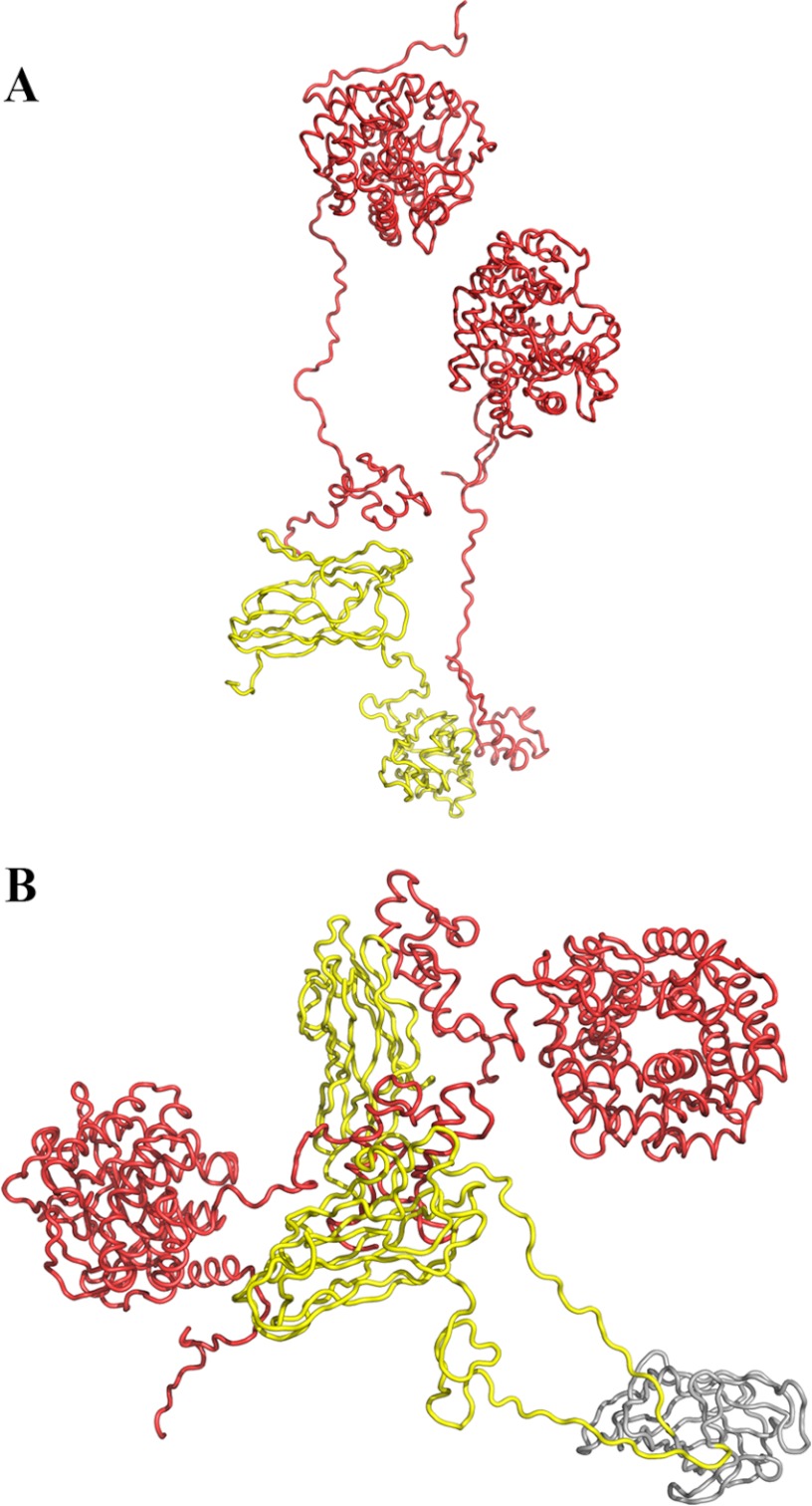
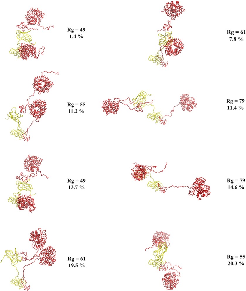
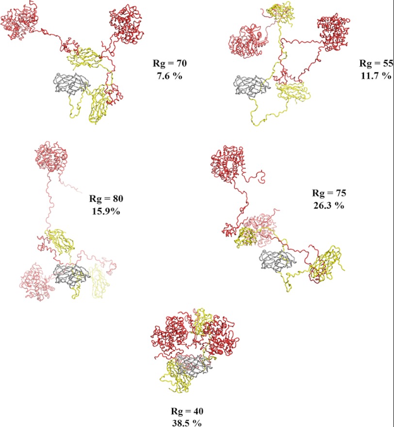
Clostridium thermocellum produces the prototypical cellulosome, a large multienzyme complex that efficiently hydrolyzes plant cell wall polysaccharides into fermentable sugars. This ability has garnered great interest in its potential application in biofuel production. The core non-catalytic scaffoldin subunit, CipA, bears nine type I cohesin modules that interact with the type I dockerin modules of secreted hydrolytic enzymes and promotes catalytic synergy. Because the large size and flexibility of the cellulosome preclude structural determination by traditional means, the structural basis of this synergy remains unclear. Small angle x-ray scattering has been successfully applied to the study of flexible proteins. Here, we used small angle x-ray scattering to determine the solution structure and to analyze the conformational flexibility of two overlapping N-terminal cellulosomal scaffoldin fragments comprising two type I cohesin modules and the cellulose-specific carbohydrate-binding module from CipA in complex with Cel8A cellulases. The pair distribution functions, ab initio envelopes, and rigid body models generated for these two complexes reveal extended structures. These two N-terminal cellulosomal fragments are highly dynamic and display no preference for extended or compact conformations. Overall, our work reveals structural and dynamic features of the N terminus of the CipA scaffoldin that may aid in cellulosome substrate recognition and binding.
Figures
Similar articles
-
Scaffoldin conformation and dynamics revealed by a ternary complex from the Clostridium thermocellum cellulosome.J Biol Chem. 2012 Aug 3;287(32):26953-61. doi: 10.1074/jbc.M112.343897. Epub 2012 Jun 15. J Biol Chem. 2012. PMID: 22707718 Free PMC article.
-
Insights into higher-order organization of the cellulosome revealed by a dissect-and-build approach: crystal structure of interacting Clostridium thermocellum multimodular components.J Mol Biol. 2010 Mar 5;396(4):833-9. doi: 10.1016/j.jmb.2010.01.015. Epub 2010 Jan 11. J Mol Biol. 2010. PMID: 20070943
-
In vitro reconstitution of the complete Clostridium thermocellum cellulosome and synergistic activity on crystalline cellulose.Appl Environ Microbiol. 2012 Jun;78(12):4301-7. doi: 10.1128/AEM.07959-11. Epub 2012 Apr 20. Appl Environ Microbiol. 2012. PMID: 22522677 Free PMC article.
-
The cellulosome: an exocellular, multiprotein complex specialized in cellulose degradation.Crit Rev Biochem Mol Biol. 1996 Jun;31(3):201-36. doi: 10.3109/10409239609106584. Crit Rev Biochem Mol Biol. 1996. PMID: 8817076 Review.
-
Cellulosomes: microbial nanomachines that display plasticity in quaternary structure.Mol Microbiol. 2007 Mar;63(6):1568-76. doi: 10.1111/j.1365-2958.2007.05640.x. Mol Microbiol. 2007. PMID: 17367380 Review.
Cited by
-
Mapping the deformability of natural and designed cellulosomes in solution.Biotechnol Biofuels Bioprod. 2022 Jun 20;15(1):68. doi: 10.1186/s13068-022-02165-3. Biotechnol Biofuels Bioprod. 2022. PMID: 35725490 Free PMC article.
-
The structure of the catalytic domain of a plant cellulose synthase and its assembly into dimers.Plant Cell. 2014 Jul;26(7):2996-3009. doi: 10.1105/tpc.114.126862. Epub 2014 Jul 10. Plant Cell. 2014. PMID: 25012190 Free PMC article.
-
Stoichiometric Assembly of the Cellulosome Generates Maximum Synergy for the Degradation of Crystalline Cellulose, as Revealed by In Vitro Reconstitution of the Clostridium thermocellum Cellulosome.Appl Environ Microbiol. 2015 Jul;81(14):4756-66. doi: 10.1128/AEM.00772-15. Epub 2015 May 8. Appl Environ Microbiol. 2015. PMID: 25956772 Free PMC article.
-
Dual binding in cohesin-dockerin complexes: the energy landscape and the role of short, terminal segments of the dockerin module.Sci Rep. 2018 Mar 22;8(1):5051. doi: 10.1038/s41598-018-23380-9. Sci Rep. 2018. PMID: 29568013 Free PMC article.
-
Research progress and the biotechnological applications of multienzyme complex.Appl Microbiol Biotechnol. 2021 Mar;105(5):1759-1777. doi: 10.1007/s00253-021-11121-4. Epub 2021 Feb 10. Appl Microbiol Biotechnol. 2021. PMID: 33564922 Review.
References
-
- Brett C. T., Waldren K. (1996) Physiology and Biochemistry of Plant Cell Walls: Topics in Plant Functional Biology, 2nd Ed., Chapman & Hall, London
-
- Himmel M. E., Bayer E. A. (2009) Lignocellulose conversion to biofuels: current challenges, global perspectives. Curr. Opin. Biotechnol. 20, 316–317 - PubMed
-
- Jordan D. B., Bowman M. J., Braker J. D., Dien B. S., Hector R. E., Lee C. C., Mertens J. A., Wagschal K. (2012) Plant cell walls to ethanol. Biochem. J. 442, 241–252 - PubMed
-
- Warren R. A. (1996) Microbial hydrolysis of polysaccharides. Annu. Rev. Microbiol. 50, 183–212 - PubMed
-
- Bayer E. A., Belaich J. P., Shoham Y., Lamed R. (2004) The cellulosomes: multienzyme machines for degradation of plant cell wall polysaccharides. Annu. Rev. Microbiol. 58, 521–554 - PubMed
Publication types
MeSH terms
Substances
LinkOut - more resources
Full Text Sources
Other Literature Sources
