. 2013 Mar 15;288(11):7536-7548.
doi: 10.1074/jbc.M112.439273. Epub 2013 Jan 22.

The HsiB1C1 (TssB-TssC) complex of the Pseudomonas aeruginosa type VI secretion system forms a bacteriophage tail sheathlike structure

Affiliations

The HsiB1C1 (TssB-TssC) complex of the Pseudomonas aeruginosa type VI secretion system forms a bacteriophage tail sheathlike structure

Nadine S Lossi et al. J Biol Chem. .

Abstract

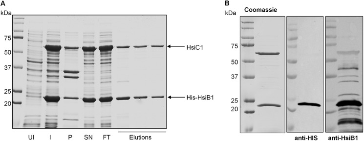
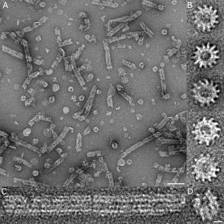
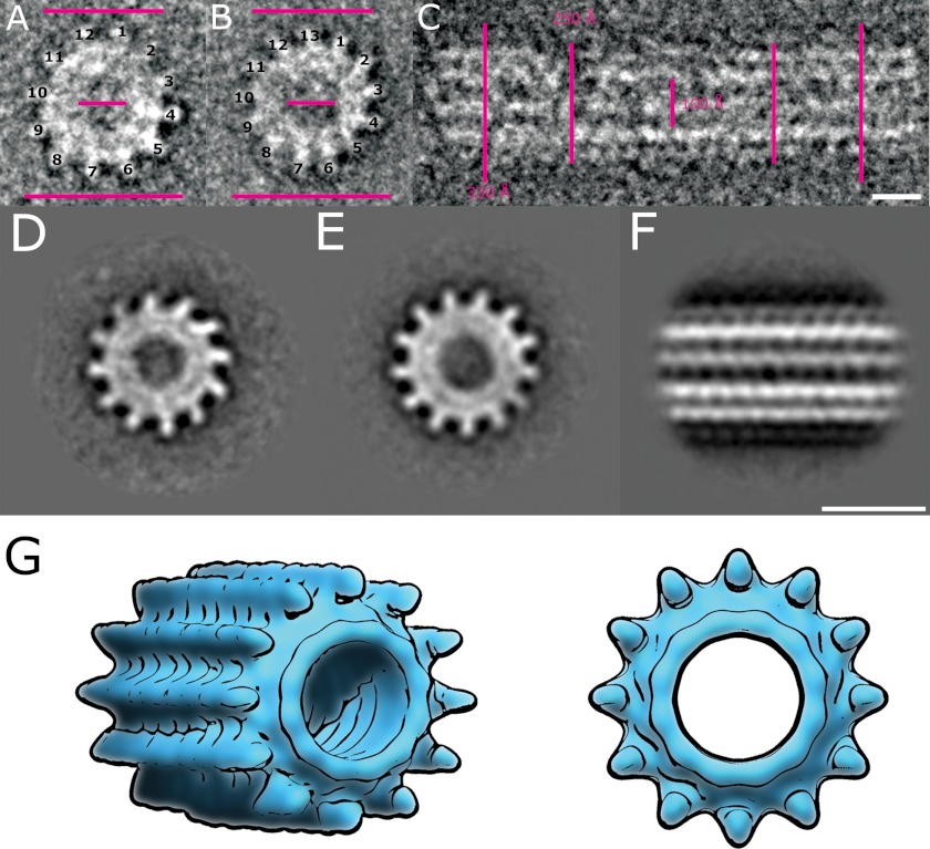
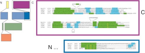
Protein secretion systems in Gram-negative bacteria evolved into a variety of molecular nanomachines. They are related to cell envelope complexes, which are involved in assembly of surface appendages or transport of solutes. They are classified as types, the most recent addition being the type VI secretion system (T6SS). The T6SS displays similarities to bacteriophage tail, which drives DNA injection into bacteria. The Hcp protein is related to the T4 bacteriophage tail tube protein gp19, whereas VgrG proteins structurally resemble the gp27/gp5 puncturing device of the phage. The tube and spike of the phage are pushed through the bacterial envelope upon contraction of a tail sheath composed of gp18. In Vibrio cholerae it was proposed that VipA and VipB assemble into a tail sheathlike structure. Here we confirm these previous data by showing that HsiB1 and HsiC1 of the Pseudomonas aeruginosa H1-T6SS assemble into tubules resulting from stacking of cogwheel-like structures showing predominantly 12-fold symmetry. The internal diameter of the cogwheels is ~100 Å, which is large enough to accommodate an Hcp tube whose external diameter has been reported to be 85 Å. The N-terminal 212 residues of HsiC1 are sufficient to form a stable complex with HsiB1, but the C terminus of HsiC1 is essential for the formation of the tubelike structure. Bioinformatics analysis suggests that HsiC1 displays similarities to gp18-like proteins in its C-terminal region. In conclusion, we provide further structural and mechanistic insights into the T6SS and show that a phage sheathlike structure is likely to be a conserved element across all T6SSs.


Figures

FIGURE 1.
FIGURE 1.
HsiB1 forms a stable complex with HsiC1. A, co-purification of HsiB1 and HsiC1. HsiB1 was expressed with an N-terminal His tag from pET-B1C1, which also expressed untagged HsiC1. His-HsiB1 was purified by Ni+ affinity chromatography in complex with untagged HsiC1. Samples were analyzed for the presence of both HsiB1 and HsiC1 by SDS-PAGE followed by Coomassie staining. UI, uninduced cells; I, 16 h after protein induction; SN, supernatant or soluble fraction; P, pellet or insoluble fraction; FT, flow-through; Elutions, fractions collected after elution of proteins by imidazole gradient. B, identification of components of the purified HsiB1C1 complex. The purified protein complex was analyzed by SDS-PAGE followed by Coomassie staining (left panel). HsiB1 was further identified by immunoblotting using anti-HsiB1 (middle panel) and monoclonal anti-His antibody (right panel), respectively. The molecular mass is indicated on the left in kDa. Note that the identity of the two apparent bands was also confirmed by mass spectrometry; the lower band is HsiB1 and the upper band is HsiC1 (not shown).
FIGURE 2.
FIGURE 2.
The interaction between HsiB1 and HsiC1 is specific. Bacterial two-hybrid analysis of possible interactions among HsiB1, HsiC1, HsiB2, and HsiC2 is shown. Various combinations of recombinant pKT25 and pUT18C plasmids harboring proteins of interest were co-transformed into E. coli DHM1, and β-galactosidase activity of co-transformants was measured after plating on MacConkey agar plates. Plasmid combinations are annotated in the order pKT25/pUT18C fusion. Experiments were carried out in triplicates, and error bars represent the S.D. Zip, leucine zipper domain of the yeast transcription factor GCN4 (positive control); T18, empty vector pUT18C; T25, empty vector pKT25. For T25/T18 fusion proteins, B1 represents HsiB1, C1 represents HsiC1, B2 represents HsiB2, and C2 represents HsiC2. A, images of colonies formed by co-transformants on MacConkey Agar plates (dark red colonies indicate a positive interaction). Plasmid combinations are indicated in the top left corner of every panel in the order T25/T18 fusion protein. The strength of interaction was investigated by measuring the β-galactosidase activity of cells in the respective colonies, and the average activity in Miller units is indicated in the bottom right corner of each image and is represented in B. B, graphic representation of the β-galactosidase activity of co-transformants after incubation on MacConkey agar. Experiments were carried out in triplicates, and error bars represent the S.D. Plasmid combinations are indicated in the order pKT25/pUT18C and abbreviations apply as described above.
FIGURE 3.
FIGURE 3.
HsiC2 cannot fulfill the function of HsiC1. The H1-T6SS component hsiC1 was deleted in PAKΔretS, resulting in PAKΔretSΔhsiC1. The genes hsiC1 and hsiC2 were integrated into the chromosome of PAKΔretSΔhsiC1 using mini-CTX-C1 (PAKΔretSΔhsiC1+C1) and mini-CTX-C2 (PAKΔretSΔhsiC1+C2), respectively. A, expression of HsiC2 does not restore a functional H1-T6SS in PAKΔretSΔhsiC1. The secretion profile of PAKΔretSΔhsiC1, PAKΔretSΔhsiC1+C1, and PAKΔretSΔhsiC1+C2 was compared with that of PAKΔretS (H1-T6SS induced). Strains were grown to late exponential phase before culture supernatants were separated from bacterial cells, and secretion of VgrG1a, Hcp1, and Tse3 was monitored by Western blotting using polyclonal antibodies directed against VgrG1a/c (second panel), Hcp1 (third panel), and Tse3 (fourth panel). RNA polymerase was monitored in both whole cell lysates and culture supernatants using monoclonal antibody directed against the β-subunit of RNA polymerase (RNAP) (150 kDa). The absence of RNA polymerase in the culture supernatant ensured the absence of cell lysis. Anti-VgrG1a antibody also detects VgrG1b in whole cell extract that is secreted independently of a functional H1-T6SS and is indicated by *. Stability of the sheath component HsiB1 was monitored in whole cell lysates using polyclonal anti-HsiB1 antibody (first panel). B, impact of hsiC1 or hsiC2 expression in PAKΔretSΔhsiC1 on complementing H1-T6SS-mediated bacterial killing. Overnight cultures of the P. aeruginosa strains PAK (H1-T6SS-negative), PAKΔretS (constitutive H1-T6SS expression), PAKΔretSΔhsiC1, PAKΔretSΔhsiC1+C1, and PAKΔretSΔhsiC1+C2 were mixed with equivalent numbers of E. coli carrying a plasmid expressing β-galactosidase. Mixed cultures were spotted onto LB agar and incubated for 5 h. Dilution series of recovered bacteria ranging from 0 (undiluted) to −7 (107-fold diluted) were spotted onto LB containing X-gal. The level of visible blue color indicates survival of E. coli. C, semiquantitative analysis of bacterial killing. Graphic representation of the β-galactosidase activity recovered from bacterial patches is shown in B. Bacteria were recovered from the patches of the 10−2 dilution, and β-galactosidase activity was determined using ortho-nitrophenyl β-galactoside as a substrate. Error bars represent S.D. of triplicates of three independent patches. ΔretS, PAKΔretS; ΔΔhsiC1, PAKΔretSΔhsiC1; ΔΔhsiC1+C1, PAKΔretSΔhsiC1; ΔΔhsiC1+C2, PAKΔretSΔhsiC1+C2.
FIGURE 4.
FIGURE 4.
Negative stain electron microscopy images of the HsiB1C1 complex shows tubular and cogwheel structures. A, micrograph of negatively stained HsiB1C1 complexes showing both tubular and cogwheel-like structures. The scale bar corresponds to 100 nm. B, examples of raw images of cogwheels exhibiting both 13- and 12-mer arrangements with a central hole of ∼100 Å. C, examples of tubules that show a four-layered striation with discontinuous staining. D, oblique view of a short tubule structure that clearly shows the central channel. The scale bar corresponds to 10 nm (applies also to B and C).
FIGURE 5.
FIGURE 5.
Dimensions and image processing of the HsiB1C1 complex. A and B, top view of negatively stained HsiB1C1 cogwheel structures showing 12- and 13-fold symmetry, respectively. C, side view of single negatively stained HsiB1C1 tubule structure. The bars in both panels correspond to 250, 100, and 300 Å, respectively, and the scale bars correspond to 10 nm. D and E, class sums of negatively stained HsiB1C1 cogwheel structures showing 12- and 13-fold symmetry, respectively. F, total sum of 300 individual raw images of mixed tubular assemblies and shortened tubular structures treated as single particles. A four-layered striated structure is consistent with cogwheel structures in projection. The scale bar corresponds to 35 nm. G, sketch of a cogwheel and tubule with a 12-fold symmetry.
FIGURE 6.
FIGURE 6.
Structural conservation between HsiC and phage tail sheath proteins. On the left is a topology diagram of the domain organization of gp18 proteins (adapted from Leiman and Shneider (57)). The C-terminal half of HsiC shows homology to the C-terminal part of gp18, which is framed in black. On the right is a structure-based sequence alignment of HsiC1 (top sequence) to the bacteriophage gp18 proteins DSY3957 (second sequence), Lin1278 (third sequence), and T4 gp18 (bottom sequence). Predicted (HsiC1 and Protein Data Bank code 3FOA beyond residue 510) and observed (gp18 proteins) secondary structure elements are highlighted in green (helices) and cyan (strands). The colored boxes correspond to the domains framed in black on the left. Residues conserved in at least three sequences are shown in orange. Despite the marginal sequence similarity, the secondary structure elements are largely conserved.
FIGURE 7.
FIGURE 7.
The N-terminal 212 residues of HsiC1 are sufficient for interaction with HsiB1. HsiC1(1–212) is sufficient for interaction with HsiB1 as determined by BTH. Various combinations of recombinant pKT25 and pUT18C plasmids harboring proteins of interest were co-transformed into E. coli DHM1, and β-galactosidase activity of co-transformants was measured after plating on MacConkey agar plates. Plasmid combinations are annotated in the order pKT25/pUT18C fusion. Experiments were carried out in triplicates, and error bars represent the S.D. zip, leucine zipper domain of the yeast transcription factor GCN4 (positive control); T18, empty vector pUT18C; T25, empty vector pKT25. For the T25/T18 fusion proteins, B1 represents HsiB1, C1 represents HsiC1, and C1N represents HsiC1(1–212). A, images of colonies formed by co-transformants on MacConkey Agar plates (dark red colonies indicate a positive interaction). Plasmid combinations are indicated in the top left corner of every image in the order T25/T18 fusion protein. The strength of interaction was investigated by measuring the β-galactosidase activity of cells in the respective colonies, and the average activity in Miller Units is indicated in the bottom left corner of each image and is represented in B. B, graphic representation of the β-galactosidase activity of co-transformants after incubation on MacConkey agar. Experiments were carried out in triplicates, and error bars represent the S.D. Plasmid combinations are indicated in the order pKT25/pUT18C, and abbreviations apply as described above.
FIGURE 8.
FIGURE 8.
The N-terminal 212 residues of HsiC1 are sufficient for complex formation with HsiB1. Co-purification of HsiB1 and HsiC1(1–212) (indicated as HsiC1N) is shown. HsiB1 was expressed with an N-terminal His tag from pET-B1C1N, which also expressed the untagged N-terminal region of HsiC1 (HsiC1(1–212)). His-HsiB1 was purified by Ni+ affinity chromatography in complex with untagged HsiC1(1–212). A, co-purification of HsiB1 and HsiC1(1–212). Fractions collected after elution of proteins by imidazole gradient were analyzed by SDS-PAGE followed by Coomassie staining. Uninduced (UI) and induced (I) whole cell lysates were analyzed. SN, supernatant or soluble fraction; P, pellet or insoluble fraction; FT, flow-through; Elutions, fractions collected after elution of proteins by imidazole gradient. B, pooled fractions analyzed in A containing both HsiB1 and HsiC1(1–212) were collected and further purified by size exclusion chromatography. The resulting protein complex was analyzed by SDS-PAGE and Coomassie staining. Analysis of the two apparent bands by mass spectrometry identified the lower band as His-HsiB1 and the upper band as HsiC1(1–212). The molecular mass is indicated on the left of each image in kDa.

Similar articles

Cited by

References

    1. Silverman J. M., Brunet Y. R., Cascales E., Mougous J. D. (2012) Structure and regulation of the type VI secretion system. Annu. Rev. Microbiol. 66, 453–472 - PMC - PubMed
    1. Bleves S., Viarre V., Salacha R., Michel G. P., Filloux A., Voulhoux R. (2010) Protein secretion systems in Pseudomonas aeruginosa: a wealth of pathogenic weapons. Int. J. Med. Microbiol. 300, 534–543 - PubMed
    1. Jones A. M., Dodd M. E., Morris J., Doherty C., Govan J. R., Webb A. K. (2010) Clinical outcome for cystic fibrosis patients infected with transmissible Pseudomonas aeruginosa: an 8-year prospective study. Chest 137, 1405–1409 - PubMed
    1. Mougous J. D., Cuff M. E., Raunser S., Shen A., Zhou M., Gifford C. A., Goodman A. L., Joachimiak G., Ordoñez C. L., Lory S., Walz T., Joachimiak A., Mekalanos J. J. (2006) A virulence locus of Pseudomonas aeruginosa encodes a protein secretion apparatus. Science 312, 1526–1530 - PMC - PubMed
    1. Filloux A., Hachani A., Bleves S. (2008) The bacterial type VI secretion machine: yet another player for protein transport across membranes. Microbiology 154, 1570–1583 - PubMed

Publication types

MeSH terms

Substances

LinkOut - more resources