. 2013 Mar 8;288(10):7137-46.
doi: 10.1074/jbc.M112.408765. Epub 2013 Jan 22.

Forskolin-inducible cAMP pathway negatively regulates T-cell proliferation by uncoupling the interleukin-2 receptor complex

Affiliations

Forskolin-inducible cAMP pathway negatively regulates T-cell proliferation by uncoupling the interleukin-2 receptor complex

Georgialina Rodriguez et al. J Biol Chem. .

Abstract

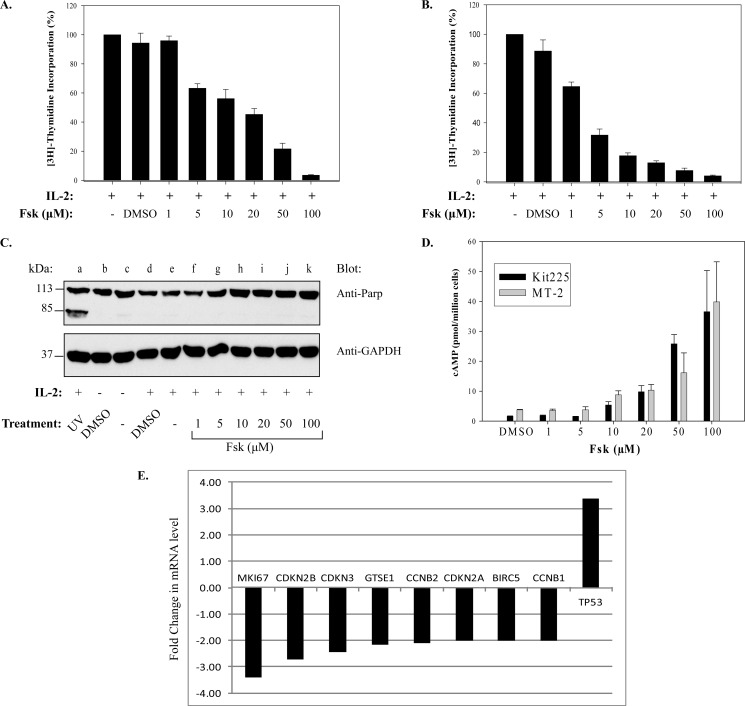
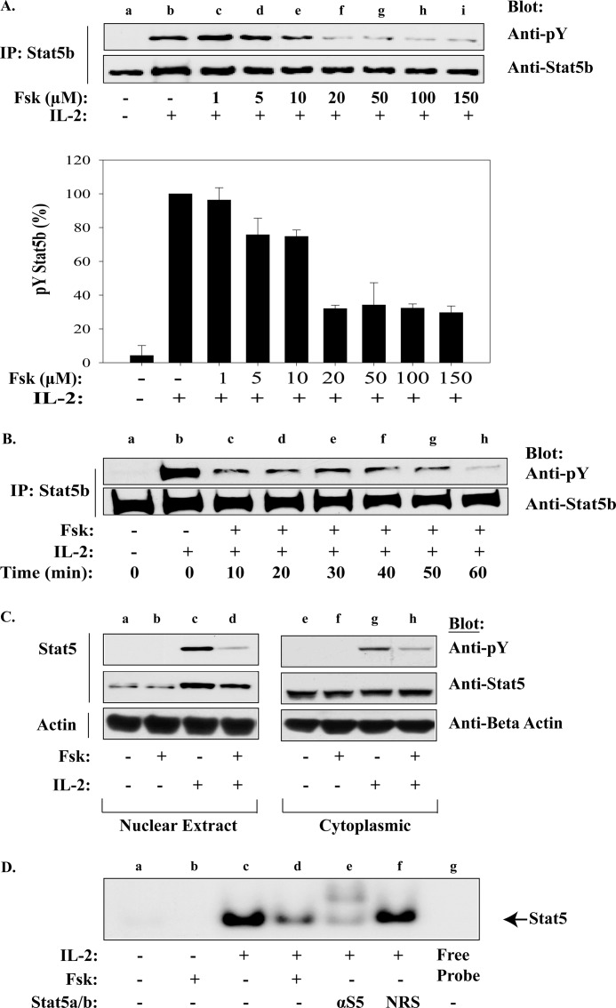
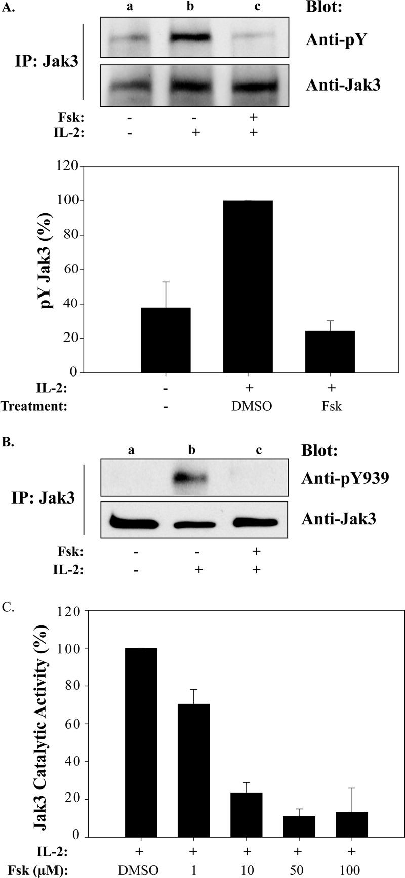
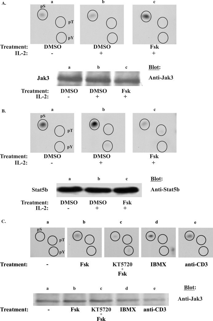
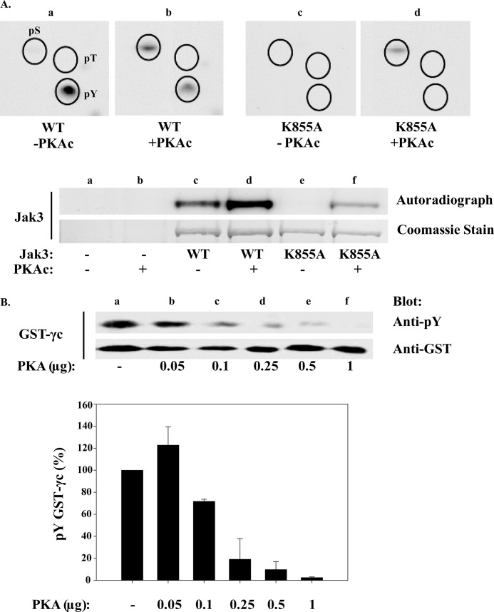
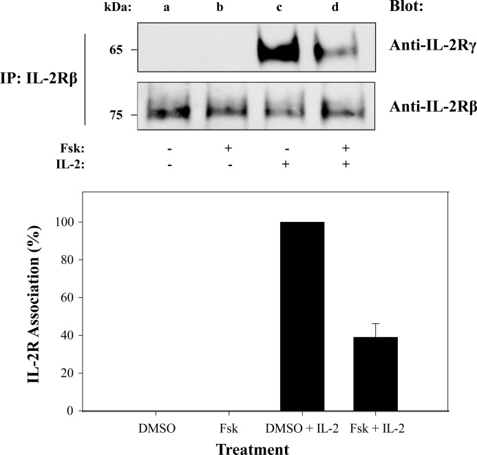
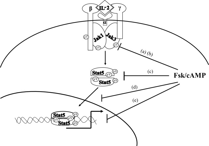
Cytokine-mediated regulation of T-cell activity involves a complex interplay between key signal transduction pathways. Determining how these signaling pathways cross-talk is essential to understanding T-cell function and dysfunction. In this work, we provide evidence that cross-talk exists between at least two signaling pathways: the Jak3/Stat5 and cAMP-mediated cascades. The adenylate cyclase activator forskolin (Fsk) significantly increased intracellular cAMP levels and reduced proliferation of the human T-cells via inhibition of cell cycle regulatory genes but did not induce apoptosis. To determine this inhibitory mechanism, effects of Fsk on IL-2 signaling was investigated. Fsk treatment of MT-2 and Kit 225 T-cells inhibited IL-2-induced Stat5a/b tyrosine and serine phosphorylation, nuclear translocation, and DNA binding activity. Fsk treatment also uncoupled IL-2 induced association of the IL-2Rβ and γc chain, consequently blocking Jak3 activation. Interestingly, phosphoamino acid analysis revealed that Fsk-treated cells resulted in elevated serine phosphorylation of Jak3 but not Stat5, suggesting that Fsk can negatively regulate Jak3 activity possibly mediated through PKA. Indeed, in vitro kinase assays and small molecule inhibition studies indicated that PKA can directly serine phosphorylate and functionally inactivate Jak3. Taken together, these findings suggest that Fsk activation of adenylate cyclase and PKA can negatively regulate IL-2 signaling at multiple levels that include IL-2R complex formation and Jak3/Stat5 activation.


Figures

FIGURE 1.
FIGURE 1.
Fsk-cAMPi inhibits cellular proliferation of human T-cell lines and cell cycle genes. Kit 225 (A) or MT-2 cells (B) were untreated or pretreated with vehicle (1% DMSO) or increasing concentrations of Fsk (1, 5, 10, 25, 50, or 100 μm) prior to stimulation with IL-2 for 20 h. Cells were pulsed with [3H]thymidine for an additional 4 h, and radiolabeled DNA was counted as described under “Experimental Procedures.” Data represent the mean ± S.D. (n = 4). C, MT-2 cells were treated as described above with Fsk (lanes f–k) and IL-2 (lanes d–k). Control cells received either no treatment (lane c), 1% DMSO without IL-2 (lane b) or with IL-2 (lane d). MT-2 cells treated with IL-2 and then exposed to UV light were used as a positive control for apoptosis (lane a). PARP cleavage and GAPDH were detected by Western blot analysis. A representative blot from three independent experiments is shown. D, Kit 225 and MT-2 cells were treated with 1% DMSO or increasing concentrations of Fsk (1–100 μm) for 20 min. cAMP concentrations were determined in triplicates by ELISA. Data represent the mean ± S.D. Each experiment was repeated three times, and representative data from one experiment are shown. E, Kit 225 cells were treated with vehicle (1% DMSO) or 100 μm Fsk for 1 h prior to stimulation with IL-2 for 30 h at 37 °C. cDNA obtained from treated cells was amplified using a human cell cycle PCR array. Quantification based on real-time monitoring of gene amplification was determined. Treatments were performed in duplicate, and data were analyzed using the ΔΔCt method normalized to an average of 4 housekeeping genes (B2M, RPL13A, GAPDH, and ACTB). Genes with Ct > 32 were excluded in the analysis.
FIGURE 2.
FIGURE 2.
Fsk-cAMPi inhibits Stat5b activation, nuclear translocation, and DNA binding activity. A, MT-2 cells were untreated (lane a) or pretreated with the indicated concentrations of Fsk for 40 min prior to stimulation with IL-2 for 10 min at 37 °C (lanes b–i). Immunoprecipitated Stat5b was Western blotted with anti-phosphotyrosine (pY; upper panel) or anti-Stat5b (lower panel). Representative data from three independent experiments are shown. Densitometric analysis of phosphotyrosine Stat5b was normalized to total Stat5b and graphed (percentage of phosphotyrosine Stat5b) (lane b). Data represent the mean ± S.D. from three independent experiments. B, MT-2 cells were treated as above with vehicle (lanes a and b) or pretreated with 100 μm Fsk for the times indicated before IL-2 stimulation for 10 min at 37 °C (lanes c–h). Immunoprecipitated Stat5b was Western blotted with anti-phosphotyrosine (upper panel) or anti-Stat5b (lower panel). C, MT-2 cells were either untreated or pretreated with 100 μm Fsk for 40 min prior to IL-2 stimulation. Immunoprecipitated nuclear (lanes a–d) and cytoplasmic (lanes e–h) localized Stat5b proteins were separated by SDS-PAGE and Western blotted using antibodies for phosphotyrosine, Stat5, or β-actin (internal standard). D, EMSA analysis was performed on MT-2 cells treated as in C. Nuclear proteins were isolated and incubated with [γ-32P]ATP-labeled β-casein promoter alone (lanes a–d), with anti-Stat5 antibody (lane e) or normal rabbit serum (NRS; lane f) or free probe without cell lysate (lane g). Stat5 migration is indicated by the arrow.
FIGURE 3.
FIGURE 3.
Fsk-cAMPi inhibits IL-2-induced Jak3 activation and catalytic activity. A, MT-2 cells were left untreated (lane a) or treated with vehicle (1% DMSO) (lane b) or 100 μm Fsk for 40 min (lane c) prior to stimulation with 100 nm IL-2 for 10 min (lanes b and c) at 37 °C. Immunoprecipitated Jak3 protein was Western blotted using anti-phosphotyrosine (pY) antibody (upper panel) or total Jak3 (lower panel). Densitometric analyses were carried out (n = 3), and tyrosine phosphorylation levels of Jak3 were normalized to total Jak3 protein and plotted (lower panel). B, cells were treated as described in A and Western blotted with anti-phosphotyrosine 939 (pY939). C, MT-2 cells were treated with increasing concentrations of Fsk for 40 min prior to stimulation with 100 nm IL-2 for 10 min. Immunocaptured Jak3 was used for in vitro kinase assays with GST-γc as a substrate and Western blotted with anti-phosphotyrosine or anti-GST antibodies. Densitometric analysis was performed, and tyrosine phosphorylation of GST-γc was normalized to total GST expression. The results are presented as percentage of IL-2 activated Jak3 activity in the absence or presence of Fsk.
FIGURE 4.
FIGURE 4.
Fsk and IBMX induce Jak3 serine phosphorylation but blocks IL-2 inducible Stat5b serine phosphorylation. A, MT-2 cells were metabolically labeled with [32P]orthophosphate and treated with 1% DMSO (panel a), IL-2 for 10 min (panel b), or 100 μm Fsk followed by 10 min of IL-2 stimulation (panel c). Jak3 or Stat5b (B) were immunoprecipitated and Western blotted for protein. Bands corresponding to Jak3 or Stat5b were excised and subjected to phosphoamino acid analysis (n = 3 and n = 2, respectively) to determine the global phosphorylation status of serine (pS), threonine (pT), and tyrosine (pT) residues within these residues. Comparable amounts of Jak3 (A, lower panel) and Stat5b (B, lower panel) were determined using Western blot analysis to ensure equal protein loading. C, Kit 225 cells were similarly labeled and treated with 1% DMSO (panel a), 100 μm Fsk for 1 h (panel b), Fsk and 10 μm KT5720 (KT) (panel c), 1 mm IBMX for 15 min (panel d), or 5 μg of anti-CD3 for 10 min (panel e). Phosphoamino acid analysis was assessed for Jak3 phosphorylation status (upper panel, n = 2). The lower panel indicates the separation of Jak3 immunoprecipitates prior to phosphoamino acid analysis. The immunoprecipitates were electrophorized and autoradiographed (upper bands) or Western blotted using anti-Jak3 antibody (lower bands).
FIGURE 5.
FIGURE 5.
PKA directly phosphorylates Jak3 serine residues and disrupts its kinase activity. A, Hek 293 cells were transfected with wild type (WT) or catalytically inactive (K855A) Jak3. Cells were then lysed, and Jak3 immunoprecipitates used for a [γ-32P]ATP in vitro kinase assay in the presence or absence of purified PKAc and subjected to phosphoamino acid analysis (upper panels a–d). The lower panel indicates the Coomassie Blue stain and autoradiograph of reactions separated by SDS-PAGE; Hek293 cells carrying an empty vector unexposed (lane a) or exposed to PKAc (lane b), WT Jak3 in the absence (lane c) and presence (lane d) of PKAc, and kinase dead K855A Jak3 with (lane e) or without (lane f) PKAc. B, Jak3 was immunopurified from MT-2 cells and subsequently incubated without (lane a) or with increasing amounts of purified PKAc for 30 min (lanes b–f). Jak3-bound beads were then washed free of PKAc and used in a second in vitro kinase assay with GST-γc substrate. The upper panel shows Western blots of the latter in vitro kinase reaction using anti-phosphotyrosine antibody to assess γc tyrosine phosphorylation and reblotted for total GST (lower panel). Each error bar represents the mean ± S.D. of three independent experiments. γc tyrosine phosphorylation normalized to GST is plotted below.
FIGURE 6.
FIGURE 6.
cAMP disrupts IL-2 receptor β and γ chain association. MT-2 cells were treated with 1% DMSO (lanes a and c) or 100 μm Fsk for 1 h (lanes b and d) followed by IL-2 stimulation for 15 min (lanes c and d). Cells were lysed, immunoprecipitated for IL-2Rβ, separated by SDS-PAGE, and Western blotted using antibodies against γc or IL-2Rβ (upper panel). IL-2Rγ band intensities were normalized to IL-2Rβ levels using densitometric analysis and presented as percentage of IL-2R association (lower panel). Representative data from three independent experiments are shown.
FIGURE 7.
FIGURE 7.
Model for the putative mechanism by which Fsk-cAMPi negatively regulates IL-2R signaling in human T-cells. Multiple targets for Fsk-cAMPi regulation are shown. Fsk prevents the association of IL-2Rγ and β chain (a) as well as Jak3 activation and kinase activity (b). Stat5 activation (c), nuclear translocation (d), and DNA binding (e) are prevented, resulting in severe reduction in IL-2R signaling and T lymphocyte proliferation.

Similar articles

Cited by

References

    1. Leonard W. J. (1996) STATs and cytokine specificity. Nat. Med. 2, 968–969 - PubMed
    1. Weaver A. M., Silva C. M. (2006) Modulation of signal transducer and activator of transcription 5b activity in breast cancer cells by mutation of tyrosines within the transactivation domain. Mol. Endocrinol. 20, 2392–2405 - PubMed
    1. Lin J. X., Mietz J., Modi W. S., John S., Leonard W. J. (1996) Cloning of human Stat5B. Reconstitution of interleukin-2-induced Stat5A and Stat5B DNA binding activity in COS-7 cells. J. Biol. Chem. 271, 10738–10744 - PubMed
    1. Lin J. X., Leonard W. J. (2000) The role of Stat5a and Stat5b in signaling by IL-2 family cytokines. Oncogene 19, 2566–2576 - PubMed
    1. Park S. H., Yamashita H., Rui H., Waxman D. J. (2001) Serine phosphorylation of GH-activated signal transducer and activator of transcription 5a (STAT5a) and STAT5b: impact on STAT5 transcriptional activity. Mol. Endocrinol. 15, 2157–2171 - PubMed

Publication types

MeSH terms