. 2013 Mar 8;288(10):6849-63.
doi: 10.1074/jbc.M112.405530. Epub 2013 Jan 22.

Streptococcus pneumoniae endopeptidase O (PepO) is a multifunctional plasminogen- and fibronectin-binding protein, facilitating evasion of innate immunity and invasion of host cells

Affiliations

Streptococcus pneumoniae endopeptidase O (PepO) is a multifunctional plasminogen- and fibronectin-binding protein, facilitating evasion of innate immunity and invasion of host cells

Vaibhav Agarwal et al. J Biol Chem. .

Abstract

Streptococcus pneumoniae infections remain a major cause of morbidity and mortality worldwide. Therefore a detailed understanding and characterization of the mechanism of host cell colonization and dissemination is critical to gain control over this versatile pathogen. Here we identified a novel 72-kDa pneumococcal protein endopeptidase O (PepO), as a plasminogen- and fibronectin-binding protein. Using a collection of clinical isolates, representing different serotypes, we found PepO to be ubiquitously present both at the gene and protein level. In addition, PepO protein was secreted in a growth phase-dependent manner to the culture supernatants of the pneumococcal isolates. Recombinant PepO bound human plasminogen and fibronectin in a dose-dependent manner and plasminogen did not compete with fibronectin for binding PepO. PepO bound plasminogen via lysine residues and the interaction was influenced by ionic strength. Moreover, upon activation of PepO-bound plasminogen by urokinase-type plasminogen activator, generated plasmin cleaved complement protein C3b thus assisting in complement control. Furthermore, direct binding assays demonstrated the interaction of PepO with epithelial and endothelial cells that in turn blocked pneumococcal adherence. Moreover, a pepO-mutant strain showed impaired adherence to and invasion of host cells compared with their isogenic wild-type strains. Taken together, the results demonstrated that PepO is a ubiquitously expressed plasminogen- and fibronectin-binding protein, which plays role in pneumococcal invasion of host cells and aids in immune evasion.


Figures
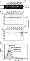
FIGURE 1.
FIGURE 1.
Distribution of pepO gene and its expression in various pneumococcal isolates. A, agarose gel electrophoresis image showing the amplified product of the pepO gene (1893 bp) in clinical isolates. B, immunoblots demonstrating the presence of PepO in the bacterial whole cell lysates of pneumococcal isolates. The molecular weight protein marker was used as a reference. C, representative flow cytometry data for the surface presentation of PepO on S. pneumoniae NTC10319 serotype 35A.
FIGURE 2.
FIGURE 2.
Secretion of PepO by pneumococci. A, immunoblot showing the presence of PepO in the culture supernatant of various isolates of S. pneumoniae. Mid-logarithmic phase culture supernatant of pneumococcal isolates were collected and subjected to SDS-PAGE. Presence of PepO protein in the culture supernatant was analyzed using rabbit anti-PepO polyclonal Abs. B and C, growth phase-dependent secretion of PepO by S. pneumoniae strain NCTC10319 serotype 35A. Bacteria were cultured in THY media and the culture supernatant was collected at regular intervals (B) and separated by 10% SDS-PAGE (C). After blotting, the PepO protein was detected using rabbit anti-PepO polyclonal Abs. THY media containing no bacteria was used as control and the molecular weight protein marker was used as a reference.
FIGURE 3.
FIGURE 3.
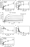
Plasminogen binding to PepO. Microtiter plates were coated with either PepO (5 μg/ml) (A) or plasminogen (5 μg/ml) (B) and increasing amounts of plasminogen or PepO was added. Binding was detected using specific polyclonal Abs. BSA was used as negative control. Statistical significance was calculated using two-way analysis of variance and Bonferroni post test. C, binding of PepO to immobilized plasminogen as analyzed by surface plasmon resonance. Increasing concentrations of PepO (22–695 nm) were injected onto plasminogen-coated CM5 sensor chip. The amount of PepO associating with the plasminogen was measured in response units. Representative sensorgrams are presented. D, microtiter plates were coated with PepO and the effect of different concentrations of NaCl on binding of plasminogen (5 μg/ml) to PepO was analyzed. Amount of plasminogen bound in the absence of NaCl was set at 100%. Specific polyclonal Abs detected bound plasminogen. BSA was used as negative control. One-way analysis of variance and Dunnett's post test were performed to calculate the statistical difference compared with the binding at 150 mm NaCl. E, the inhibitory effect of the lysine analog ϵ-ACA for binding of plasminogen to PepO was evaluated. Specific polyclonal Abs detected bound plasminogen. The signal obtained in the absence of ϵ-ACA was set to 100% and one-way analysis of variance and Dunnett's post test was performed to calculate statistical difference compared with the binding in the absence of ϵ-ACA. F, binding of plasminogen to PepO from human serum was assessed. Microtiter plates were coated with PepO (5 μg/ml) and increasing amounts of serum was added. Bound plasminogen was detected using specific polyclonal Abs. Gelatin was used as negative control. Statistical significance was calculated using two-way analysis of variance test. The data represents the mean ± S.D. of three independent experiments performed in duplicates. ns, not significant; *, p < 0.05; **, p < 0.01; ***, p < 0.001.
FIGURE 4.
FIGURE 4.
PepO binds fibronectin. Microtiter plates were coated with either PepO (5 μg/ml) (A) or fibronectin (5 μg/ml) (B) and increasing amounts of fibronectin or PepO were added. Binding was detected using specific polyclonal Abs. BSA was used as negative control. Statistical significance was calculated using two-way analysis of variance and Bonferroni post test. The data represents the mean ± S.D. of three independent experiments performed in duplicates. ns, not significant; **, p < 0.01; ***, p < 0.001. C, binding of PepO to immobilized fibronectin as analyzed by surface plasmon resonance. Increasing concentrations of PepO (22–695 nm) were injected onto fibronectin-coated CM5 sensor chip. The amount of PepO associating with the fibronectin was measured in response units. Representative sensorgrams are presented.
FIGURE 5.
FIGURE 5.
Binding of plasminogen and fibronectin to PepO. PepO was immobilized on microtiter plates. A constant amount of plasminogen (5 μg/ml) together with increasing amounts of fibronectin (A) or constant amount of fibronectin (5 μg/ml) with increasing amounts of plasminogen (B) was added. Bound fibronectin and plasminogen were detected using specific Abs. Data presented are from three independent duplicate experiments ± S.D. One-way analysis of variance and Dunnett's post test was used to calculate the statistical significance between the binding in the absence and the presence of proteins. *, p < 0.05; ***, p < 0.001.
FIGURE 6.
FIGURE 6.
Plasminogen bound to PepO is functionally active. A, PepO (5 μg/ml) was immobilized on a microtiter plate. After blocking it was incubated with plasminogen (Plg, 1 μg/well) in the absence or presence of the activator uPA together with the chromogenic substrate S-2251. Measuring the absorbance at 405 nm assessed conversion of the substrate by the generated plasmin. The mean values of three independent experiments and the S.D. values are indicated. B, degradation of the natural substrate fibrinogen (Fbg) by plasmin(ogen) bound to immobilized PepO. Plasminogen (5 μg/well) was bound to immobilized PepO (5 μg/well) and after washing, uPA (10 units/well) together with fibrinogen (5 μg/well) were added. At the indicated time points, sample aliquots were removed, separated by SDS-PAGE, transferred to a membrane, and degradation of fibrinogen was assayed by Western blotting using a rabbit fibrinogen antiserum and a peroxidase-conjugated secondary Abs. Fibrinogen incubated in the absence or presence of uPA alone were used as control. Representative data from three independent experiments are shown. PepO-bound plasminogen cleaves complement protein C3b (C), ECM components fibronectin (Fbn) (D), or laminin (E). Microtiter plates coated with PepO (5 μg/well) were incubated with plasminogen (5 μg/well) followed by addition of uPA (10 units/well) and 125I-C3b, fibronectin, or laminin (100 kcpm) and incubation at 37 °C. Samples were taken at the indicated time points for C3b degradation and after an 18-h incubation for fibronectin and laminin and then separated by SDS-PAGE. For C3b degradation, the positive control (+ve Ctrl) contained Factor H mixed with Factor I, 125I-labeled C3b, whereas in the negative control (−ve Ctrl) Factor I was omitted. An arrow marks the cleavage products of C3b. Representative data from three independent experiments are shown.
FIGURE 7.
FIGURE 7.
Effect of PepO deficiency on pneumococcal binding to fibronectin and plasminogen. Immunoblot analysis of PepO production in the wild-type (WT) strains D39 and NCTC10319 or their respective isogenic pepO-mutants (ΔpepO) using whole bacterial lysate (A) or culture supernatant (B). The presence of PepO was detected using rabbit anti-PepO polyclonal Abs. Pneumococcal phosphoglycerate kinase (PGK) was used as a loading control. C, binding of pneumococci to immobilized fibronectin. Microtiter plates were coated with fibronectin (5 μg/ml) and binding of FITC-labeled S. pneumoniae D39, NCTC10319 wild-type, or their respective isogenic pepO-mutants was assessed. Binding was measured at 485/535 (excitation/emission). The data represents the mean ± S.D. of three independent experiments performed in duplicates. Statistical significance was calculated using two-way analysis of variance and Bonferroni post-test. ***, p < 0.001. D, binding of plasminogen to pneumococci. Microtiter plates were coated with 50 μl of 108 cfu/ml of S. pneumoniae D39, NCTC10319 wild-type, or their respective isogenic pepO-mutants strains and binding of plasminogen (0, 2.5, 5 and 10 μg/ml) was assessed. Bound plasminogen was detected using specific polyclonal Abs. The data represent the mean ± S.D. of three independent experiments performed in duplicates. Statistical significance was calculated using one-way analysis of variance and Tukey post test. *, p < 0.05; ***, p < 0.001.
FIGURE 8.
FIGURE 8.
PepO mediates pneumococcal adherence and invasion of host epithelial cells. A and B, binding of recombinant PepO to A549 epithelial cells. Host cells were incubated with increasing amounts of recombinant PepO, after washing the cells, binding was detected with rabbit anti-PepO Abs followed by Alexa 488-conjugated secondary Abs. Binding of protein was quantified by flow cytometry (A) or detected by immunofluorescence microscopy (B). Representative flow cytometry data from three independent experiments are shown. Bar represents 10 μm. C, adherence of pneumococci NCTC10319 and D39 and their respective isogenic pepO-mutants was determined by counting the cfu per well obtained from sample aliquot plated onto the blood agar plate after 3 h of infection. D, invasion and intracellular survival of pneumococci were determined by the antibiotic protection assay. E, recombinant PepO protein inhibits pneumococcal adherence to A549 cells. Epithelial cells were incubated with recombinant PepO for 30 min prior to infections. The total number of bacteria associated with host cells was determined after removing unbound extracellular bacteria and plating the cells on blood agar plates. The data represents the mean ± S.D. of three independent experiments performed in duplicates. Statistical significance was calculated using Student's t test. ns, not significant; **, p < 0.01; ***, p < 0.001.
FIGURE 9.
FIGURE 9.
PepO mediates pneumococcal adherence and invasion of host endothelial cells. A, binding of recombinant PepO to endothelial cells (HUVEC). HUVEC were incubated with increasing amounts of recombinant PepO and after washing, binding was detected with rabbit anti-PepO Abs followed by Alexa 488-conjugated secondary Abs. Representative flow cytometry data from two independent experiments are shown. B, adherence of D39 and its isogenic pepO-mutants was determined by counting the cfu per well obtained from the sample aliquot plated onto the blood agar plate after 3 h of infection. The effect of recombinant PepO protein on wild-type D39 adherence to HUVEC was determined after incubation of cells with PepO (50 μg/ml) for 30 min prior to infections. The total number of bacteria associated with host cells was determined after removing unbound extracellular bacteria and plating the cells on blood agar plates. C, invasion and intracellular survival of wild-type and the PepO-deficient D39 strain were determined by the antibiotic protection assay. D, S. pneumoniae D39 and its isogenic pepO-mutants strains (10 μl, 1000 cfu) were added to refludan-treated whole blood (250 μl) and then gently mixed for 3 h at 37 °C. The bacterial survival was determined by plating onto blood agar plates and determining the number of cfu obtained. The data represents the mean ± S.D. of three independent experiments performed in duplicates. Statistical significance was calculated using Student's t test. ns, not significant; *, p < 0.05; ***, p < 0.001.

References

    1. Cartwright K. (2002) Pneumococcal disease in Western Europe. Burden of disease, antibiotic resistance and management. Eur. J. Pediatr. 161, 188–195 - PubMed
    1. File T. M. (2003) Community-acquired pneumonia. Lancet 362, 1991–2001 - PMC - PubMed
    1. O'Brien K. L., Wolfson L. J., Watt J. P., Henkle E., Deloria-Knoll M., McCall N., Lee E., Mulholland K., Levine O. S., Cherian T. (2009) Burden of disease caused by Streptococcus pneumoniae in children younger than 5 years. Global estimates. Lancet 374, 893–902 - PubMed
    1. Bergmann S., Hammerschmidt S. (2006) Versatility of pneumococcal surface proteins. Microbiology 152, 295–303 - PubMed
    1. Kadioglu A., Weiser J. N., Paton J. C., Andrew P. W. (2008) The role of Streptococcus pneumoniae virulence factors in host respiratory colonization and disease. Nat. Rev. Microbiol. 6, 288–301 - PubMed

Publication types

MeSH terms

LinkOut - more resources